Currently at ]Physics and Materials Science Research Unit, University of Luxembourg, L-1511 Luxembourg, Grand Duchy of Luxembourg.
Supraferromagnetic correlations in clusters of magnetic nanoflowers
Abstract
Magnetic nanoflowers are densely packed aggregates of superferromagnetically coupled iron oxide nanocrystallites, which excel during magnetic hyperthermia experiments. Here, we investigate the nature of the moment coupling within a powder of such nanoflowers using spin-resolved small-angle neutron scattering. Within the powder the nanoparticles are agglomerated to clusters, and we can show that the moments of neighboring nanoflowers tend to align parallel to each other. Thus, the whole system resembles a hierarchical magnetic nanostructure consisting of three distinct levels, i.e. (i) the ferrimagnetic nanocrystallites as building blocks, (ii) the superferromagnetic nanoflowers, and (iii) the supraferromagnetic clusters of nanoflowers. We surmise that such a supraferromagnetic coupling explains the enhanced magnetic hyperthermia performance in case of interacting nanoflowers.
The working principle of magnetic hyperthermia (MHT) is to administer a moderate quantity of magnetic nanoparticles within tumors and to heat them up by applying alternating magnetic fields with clinically acceptable parameters (i.e. comparatively high frequencies but low amplitudes Pankhurst et al. (2009); Southern and Pankhurst (2018)) to kill the tumors. Additionally, a magneto-mechanical actuation of the embedded particles may disrupt the cytoskeleton and lead to cell death. Zhang et al. (2014); Master et al. (2016) In physiological environment nanoparticles usually agglomerate, which can significantly modify their magnetic properties compared to the dilute non-interacting case Eberbeck et al. (2006); Gutierrez et al. (2019) and which in turn may alter their heating behavior. Di Corato et al. (2014); Périgo et al. (2015); Sanz et al. (2016); Déjardin et al. (2017) Depending on the characteristics of the individual particles and the field parameters, such a clustering can either improve or impair the MHT performance. Mehdaoui et al. (2013); Sadat et al. (2014); Blanco-Andujar et al. (2015); Andreu et al. (2015); Coral et al. (2016) In fact it was observed for so-called nanoflowers, which are densely packed aggregates of iron oxide crystallites, that they excel during MHT experiments compared to the single-crystals Lartigue et al. (2012) and other systems such as magnetosomes. Dutz and Hergt (2014) This intriguing result motivated numerous studies regarding synthesis and characterization of such flower-shaped particles. Kostopoulou et al. (2014); Sakellari et al. (2016); Gavilán et al. (2017a, b); Wetegrove et al. (2019); Shaw et al. (2019) It can be shown that an exchange coupling between the cores leads to a superferromagnetic magnetization state Alonso et al. (2010) within the individual nanoflowers, Dutz (2016) but with a significant internal spin disorder caused by the high defect density, e.g. due to the grain boundaries. Döbrich et al. (2009) It is speculated that such a disordered state enables an increased excitation of the moments Bender et al. (2018a, b), similar to other defect-rich particles. Lak et al. (2018) When introduced into tumors, it is safe to assume that the nanoflowers will agglomerate to clusters, and thus interparticle interactions will be relevant. Kuznetsov (2019) In Bender et al. (2018c) we could show for homogeneous superparamagnetic nanoparticles a predominance for antiferromagnetic-like moment correlations within particle clusters via polarized small-angle neutron scattering (SANS). In this work we use the same approach to determine the nature of the moment coupling within a powder of iron oxide nanoflowers.
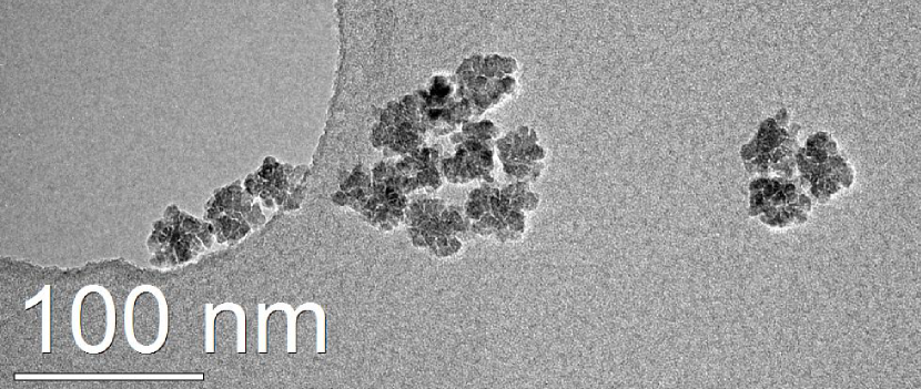
The synomag-D nanoflowers were supplied by micromod Partikeltechnologie GmbH, Germany, which consist predominately of , and are coated with dextran. A detailed study of these particles can be found in Bender et al. (2018a) which showed that they are around 39 nm in size and consist of crystallites with sizes ranging from nm. Transmission electron microscopy (TEM) images were taken with a FEI Titan 80-300 TEM, for which the sample was prepared by putting a small droplet of the dilute dispersion of the particles on a carbon-coated copper grid. Figure 1 shows a typical TEM image of the nanoflowers, in which they are agglomerated to small clusters of 3 and 9 particles, respectively. As can be seen, the nanoparticles are irregular in shape, and around 30-40 nm in size.
The polarized SANS experiment of the nanoflower powder Bender et al. (2017a) was performed with the instrument D33 at the Institut Laue-Langevin (ILL), Grenoble (France) Dewhurst et al. (2016) at room-temperature using a mean wavelength of nm (%) and a detector distance of 10.3 m. We employed longitudinal neutron-spin analysis (POLARIS) to collect the four spin-resolved intensities , , and , where denotes the polarization state spin-up. This approach enables the separation of nuclear and magnetic scattering contributions, and was applied in several studies to investigate magnetic nanoparticle ensembles. Krycka et al. (2010); Bender et al. (2018c); Orue et al. (2018) A homogeneous magnetic field H was applied perpendicular to the neutron beam () with a field amplitude of mT, which was necessary to maintain the neutron beam polarization.
Figs. 2(a) and (b) display the 2D scattering patterns of the non-spin flip (nsf) cross section and of the spin-flip (sf) cross section , respectively. For the geometry the nsf cross sections , can be written as:
| (1) |
where is the angle between the scattering vector and the magnetic field and , with being the Bohr magneton. Hence, in Figs. 2(a) and (b) the field was applied along . Moreover, and are the Fourier transforms of the nuclear scattering length density and of the magnetization vector field in the -, - and -directions, respectively, and the index ∗ denotes the complex conjugate. One remarkable advantage of POLARIS, is that the purely nuclear scattering can be accessed without further assuming a saturated magnetic system (absence of misaligned moments). To be precise, the purely nuclear cross section can be determined, in case of isotropic structures, from the sector parallel to of the nsf intensities.
The sf intensities, on the other hand, are of purely magnetic origin. We assume for our sample that chiral scattering terms can be neglected Mühlbauer et al. (2019), and thus we can write , with (for ): Honecker et al. (2010)
| (2) |
The nsf intensity in Fig. 2(a) exhibits basically no anisotropy, indicating the dominance of the isotropic nuclear scattering, and thus verifying a randomly oriented microstructure. The purely nuclear 1D cross section is determined from the sector parallel to magnetic field of and is plotted in Fig. 2(c). As can be seen, exhibits a peak at around . For particle ensembles the total nuclear cross section is usually written as , where is the particle form factor and the structure factor arising from the particle arrangement. Pedersen (1997) For comparison, in Fig. 2(c) we also plot the purely nuclear scattering cross section of the same nanoflowers in dilute colloidal dispersion from Bender et al. (2018a). In this case there is no significant structure formation and thus . The observed peak for of the powder can be thus attributed to inter-particle correlations, and which implies an average center-to-center distance between the nanoflowers of (nearest neighbor correlations) Li, Senesi, and Lee (2016); Alba Venero et al. (2016). This estimation is in good agreement with our previous analysis in Bender et al. (2018a), where we determined an average particle size of around 39 nm. For (i.e. the interparticle length scale) the forward scattering intensity increases, which indicates the presence of larger structures within the samples. Li, Senesi, and Lee (2016) Thus we can conclude that no long-range order exists but that the nanoflowers within the powder are agglomerated to large clusters with average sizes outside the minimal -resolution, i.e. average cluster sizes of .
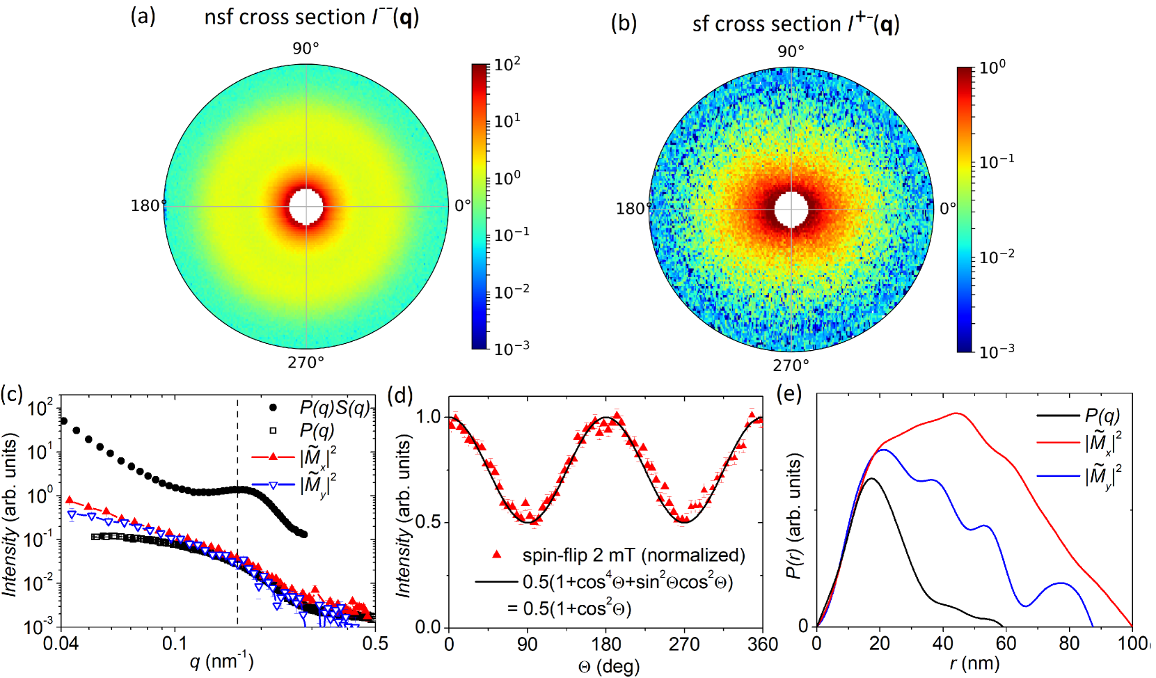
The sf intensity in Fig. 2(b) exhibits a well-pronounced anisotropy, and in Fig. 2(d) we plot integrated over the whole -range as a function of . The functional form is well described by the trigonometric terms from Eq. Supraferromagnetic correlations in clusters of magnetic nanoflowers without the linear term, which implies equal magnetization along the -, - and -direction and a zero net magnetization. This is expected because the sample was in the demagnetized state (i.e., the powder was not exposed to a magnetic field prior to the polarized SANS experiment) and 2 mT is not sufficient to significantly align the moments (as a reminder, the low magnetic field had to be applied to remain the polarization of the neutron beam). In Fig. 2(c) we plot determined perpendicular to the field direction, i.e. , and the difference between and , i.e. . Both cross sections are in the high -range (i.e. the intraparticle -range) basically identical to each other and to the nuclear particle form factor . This confirms the superferromagnetic magnetization state within the individual nanoflowers. In the interparticle -range (), however, both and start to deviate from and increase strongly with decreasing . Additionally it can be observed in Fig. 2(c) that in the low -range, significantly deviates from . This can be attributed to the anisotropy of the magnetic structure factor and indicates a disordered microstructure without a short range pseudo-crystalline order. Honecker, Barquín, and Bender (2019) The deviation of both magnetic contributions and from is an evidence for interparticle moment correlations between neighboring nanoflowers. To reveal the nature of these interactions we extracted the underlying magnetic correlation functions from the scattering intensities by indirect Fourier transforms. Bender et al. (2017b) As can be seen in Fig. 2(e), for the two magnetic contributions and we obtain positive values for for length scales well above the nanoflower size (), which indicates positive correlations between the moments of neighboring nanoflowers. This can be interpreted as evidence for a supraferromagnetic magnetization state within the clusters of these superferromagnetic nanoflowers.
We performed a spin-resolved SANS study on a powder of iron oxide nanoflowers, which enables the separation of nuclear and magnetic scattering contributions. Analysis of the nuclear SANS data shows that the nanoflowers are agglomerated to large clusters. The magnetic scattering contributions then indicate that the moments between neighboring particles are preferentially aligned parallel to each other. We interpret this as evidence for a supraferromagnetic magnetization state within the clusters of nanoflowers. Considering that the nanoflowers itself are aggregates of superferromagnetically coupled crystallites, the whole system can be thus regarded as a hierarchical magnetic nanostructure consisting of three distinct levels, i.e. (i) the ferrimagnetic nanocrystallites as building blocks, (ii) the superferromagnetic nanoflowers, and (iii) the supraferromagnetic clusters of nanoflowers. It can be assumed that such supraferromagnetic correlations increase the low-field susceptibility of the ensemble and thus its MHT performance compared to the dilute, non-interacting ensemble. Indeed, we surmise that our observation explains the intriguing result in Sakellari et al. (2016) where for colloidal dispersions of 50-nm nanoflowers an increased heating with increasing particle concentration was detected, which is in contrast to other nanoparticle ensembles for which usually increasing interactions result in a decrease of the MHT performance. Urtizberea et al. (2010); Serantes et al. (2010) Considering that in physiological environments usually a clustering of immersed nanoparticles occurs it is a promising result for such nanoflowers that their exceptional heating behavior can be even further enhanced by cluster formation. For further studies we propose a systematic investigation of the relations between cluster size and MHT performance for embedded nanoflowers, ideally accompanied by polarized SANS studies to probe the interparticle moment correlations.
We want to thank the Institut Laue-Langevin for provision of beamtime at the instrument D33 and David González-Alonso for his help during the experiment. This project has received funding from the European Commission Framework Programme 7 under Grant Agreement No. 604448 (NanoMag), the National Research Fund of Luxembourg (CORE SANS4NCC grant) and the Spanish Government (MAT2017-83631-C3-R).
References
- Pankhurst et al. (2009) Q. Pankhurst, N. Thanh, S. Jones, and J. Dobson, “Progress in applications of magnetic nanoparticles in biomedicine,” J. Phys. D: Appl. Phys. 42, 224001 (2009).
- Southern and Pankhurst (2018) P. Southern and Q. A. Pankhurst, “Commentary on the clinical and preclinical dosage limits of interstitially administered magnetic fluids for therapeutic hyperthermia based on current practice and efficacy models,” Int. J. Hyperth. 34, 671–686 (2018).
- Zhang et al. (2014) E. Zhang, M. F. Kircher, M. Koch, L. Eliasson, S. N. Goldberg, and E. Renström, “Dynamic magnetic fields remote-control apoptosis via nanoparticle rotation,” ACS Nano 8, 3192–3201 (2014).
- Master et al. (2016) A. M. Master, P. N. Williams, N. Pothayee, N. Pothayee, R. Zhang, H. M. Vishwasrao, Y. I. Golovin, J. S. Riffle, M. Sokolsky, and A. V. Kabanov, “Remote actuation of magnetic nanoparticles for cancer cell selective treatment through cytoskeletal disruption,” Sci. Rep. 6, 33560 (2016).
- Eberbeck et al. (2006) D. Eberbeck, F. Wiekhorst, U. Steinhoff, and L. Trahms, “Aggregation behaviour of magnetic nanoparticle suspensions investigated by magnetorelaxometry,” J. Phys.: Condens. Matter 18, S2829 (2006).
- Gutierrez et al. (2019) L. Gutierrez, M. Moros, E. Mazario, S. de Bernardo, J. M. de la Fuente, M. del Puerto Morales, and G. Salas, “Aggregation effects on the magnetic properties of iron oxide colloids,” Nanotechnology 30, 112001 (2019).
- Di Corato et al. (2014) R. Di Corato, A. Espinosa, L. Lartigue, M. Tharaud, S. Chat, T. Pellegrino, C. Ménager, F. Gazeau, and C. Wilhelm, “Magnetic hyperthermia efficiency in the cellular environment for different nanoparticle designs,” Biomaterials 35, 6400–6411 (2014).
- Périgo et al. (2015) E. A. Périgo, G. Hemery, O. Sandre, D. Ortega, E. Garaio, F. Plazaola, and F. J. Teran, “Fundamentals and advances in magnetic hyperthermia,” Appl. Phys. Rev. 2, 041302 (2015).
- Sanz et al. (2016) B. Sanz, M. P. Calatayud, E. De Biasi, E. Lima Jr, M. V. Mansilla, R. D. Zysler, M. R. Ibarra, and G. F. Goya, “In silico before in vivo: how to predict the heating efficiency of magnetic nanoparticles within the intracellular space,” Sci. Rep. 6, 38733 (2016).
- Déjardin et al. (2017) J.-L. Déjardin, F. Vernay, M. Respaud, and H. Kachkachi, “Effect of dipolar interactions and dc magnetic field on the specific absorption rate of an array of magnetic nanoparticles,” J. Appl. Phys. 121, 203903 (2017).
- Mehdaoui et al. (2013) B. Mehdaoui, R. Tan, A. Meffre, J. Carrey, S. Lachaize, B. Chaudret, and M. Respaud, “Increase of magnetic hyperthermia efficiency due to dipolar interactions in low-anisotropy magnetic nanoparticles: Theoretical and experimental results,” Phys. Rev. B 87, 174419 (2013).
- Sadat et al. (2014) M. Sadat, R. Patel, J. Sookoor, S. L. Bud’ko, R. C. Ewing, J. Zhang, H. Xu, Y. Wang, G. M. Pauletti, D. B. Mast, and D. Shi, “Effect of spatial confinement on magnetic hyperthermia via dipolar interactions in nanoparticles for biomedical applications,” Mater. Sci. Eng., C 42, 52–63 (2014).
- Blanco-Andujar et al. (2015) C. Blanco-Andujar, D. Ortega, P. Southern, Q. Pankhurst, and N. Thanh, “High performance multi-core iron oxide nanoparticles for magnetic hyperthermia: microwave synthesis, and the role of core-to-core interactions,” Nanoscale 7, 1768–1775 (2015).
- Andreu et al. (2015) I. Andreu, E. Natividad, L. Solozabal, and O. Roubeau, “Nano-objects for addressing the control of nanoparticle arrangement and performance in magnetic hyperthermia,” ACS Nano 9, 1408–1419 (2015).
- Coral et al. (2016) D. F. Coral, P. Mendoza Zélis, M. Marciello, M. d. P. Morales, A. Craievich, F. H. Sánchez, and M. B. Fernández van Raap, “Effect of nanoclustering and dipolar interactions in heat generation for magnetic hyperthermia,” Langmuir 32, 1201–1213 (2016).
- Lartigue et al. (2012) L. Lartigue, P. Hugounenq, D. Alloyeau, S. P. Clarke, M. Lévy, J.-C. Bacri, R. Bazzi, D. F. Brougham, C. Wilhelm, and F. Gazeau, “Cooperative organization in iron oxide multi-core nanoparticles potentiates their efficiency as heating mediators and mri contrast agents,” ACS Nano 6, 10935–10949 (2012).
- Dutz and Hergt (2014) S. Dutz and R. Hergt, “Magnetic particle hyperthermia—a promising tumour therapy?” Nanotechnology 25, 452001 (2014).
- Kostopoulou et al. (2014) A. Kostopoulou, K. Brintakis, M. Vasilakaki, K. Trohidou, A. Douvalis, A. Lascialfari, L. Manna, and A. Lappas, “Assembly-mediated interplay of dipolar interactions and surface spin disorder in colloidal maghemite nanoclusters,” Nanoscale 6, 3764–3776 (2014).
- Sakellari et al. (2016) D. Sakellari, K. Brintakis, A. Kostopoulou, E. Myrovali, K. Simeonidis, A. Lappas, and M. Angelakeris, “Ferrimagnetic nanocrystal assemblies as versatile magnetic particle hyperthermia mediators,” Mater. Sci. Eng., C 58, 187–193 (2016).
- Gavilán et al. (2017a) H. Gavilán, A. Kowalski, D. Heinke, A. Sugunan, J. Sommertune, M. Varón, L. K. Bogart, O. Posth, L. Zeng, D. González-Alonso, C. Balceris, J. Fock, E. Wetterskog, C. Frandsen, N. Gehrke, C. Grüttner, A. Fornara, F. Ludwig, S. Veintemillas-Verdaguer, C. Johansson, and M. P. Morales, “Colloidal flower-shaped iron oxide nanoparticles: Synthesis strategies and coatings,” Part. Part. Syst. Charact. 34, 1700094 (2017a).
- Gavilán et al. (2017b) H. Gavilán, E. H. Sánchez, M. E. Brollo, L. Asín, K. K. Moerner, C. Frandsen, F. J. Lázaro, C. J. Serna, S. Veintemillas-Verdaguer, M. P. Morales, and L. Gutiérrez, “Formation mechanism of maghemite nanoflowers synthesized by a polyol-mediated process,” ACS Omega 2, 7172–7184 (2017b).
- Wetegrove et al. (2019) M. Wetegrove, K. Witte, W. Bodnar, D.-E. Pfahl, A. Springer, N. Schell, F. Westphal, and E. Burkel, “Formation of maghemite nanostructures in polyol: tuning the particle size via the precursor stoichiometry,” CrystEngComm 21, 1956–1966 (2019).
- Shaw et al. (2019) S. Shaw, A. Biswas, A. Gangwar, P. Maiti, C. Prajapat, S. S. Meena, and N. Prasad, “Synthesis of exchange coupled nanoflowers for efficient magnetic hyperthermia,” J. Magn. Magn. Mater. 484, 437–444 (2019).
- Alonso et al. (2010) J. Alonso, M. Fdez-Gubieda, J. Barandiarán, A. Svalov, L. F. Barquín, D. A. Venero, and I. Orue, “Crossover from superspin glass to superferromagnet in nanostructured thin films (),” Phys. Rev. B 82, 054406 (2010).
- Dutz (2016) S. Dutz, “Are magnetic multicore nanoparticles promising candidates for biomedical applications?” IEEE Trans. Magn. 52, 1–3 (2016).
- Döbrich et al. (2009) F. Döbrich, M. Elmas, A. Ferdinand, J. Markmann, M. Sharp, H. Eckerlebe, J. Kohlbrecher, R. Birringer, and A. Michels, “Grain-boundary-induced spin disorder in nanocrystalline gadolinium,” J. Phys.: Condens. Matter 21, 156003 (2009).
- Bender et al. (2018a) P. Bender, J. Fock, C. Frandsen, M. F. Hansen, C. Balceris, F. Ludwig, O. Posth, E. Wetterskog, L. K. Bogart, P. Southern, W. Szczerba, L. Zeng, K. Witte, C. Grüttner, F. Westphal, D. Honecker, D. González-Alonso, L. Fernández Barquín, and C. Johansson, “Relating magnetic properties and high hyperthermia performance of iron oxide nanoflowers,” J. Phys. Chem. C 122, 3068–3077 (2018a).
- Bender et al. (2018b) P. Bender, J. Fock, M. Hansen, L. Bogart, P. Southern, F. Ludwig, F. Wiekhorst, W. Szczerba, L. Zeng, D. Heinke, N. Gehrke, M. T. Fernández Díaz, D. González-Alonso, J. I. Espeso, J. Rodríguez Fernández, and C. Johansson, “Influence of clustering on the magnetic properties and hyperthermia performance of iron oxide nanoparticles,” Nanotechnology 29, 425705 (2018b).
- Lak et al. (2018) A. Lak, M. Cassani, B. T. Mai, N. Winckelmans, D. Cabrera, E. Sadrollahi, S. Marras, H. Remmer, S. Fiorito, L. Cremades-Jimeno, F. J. Litterst, F. Ludwig, M. Liberato, F. J. Teran, S. Bals, and T. Pellegrino, “ deficiencies, subdomains, and structural defects favor magnetic hyperthermia performance of iron oxide nanocubes into intracellular environment,” Nano Lett. 18, 6856–6866 (2018).
- Kuznetsov (2019) A. A. Kuznetsov, “Zero-field and field-induced interactions between multicore magnetic nanoparticles,” Nanomaterials 9, 718 (2019).
- Bender et al. (2018c) P. Bender, E. Wetterskog, D. Honecker, J. Fock, C. Frandsen, C. Moerland, L. K. Bogart, O. Posth, W. Szczerba, H. Gavilán, R. Costo, M. T. Fernández-Díaz, D. González-Alonso, L. Fernández Barquín, and C. Johansson, “Dipolar-coupled moment correlations in clusters of magnetic nanoparticles,” Phys. Rev. B 98, 224420 (2018c).
- Bender et al. (2017a) P. Bender, L. Fernández Barquín, M.-L. Fdez-Gubieda, D. González-Alonso, D. Honecker, L. Marcano, and W. Szczerba, “Spin correlation in clusters of magnetic nanoparticles.” Institut Laue-Langevin (ILL) (2017a), 10.5291/ILL-DATA.5-53-267.
- Dewhurst et al. (2016) C. D. Dewhurst, I. Grillo, D. Honecker, M. Bonnaud, M. Jacques, C. Amrouni, A. Perillo-Marcone, G. Manzin, and R. Cubitt, “The small-angle neutron scattering instrument D33 at the Institut Laue–Langevin,” J. Appl. Crystallogr. 49, 1–14 (2016).
- Krycka et al. (2010) K. L. Krycka, R. A. Booth, C. R. Hogg, Y. Ijiri, J. A. Borchers, W. Chen, S. M. Watson, M. Laver, T. R. Gentile, L. R. Dedon, S. Harris, J. J. Rhyne, and S. A. Majetich, “Core-shell magnetic morphology of structurally uniform magnetite nanoparticles,” Phys. Rev. Lett. 104, 207203 (2010).
- Orue et al. (2018) I. Orue, L. Marcano, P. Bender, A. García-Prieto, S. Valencia, M. A. Mawass, D. Gil-Cartón, D. Alba Venero, D. Honecker, A. García-Arribas, L. Fernández Barquín, A. Muela, and M. L. Fdez-Gubieda, “Configuration of the magnetosome chain: a natural magnetic nanoarchitecture,” Nanoscale 10, 7407–7419 (2018).
- Mühlbauer et al. (2019) S. Mühlbauer, D. Honecker, É. A. Périgo, F. Bergner, S. Disch, A. Heinemann, S. Erokhin, D. Berkov, C. Leighton, M. R. Eskildsen, and A. Michels, “Magnetic small-angle neutron scattering,” Rev. Mod. Phys. 91, 015004 (2019).
- Honecker et al. (2010) D. Honecker, A. Ferdinand, F. Döbrich, C. D. Dewhurst, A. Wiedenmann, C. Gómez-Polo, K. Suzuki, and A. Michels, “Longitudinal polarization analysis in small-angle neutron scattering,” Eur. Phys. J. B 76, 209–213 (2010).
- Pedersen (1997) J. S. Pedersen, “Analysis of small-angle scattering data from colloids and polymer solutions: modeling and least-squares fitting,” Adv. Colloid Interface Sci. 70, 171 – 210 (1997).
- Li, Senesi, and Lee (2016) T. Li, A. J. Senesi, and B. Lee, “Small angle x-ray scattering for nanoparticle research,” Chem. Rev. 116, 11128–11180 (2016).
- Alba Venero et al. (2016) D. Alba Venero, S. Rogers, S. Langridge, J. Alonso, M. Fdez-Gubieda, A. Svalov, and L. Fernández Barquín, “Magnetic nanoscopic correlations in the crossover between a superspin glass and a superferromagnet,” J. Appl. Phys. 119, 143902 (2016).
- Honecker, Barquín, and Bender (2019) D. Honecker, L. F. Barquín, and P. Bender, “The magnetic structure factor of correlated nanoparticle moments in small-angle neutron scattering,” Preprint at https://arxiv.org/abs/1904.06243 (2019).
- Bender et al. (2017b) P. Bender, L. Bogart, O. Posth, W. Szczerba, S. Rogers, A. Castro, L. Nilsson, L. Zeng, A. Sugunan, J. Sommertune, A. Fornara, D. González-Alonso, L. Fernández Barquín, and C. Johansson, “Structural and magnetic properties of multi-core nanoparticles analysed using a generalised numerical inversion method,” Sci. Rep. 7, 45990 (2017b).
- Urtizberea et al. (2010) A. Urtizberea, E. Natividad, A. Arizaga, M. Castro, and A. Mediano, “Specific absorption rates and magnetic properties of ferrofluids with interaction effects at low concentrations,” J. Phys. Chem. C 114, 4916–4922 (2010).
- Serantes et al. (2010) D. Serantes, D. Baldomir, C. Martinez-Boubeta, K. Simeonidis, M. Angelakeris, E. Natividad, M. Castro, A. Mediano, D.-X. Chen, A. Sanchez, L. Balcells, and B. Martínez, “Influence of dipolar interactions on hyperthermia properties of ferromagnetic particles,” J Appl. Phys. 108, 073918 (2010).