Quantification of the morphological characteristics of hESC colonies
Abstract
The maintenance of the pluripotent state in human embryonic stem cells (hESCs) is critical for further application in regenerative medicine, drug testing and studies of fundamental biology. Currently, the selection of the best quality cells and colonies for propagation is typically performed by eye, in terms of the displayed morphological features, such as prominent/abundant nucleoli and a colony with a tightly packed appearance and a well-defined edge. Using image analysis and computational tools, we precisely quantify these properties using phase-contrast images of hESC colonies of different sizes (0.1 – 1.1) during days 2, 3 and 4 after plating. Our analyses reveal noticeable differences in their structure influenced directly by the colony area . Large colonies () have cells with smaller nuclei and a short intercellular distance when compared with small colonies (). The gaps between the cells, which are present in small and medium sized colonies with , disappear in large colonies () due to the proliferation of the cells in the bulk. This increases the colony density and the number of nearest neighbours.
We also detect the self-organisation of cells in the colonies where newly divided (smallest) cells cluster together in patches, separated from larger cells at the final stages of the cell cycle. This might influence directly cell-to-cell interactions and the community effects within the colonies since the segregation induced by size differences allows the interchange of neighbours as the cells proliferate and the colony grows. Our findings are relevant to efforts to determine the quality of hESC colonies and establish colony characteristics database.
keywords:
Human embryonic stem cells , Cellular characteristics , Feature extraction , Voronoi diagram , Segregation1 Introduction
Human embryonic stem cells (hESCs) are pluripotent cells, derived from the blastocyst-stage embryos, which have the capacity to differentiate and give rise to all tissues of the body. More than 20 years ago, a method to derive stem cells from human embryos was discovered and allowed the use of these cells for further research in vitro (Thomson et al., 1998). They provide an opportunity to study early human development and the processes by which pluripotency is lost and differentiation into different tissues occurs (Pera and Trounson, 2004). The specific signalling factors promoting stem cells to remain unspecialised in culture without differentiation have been highly optimised during the last two decades (Vazin and Freed, 2010). Also, protocols have been developed to differentiate hESCs towards all three germ layers for disease modelling, cell-based therapies and drug screening (Efthymiou et al., 2014). After the derivation of human induced pluripotent stem cells (hiPSCs) (Takahashi et al., 2007), which made creating patient-matched embryonic stem cell lines feasible, hESCs and hiPSCs have become an emerging model for developmental studies and personalised medicine (Soldner and Jaenisch, 2018; Odorico et al., 2004; Zhu and Huangfu, 2013).
Although the genetic and signalling pathways that control pluripotency in hESCs have been described in the last decade (Neganova et al., 2009, 2011; Dalton, 2013; Neganova et al., 2014; Zhao and Jin, 2017; Neganova et al., 2019), much less is known about the factors that control the arrangement of the cells into a pluripotent colony and how this affects pluripotency. Human ESCs grow as a multicellular colony. At the single cell level the transcription factors (TFs) associated with the maintenance of pluripotency fluctuate stochastically (Buganim et al., 2012; Li and Belmonte, 2018). These different expression states are maintained by different signalling, transcriptional, and epigenetic regulatory networks. However, pluripotency, considered as an emergent property of stem cell populations and their niches (rather than a property of single cells), is controlled by niche-mediated regulation in response to mechanical, chemical and physical stimuli (Peerani et al., 2007; Vining and Mooney, 2017). Thus, understanding how pluripotency is affected by cell segregation within the bulk of a colony is of practical importance in generating and selecting the optimum clones, and automating this for industrial-scale production.
The current paradigm in stem cell biology dictates that pluripotency is regulated by a set of TFs, such as NANOG, OCT4 and SOX2, that fluctuate stochastically along the cell cycle and show a heterogeneous expression at the single cell and colony levels (Torres-Padilla and Chambers, 2014; Ståhlberg et al., 2009; Wolff et al., 2018; Messmer et al., 2019). A quantitative analysis of the arrangement of the cells within the colonies is a prerequisite for the construction of hypothesis-driven mathematical and computational models that can provide explanations for the observed dynamics in hESC colonies and their regulation at the microenvironment level.
To characterise how hESCs regulate their assembly into a multicellular colony we performed a detailed quantitative analysis of hESC colonies of different sizes during the exponential growth phase, at days 2, 3 and 4 after plating. These quantitative properties of the colony morphologies are poorly studied, while the pluripotent regulation at the expression level captures most of the attention (Warmflash et al., 2014; Etoc et al., 2016; Nemashkalo et al., 2017; Wolff et al., 2018).
Previous works within our group have demonstrated that isolated hESCs growing on Matrigel™ with mTESR1 media are highly motile (m/h) and sensitive to the presence of nearby cells (Wadkin et al., 2017, 2018). As these single cells proliferate and possibly aggregate with other cells, colonies are formed. Within a colony, the cells collectively move in the bulk, and the whole structure remodels due to cell division and cell spreading. If the conditions for the maintenance of pluripotency are lost, the cells differentiate (Vallier et al., 2009; Neganova et al., 2009).
The collective motion of cells within colonies involves mechanical and biochemical interactions between the cells. The most important physical factors affecting the cell dynamics are the proliferation and short-range interactions with other cells, driven by the pressure flow due to the mitotic expansion. Research on self-renewal and differentiation of hESCs indicates that both processes are highly influenced by biophysical signals, such as the mechanical forces and the rigidity of the extracellular matrix (ECM) (Chen et al., 2015). Toyoda et al. (2015) demonstrated the role of local cell density and colony size, showing an increase in the correlation of differentiation efficiency of both hESC and hiPSC cell lines towards pancreatic cells. This indicates that the local density attained within the cultures should be highly relevant for the maintenance of pluripotency. Moreover, a recent study for human induced pluripotent stem cells (hiPSCs) has shown the migratory behaviours of the cells vary on different substrates (e.g., laminin, fibronectin, matrigel) due to changes in their adhesion properties, concluding that the regulation of the motility of the cells might improve the clonality of the forming colonies (Li et al., 2010; Chang et al., 2019). With the colony growth, the cells regulate each other through cell-cell and cell-media interactions (Peerani et al., 2007; Xu et al., 2010) resulting in community effects that regulate pluripotency and differentiation (Nemashkalo et al., 2017). Therefore, the results obtained at the single-cell level cannot be sufficient to deduce biological processes at the colony level as a whole.
The formation of hESC colonies in vitro is a natural process emerging when a single cell proliferates and forms a small cluster. In this work, we analyse colonies growing in feeder-free conditions (Matrigel™ ). It has been demonstrated that cells within the same colony have a higher correlation of being of the same type, e.g. pluripotent or primed towards differentiation (Nemashkalo et al., 2017), which might be achieved by the combination of endogenous signals between the cells and extrinsic factors (addition of differentiation cues).
The size and morphology of the colonies provide with preliminary information about the pluripotency status of the cells. The undifferentiated state is assessed through the specific morphology of the cells and the colonies, see Table 1, which is typically estimated visually. The morphological features of pluripotent hESCs inside a colony are: roundness, large nucleus, scant cytoplasm and prominent (highly visible) nucleoli. As the colony grows, the central part becomes more compact than the periphery. Human ESCs colonies with areas show white spaces or gaps between the cells (Amit and Itskovitz-Eldor, 2012b, a). To account for these features, we measured the individual cells by manually outlining their nucleus. This gives us the overall nuclei morphology in terms of the colony size and day after plating. Several properties, such as nucleus area and shape descriptors (aspect ratio, Feret’s diameter, circularity, roundness and solidity), defined in the supplementary material (SM)) were measured. Our results show that colonies with show distinctive features in their structural properties, such as a large nucleus cell area and a large separation between nearest neighbours. Both quantities decrease as the colony size increases, with the largest colony showing the smallest value in the mean cell nucleus area. This may be caused by the pressure build-up due to mitotic events in the bulk of the colony. To measure the segregation of the small (recently divided) cells, we introduce a segregation order parameter. Our results suggest the self-organisation of the cells in terms of their nucleus sizes, since the small cells cluster together in patches, separating the larger cells from each other. Recent results by Nemashkalo et al. (2017), using micropatterned colonies of a few cells, indicate that interactions between neighbours can lead to sustained and homogeneous signalling for differentiation. Taking these two results together, a continuous interchange of neighbours between the cells as they grow and divide, might affect their cellular communication and ultimately the pluripotent state attained by the colony as a whole.
| Type | Characteristics |
|---|---|
| Cell | Prominent nucleoli |
| Scant cytoplasm | |
| Round | |
| Small | |
| Colony | Round |
| Flat | |
| Well-defined edges | |
| Gaps between the cells () |

Computational models are helpful to quantitatively analyse and improve the understanding of the processes that underlie fate decisions in hESCs and hiPSCs. However, before establishing the appropiate protocol for in silico approaches, it is important to quantify the morphological features frequently used in the visual identification of pluripotent hESC colonies, see Table 1, in agreement with previous publications (Johkura et al., 2004; Amit and Itskovitz-Eldor, 2012a; Ullmann et al., 2006) . These give us value information about the morphological properties of the cells arranged in colonies. In the future, this information will be integrated alongside other mechanisms that determine the behaviour of the system, to build algorithms of interaction rules aiming to understand their emergent properties (Herberg and Roeder, 2015).
2 Materials and Methods
2.1 Cell culture and propagation
Human embryonic stem cells (hESCs) (H9 cell line, WiCell, Madison, WI) were passaged on 6-well plates coated with hESC-qualified Matrix at a 1:4 split ratio using an EDTA-based dissociation solution. 2 ml of mTERSR1 media was used per wel. The cells were kept in small clumps avoiding the passaging of single cells (due to low rates of survival). We aimed to plate cell aggregates of approximately 15 – 20 cells each. The culture was kept for 4 days at C with a humidified atmosphere. The colonies were imaged at day 2, 3 and 4 after plating before they reached a confluency across the well.
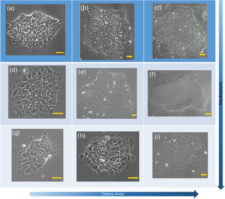
The ability of hESCs cells to form colonies depends on the cytoskeleton rearrangement, contraction of actin filaments, the interaction between the cells, and the timely function of regulatory proteins. When isolated, the cells have their cytoskeleton and lamellipodia unfolded and spreading over the substrate, see Figure 1(a). In colonies, the cells are close to each other as shown in Figure 1(b). This section of a colony contains several cells in which the nuclei, nucleoli (dark spots) and gaps (white spaces between the cells) are easily detected. Larger and denser colonies do not show gaps and the cells are closer to each other, see Figure 2.
We studied the colonies using phase contrast microscopy, since this method allows the cells to behave as naturally as possible without the need to stain the cells with fluorescent dyes which may induce photo-toxicity (Icha et al., 2017) and possible changes in cell behaviour (Lulevich et al., 2009). During the last day of image acquisition, the confluency of the cells was about , meaning that most of the colonies did not merge with each other and were quasi-bidimensional structures.
Examples images of hESC and their colonies are shown in Figure 1. The internal structure of a single isolated cell is shown in Figure 1(a) with a scheme at the bottom-left side outlining the nucleus and nucleoli. Colonies of varying size were selected with morphological features typical of undifferentiated colonies, i.e., with clear borders, containing small round cells with large nuclei and notable nucleoli (Amit and Itskovitz-Eldor, 2012a); an example is shown in Figure 1(b). Figure 1(c) shows a hESC colony with cells showing a different nucleus/cytoplasm ratio.
2.2 Imaging of hESCs

To quantify the morphological characteristics within the colony we outlined each nucleus manually and extracted several parameters such as the centroid position, nucleus area and relevant shape descriptors included in ImageJ (Schneider et al., 2012). During mitosis, the cells adopt a spherical shape, detach from the ECM, divide and reattach again, with the two new daughter cells lying in close proximity to each other. We recorded these mitotic events during the manual tracing of the cell nuclei.
We outlined the nuclei of the cells in colonies of different sizes (see Table 2 in the SM for further details). Alongside this information, the boundaries of 38 colonies were obtained using an edge detection algorithm through a canny Deriche filtering (Schneider et al., 2012), see Table 4 in the SM for more details. An example of the analysis performed on the colonies is shown in Figure 2(a). This sample has an area and it was imaged at day 3 after plating. For large colonies, we imaged the structure at low magnification (5) to account for the colony’s features, and at a higher magnification (10) focusing in the bulk, Figure 2(b), to outline the cell nuclei, Figure 2(c). Using ImageJ (Schneider et al., 2012) software (http:// rsb.info.nih.gov/ij/), we processed the outlined images and obtained the centroid position, area, perimeter and shape descriptors (aspect ratio, solidity, circularity and Feret’s diameter) of each nucleus and, at a larger scale, of each colony.
2.3 Voronoi diagram
The spatial data analyses presented in this work are based on the Voronoi diagram (VD) that divides the area in the most equalitarian fashion, in such a way that the area occupied by a cell is obtained by tracing straight lines between the position of a cell and all its neighbours and drawing a perpendicular line in the middle. These perpendicular lines form a convex polyhedron, called the Voronoi cell. Therefore the VD is the collection of Voronoi cells. The generated "cells" are not uniform in shape and their number of faces vary from one to another.
The geometric dual of the VD is called the Delaunay triangulation (DT). It connects those points of a VD that share a common border. The VD facilitates spatial analysis, e.g., the closest neighbours identification through the adjacency matrix, and is used in many fields of science, including cell biology, (Honda, 1978; Saito, 1982). We used the VD to measure the structural properties of the colonies and the DT to obtain the intercellular distances.
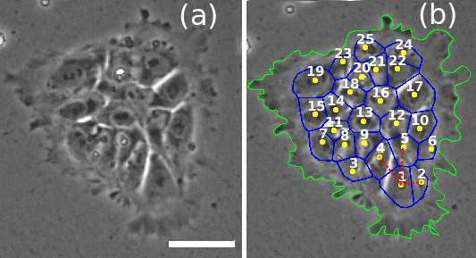
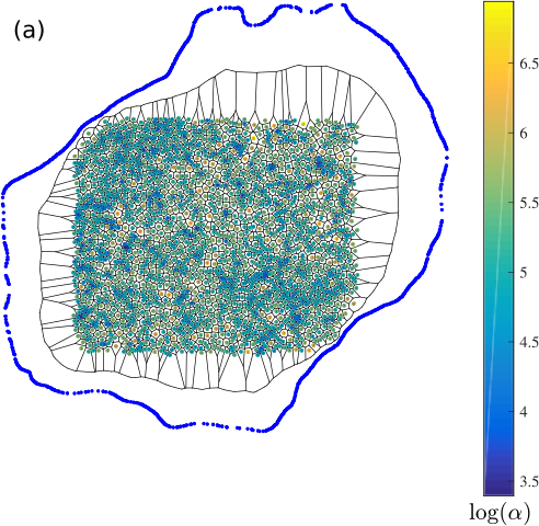
As an example, Figure 3 shows the VD for a small colony with 25 cells. The nearest neighbours of cell 1 are connected with dotted (red) lines (DT), i.e., cells 2, 4 and 5, giving the distance to the nearest neighbours. The cells 3 and 6 are the second nearest neighbours of cell 1. We performed this analysis on larger colonies, see Figure 2(a). An example of the Voronoi tessellation obtained for the region in Figure 2(b) is shown in Figure 2(c) for 1982 cells. The nuclei are coloured according to the logarithm of the nucleus area () to ease in the visualisation (see colorbar). Using the centroids as input points, we obtained the VD, shown with black continuous lines. Since the cells are round in shape (see SM, Figure 10), the VD allows accurate identification of each cell’s neighbourhood. Through the DT, we identified the nearest neighbours associated to each cell and calculated the mean number of nearest neighbours 111From now on, the angular brackets will denote the average taken over the cell population within a given colony. The bar will denote the average taken over several colonies. and the mean distance to nearest neighbours (or intracellular distance) , measured from the centroid position of the two cell pairs.
3 Results
After plating, hESCs form small clusters of several cells attached to each other and to the ECM. These are the initial seeds from which larger colonies start to grow through proliferation. The cells inside the colonies display self-propulsion, resulting in a movement of the colony as a whole through the culture. We assume events in which cells from other colonies travel through the ECM and attach to other colonies are rare for hESCs plated at low densities. Depending on the initial plating density, merging of colonies might occur after some time. In our experiments, the confluency of the colonies was less than 60% on day 4. Due to the variability in single cell movements, cell growth and mitotic events, the biophysical interactions between the cells in the colonies are not distributed uniformly. As a result, the colonies become more irregularly shaped when the number of cells increases.
3.1 Nucleus morphology
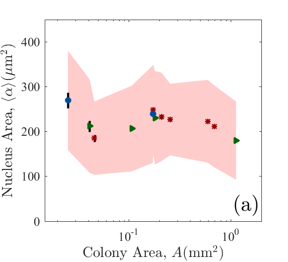
The mean nucleus area , mean number of nearest neighbours and mean intercellular distance of the colonies are shown in Figure 4 as a function of colony area . The mean cell nucleus area , Figure 4(a), shows high variability between colonies of different sizes and sampling days. The smallest colonies, with cells at day 3 ( ) and 25 – 46 cells at day 4 ( ), have the largest mean nucleus area, with m2 and m2 respectively. The small colonies analysed at day 2 with cells ( ) result in m2, a lower value than their later imaged counterparts. Although our error bars are large since we included cells undergoing mitosis, this result indicates that the cell nucleus is larger in the small colonies formed at later stages of passage, e.g day 4 colonies with a few dozen cells.
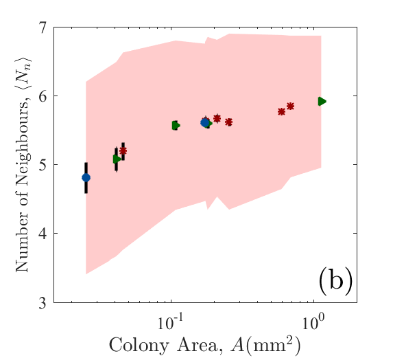
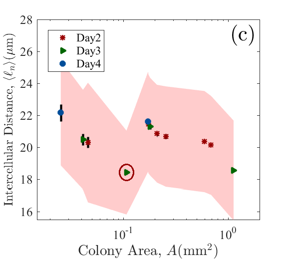
The mean number of nearest neighbours cells (day 4), 5.1 1.4 cells (day 3) and 5.2 1.4 cells (day 2) decreases with and increases with the colony area , whilst the mean intracellular distance , decreases with as expected. For small colonies at day 4 showing large cell nucleus areas, our results indicate that cell-to-cell contacts, occur on average with less than five cells that have a larger separation between them. This may influence the regulation of community effects in colonies of these sizes.
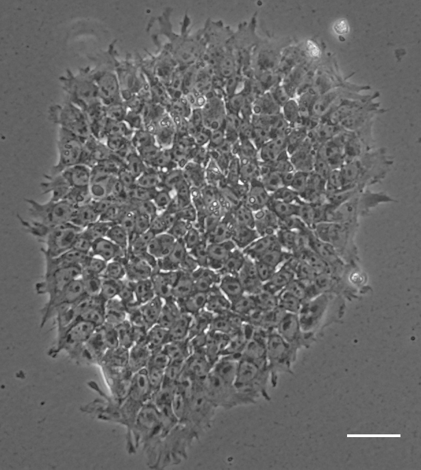
The day 3 colony highlighted with the red circle in Figure 4(c) has a particularly short intracellular distance for its area. The image of this colony, shown in Figure 5, shows several cells floating on top of the colony; this might indicate the interplay of other factors in the rearrangement of the cells in this sample.
For colonies containing between 350 and 550 cells, we obtain m2. The nucleus area is almost constant and shows only modest variability between days 2 and 3. The mean number of neighbours increases steadily with the colony area, Figure 4(b), whilst the mean intercellular distance decreases, i.e. the colony becomes more compact, Figure 4(c).
Finally, for the largest colonies analysed (the last three points to the right in Figures 4), there is a clear decrease in the mean nucleus area with colony size. The largest colony has the highest mean number of neighbours and the smallest inter-cell distances. Therefore, for large colonies (higher density) on average six neighbours are involved in cell-to-cell interactions. Visually these colonies are very dense, cells are tightly packed and there are no gaps between them.
In summary, colonies with and cells have the largest nuclei and intercellular distances with less neighbouring cells. On the other hand, the largest colony has cells with the smallest nuclei, short intercellular distances and six neighbours on average. Since the number of nearest neighbours increases with the colony area (and the number of cells) we suggest that the number of cells in a colony increases faster than its area as the cells fill the space within the colony.
We measured several other properties for the nuclei, such as their aspect ratio, perimeter, Feret’s diameter, circularity, roundness and solidity, as shown in Figure 10 and 11 in the SM. Some of these parameters have been used to characterise mouse embryonic stem cell (mESCs) colonies during differentiation (Bongiorno et al., 2018). However, for hESCs, at the single and colony level, these measurements do not show any significant change in behaviour that would indicate changes in the the morphology of the cells and colonies in terms of the days after plating and colony sizes, see Supplement Tables 3 and 4.
3.2 Probability distribution functions of nuclei area
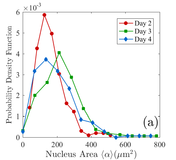
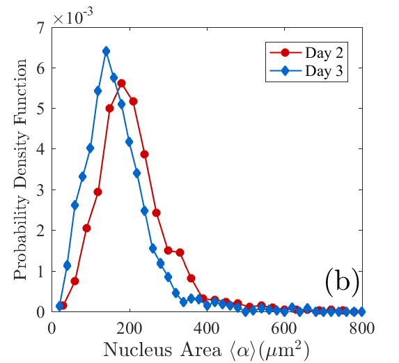
The size and shape of the cells are good indicators of their health and most importantly of their viability as a pluripotent cell for stem cell research. The averages of the quantities obtained in the previous section give a rough estimation of the behaviour of these variables in terms of the colony sizes. However, to account for the variability of the nuclei areas within a colony, we calculated the probability distribution functions PDF of , shown in Figure 6, for several samples, dividing them according to sampling day and size.
Colonies with (day 4) show an abrupt change in as a function of sampling day. Both day 3 and 4 colonies have a broader distribution, with cells having nuclei of sizes m2. The colonies at day 2 have a narrower distribution in which all nuclei have an area m2. It is important to keep in mind that day 3 and 4 colonies have half as many cells as the day 2 colonies, see Table 2 in the SM for further details. Overall, we observed a similar PDF for the smallest colonies, which becomes narrower as the cells increase their numbers. Therefore, a large nucleus in small samples may be due to a lack of compactness and pressure between the cells.
For the largest colonies analysed at days 2 and 3, with = 1257 327 and 1982 cells, respectively, the distributions become narrower as the colonies get bigger, see Figure 6(b). Occurrences of nuclei area disappear, and overall the cells become more homogeneous in size with for the day 3 colonies, see Figure 2. Under these circumstances, crowding effects due to mechanical cell competitions may take place in the bulk.
3.3 Colony morphology
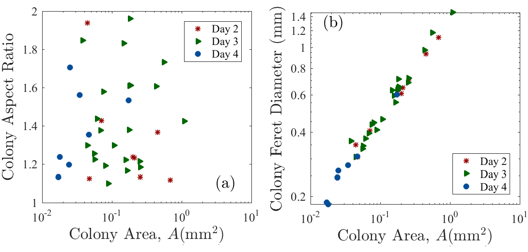
During colony formation, there are physical forces transmitted through the cells that affect the local mechanical properties and, therefore, play important roles in cellular behaviour such as adhesion properties, cell proliferation, differentiation and death (through the activation of biochemical signals) (Taylor, 2001; Ananthakrishnan and Ehrlicher, 2007; Fletcher and Mullins, 2010; Han et al., 2004). The colony shape is one of the qualitative features used to identify the best colonies and best clones. To quantify their form, we obtained the area , perimeter and shape descriptors of 38 colonies, see Supplement Table 4 in the SM. To measure changes in cell and colony morphologies as the cell numbers increase, we counted the cells in 19 colonies and added these results to the other 19 colonies analysed in the previous section.
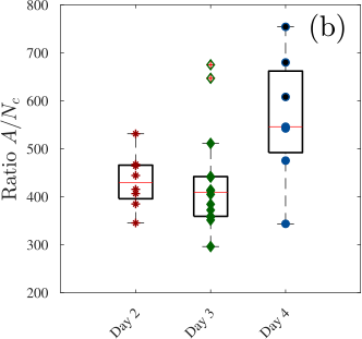
Figure 7(a) shows the the number of cells as a function of colony area . A power function trend line, , is appropriate with scaling factor and exponent ( = 0.97), see red dotted line. The exponent is approximately one, which corresponds to the cells maintaining the same nucleus area while the colony grows. The two largest colonies at day 2, with and , follow the same trend. However, small colonies from day 4 (left red circle) and some samples on day 3 (right red circle) deviate from this relationship.
We detect five small colonies, imaged at day 4 (with ), whose overall behaviour indicated exceedingly large nuclei, see Figures 1(c) and 17(d) in the SM. Those colonies are highlighted in Figure 7(b) with black points and are detected as outliers by the boxplot method. A more detailed analysis of these colonies indicates that a proportion of the cell population has undergone differentiation, see Figure 1(c).
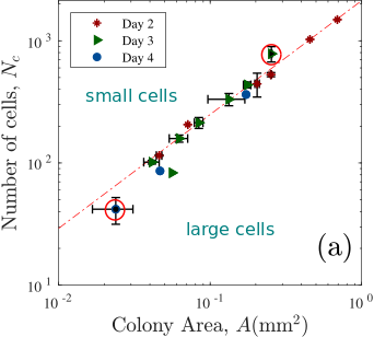
Comparing colonies of similar sizes measured at day 2, , and day 3, , we observe that the former has fewer cells ( = ) than the latter (). Therefore, there is an increment of cells in the bulk of the colony without an increase in the colony area. We can infer that the disappearance of the gaps between the cells, highly visible at day 2, is a result of newly dividing cells filling the voids. Consequently, the power-law relationship (linear on log-log scale) shown in Figure 7(a) between and holds only for colonies at day 2 and some colonies on day 3.
Colonies with show gaps between the cells. For these gaps start to disappear in the middle of the colony and are completely lost for colonies with areas . It is known that hiPSC colonies form actin (a linear polymeric micro-filament) fences encircling the colony that exerts extensive mechanical stress to enforce colony morphology and compaction (Närvä et al., 2017). We suppose that at initial stages of colony formation the cells accommodate themselves in such way that they have a higher intercellular distance between them, without being tightly packed, forming a polymeric fence around them to enforce compactness. For small and medium-sized colonies with gaps, there should be an outward pressure flow of cells at the boundary in order to accommodate newly divided cells in the bulk while keeping these spaces empty.
With the increase in cell numbers, we assume that there are more mitotic events in the colony and less time to re-organise the colony edges. Therefore it is possible that the fences formed at previous stages continue to maintain a strong adhesion at the border with the ECM, making the filling of gaps possible.
3.4 Segregation and population mixing
Segregation of cells in tissues and during pattern formation is an important phenomenon that occurs during the early phase of embryonic development, which ends with the formation of the three germ layers (Kurosaka and Kashina, 2008). The arrangement of cells in the embryo occurs due to changes in the environment (surface cues) that induce differences in adhesion properties and changes in the cytoskeleton (Fagotto, 2014). These differences in adhesion properties between neighbouring cells maintain a physical separation between different cell types, and it is one of the basic mechanisms for the pattern formation during development and wound healing (Krens and Heisenberg, 2011). Although in vivo, migration of hESCs is responsible for the segregation into physically distinct regions after a few rounds of divisions, in vitro, the presence of migratory effects is undesirable due to population mixing and loss of clonality (Chang et al., 2019).
From experiments on isolated hESCs, our measurements indicate that the cell grows until it reaches a size of m2 (unpublished results), after which it divides into two almost identical cells of sizes m2. Although hESCs form quasi-flat colonies, they show a highly dynamic behaviour in the bulk, with cells constantly migrating, interchanging neighbours and displacing each other due to mitosis. Specifically, it is at this stage that the cells detach from the culture, divide and re-attach again, resulting in an important factor that promotes cell mixing within the population which possibly affects the level of pluripotency achieved within the colony. To account for segregation in hESC colonies, we explore if the newly divided (small) cells are driven away from larger cells in the colony, which may be caused by the pushing of larger cells.
To measure if the small cells are segregated from the largest cells in the bulk of the colony, we introduce a segregation order parameter depending on the level of separation between small (type A) and large (type B) cells. Several order parameters can be introduced to characterise processes of segregation according to several segregation criteria (Rivas et al., 2011).
The VD, see Section §2.3, identifies accurately the number of nearest neighbours in each colony. We introduce a suitable segregation order parameter that depends explicitly on the number of nearest neighbours. We consider two types of particles A and B, if the system is segregated, each particle A will have more neighbours of the same type. The segregation order parameter is defined as follows,
| (1) |
where is the sum of the number of A Delaunay neighbours that B particles have, double counting the A particles that are neighbours of different B particles and is the number of nearest neighbours that each particle has on average. For a perfectly mixed system, with Delaunay neighbours, equation (1) results in . If the system is completely segregated, for example, one cluster of A particles surrounded by other of B particles, . The calculation of was performed for the largest colonies analysed each day for which the mean number of nearest neighbours exceeded , see Figure 4(b).
We use an area threshold to group the cells into two categories: type cells () and type B cells () and vary between m2 and m2. Applying equation (1) to the largest colonies measured each day, gives the results shown in Figure 8. We also show the results of the bootstrap to detect differences with a re-sampled data set. The colonies with areas (day 2) and (day 3) contain and cells respectively, panel a and b, whereas the colony with area have cells, panel c. Our results strongly suggest that, in large colonies, small cells () are segregated from larger ones. In both cases, see panels a and b in Figure 8, the curve for is above the one obtained from the bootstrap method. For m2, in Figure 8(b), reaches lower values that the bootstrap; this means that cells with m2 have less chance of having neighbours of the same size, and therefore, they are surrounded by smaller particles which are clustered together. As we increase beyond , decreases reaching the values for the random configuration. Therefore, for the largest colony, Figure 8(b), the cells are separated in a random fashion at = . However, for > , continues decreasing until it reaches , which indicates that the larger cells within the colony (mitotic events) tend occur far apart from each other. The results shown in Figure 8(c), for the colony with remain within the values obtained with the bootstrap method and the results are inconclusive, suggesting the need of larger colonies to obtain accurate measurements.

4 Discussion
We quantified the morphological and structural properties as well as the behaviour of hESCs during colony formation. Human embryonic stem cells self-organise into colonies with sharp edges and a strong adhesion at the border that promotes the maintenance of the pluripotent state by keeping the colonies tightly packed (Ginis et al., 2004; Närvä et al., 2017; Neganova et al., 2019). Our analyses reveal that the colonies change their morphological properties as the cells proliferate and the whole colony becomes larger.
Colonies with show white spaces between the cells that disappear, starting from the middle of the colony and finally at the edges, as the colony grows. There is a decrease in the nuclei size with an increase of colony size, accompanied by an increase in the number of first nearest neighbours available for each cell. Each cell within the largest colony, interacts, by contact, on average with six other cells. We suggest that this may have implications in the establishment of pluripotency on the local environment.
After these gaps are completely filled, we observe in large colonies the emergence of collective effects driven by the constant pushing and pulling of cells which drive the smallest and newly divided individuals to cluster in patches within the colony. Since the analysed colonies were grown on MatrigelTM, their migratory effects are large (Wadkin et al., 2017, 2018) and this could be a relevant factor in the spatial organisation of the cells within the colony. The continuous re-organisation of the colonies implies that neighbours are interchanged continually and consequently the cell population is continuously mixed; this directly influences the level of clonality within the colonies and the outcome of community effects that will furthermore influence the pluripotency achieved by the population (Chang et al., 2019; Nemashkalo et al., 2017).
Recent studies on hiPSCs with modified molecular regulators of cortical tension and cell-cell adhesion (through target genes ROCK1 and CDH1, respectively) have shown the emergence of distinct patterning events within hiPSC colonies through cell-driven segregation that dictated the colony organisation without the loss of pluripotency (Libby et al., 2018). Our results indicate that newly divided (small) cells are driven away from larger cells, clumped together in patches. Whether this effect is solely due to the mechanical effects (pushing) between the cells or changes in the cells’ cortical tension/adhesion properties along the cell cycle remains unknown and its elucidation requires further work.
5 Conclusion
The morphological analysis of hESC colonies is a powerful non-invasive tool to evaluate their quality and choose the best clones for medical applications, unlike invasive labelling procedures that involve genetic manipulation. Although the implementation of an algorithm for the automatic detection of cells within a colony was beyond the scope of this work, once such a method is developed, the parameters estimated throughout this paper can be easily implemented at a larger scale, to quantify accurately the parametric properties of pluripotent colonies.
Our work indicates that the mean nuclei area and mean distance between nearest neighbours might be good parameters to detect changes in the morphology of the colonies, despite the inherent variability in the cell sizes associated to the cell growth and the cell cycle. Our algorithms detect that small colonies at day 4 show distinctively larger cell nuclei and intercellular distances. These changes in their morphological properties might affect their pluripotency levels. Assuming an average hESC cycle duration of 14.6 h (Wolff et al., 2018) and an exponential colony growth starting from a single founder cell, we estimate that day 4 colonies with cells, were formed between 2–3 days before imaging. Therefore, later formed colonies have cells with changed morphological characteristics. Following this same premise, the larger colonies analysed at day 2 and 4 with thousands of cells, most certainly did not started from single founder cells. Our results suggests that this might be advantageous for the maintenance of their structural properties. The segregation of the cells inside the colony has strong biological implications in regards of the genetic and phenotypic spreading, since neighbouring individuals eventually end up in completely different locations.
Data Availability
The datasets used and/or analysed during the current study are available from the corresponding author on reasonable request.
Author Contributions
SOF, IN, NGP, RAB, ML and AS designed the research. SOF designed and developed the computational framework and analysed the data. IN conceived, planned and performed the experiments. SOF, IN and LEW wrote the manuscript. All authors contributed critically to the drafts and gave final approval for publication.
Acknowledgments
SOF acknowdledge financial support from the Consejo Nacional de Ciencia y Tecnología (CONACyT, Mexico) for the grant CVU-174695. ML acknowledges BBSRC UK (BB/I020209/1) and the H02020 ERC (614620) fellowships for providing financial support for this work. RAB wants to acknowledge financial support from CONACyT (Mexico) through the project 283279.
References
- Amit and Itskovitz-Eldor (2012a) Amit, M., Itskovitz-Eldor, J., 2012a. Chapter three - morphology of human embryonic stem cells and induced pluripotent stem cells cultured in feeder layer-free conditions, in: Amit, M., Itskovitz-Eldor, J. (Eds.), Atlas of Human Pluripotent Stem Cells: Derivation and Culture. Springer. Stem Cell Biology and Regenerative Medicine, pp. 41 – 55. URL: https://link.springer.com/book/10.1007/978-1-61779-548-0, doi:https://doi.org/10.1007/978-1-61779-548-0.
- Amit and Itskovitz-Eldor (2012b) Amit, M., Itskovitz-Eldor, J., 2012b. Chapter two - morphology of Human Embryonic and Induced Pluripotent Stem Cell Colonies cultured with feeders, in: Amit, M., Itskovitz-Eldor, J. (Eds.), Atlas of Human Pluripotent Stem Cells: Derivation and Culture. Springer. Stem Cell Biology and Regenerative Medicine, pp. 15 – 39. URL: https://link.springer.com/book/10.1007/978-1-61779-548-0, doi:https://doi.org/10.1007/978-1-61779-548-0.
- Ananthakrishnan and Ehrlicher (2007) Ananthakrishnan, R., Ehrlicher, A., 2007. The forces behind cell movement. International Journal of Biological Sciences 3, 303 – 317.
- Bongiorno et al. (2018) Bongiorno, T., Gura, J., Talwar, P., Chambers, D., Young, K.M., Arafat, D., Wang, G., Jackson-Holmes, E.L., Qiu, P., McDevitt, T.C., Sulchek, T., 2018. Biophysical subsets of embryonic stem cells display distinct phenotypic and morphological signatures. PLOS ONE 13, 1–25. URL: https://doi.org/10.1371/journal.pone.0192631, doi:10.1371/journal.pone.0192631.
- Buganim et al. (2012) Buganim, Y., Faddah, D., Cheng, A., Itskovich, E., Markoulaki, S., Ganz, K., Klemm, S., van Oudenaarden, A., Jaenisch, R., 2012. Single-cell expression analyses during cellular reprogramming reveal an early stochastic and a late hierarchic phase. Cell 150, 1209 – 1222. URL: http://www.sciencedirect.com/science/article/pii/S0092867412010215, doi:https://doi.org/10.1016/j.cell.2012.08.023.
- Chang et al. (2019) Chang, J., Kim, M.H., Agung, E., Senda, S., Kino-oka, M., 2019. Effect of migratory behaviors on human induced pluripotent stem cell colony formation on different extracellular matrix proteins. Regenerative Therapy 10, 27 – 35. URL: http://www.sciencedirect.com/science/article/pii/S2352320418300361, doi:https://doi.org/10.1016/j.reth.2018.10.004.
- Chen et al. (2015) Chen, D., Sun, Y., CX, C.D., Fu, J., 2015. Improving survival of disassociated human embryonic stem cells by mechanical stimulation using acoustic tweezing cytometry. Biophysical Journal 108, 1315 – 1317. doi:https://doi:10.1016/j.bpj.2015.01.033.
- Dalton (2013) Dalton, S., 2013. Signaling networks in human pluripotent stem cells. Current Opinion in Cell Biology 25, 241 – 246. doi:http://doi.org/10.1016/j.ceb.2012.09.005.
- Efthymiou et al. (2014) Efthymiou, A.G., Chen, G., Rao, M., Chen, G., Boehm, M., 2014. Self-renewal and cell lineage differentiation strategies in human embryonic stem cells and induced pluripotent stem cells. Expert Opinion on Biological Therapy 14, 1333 – 1344. doi:https://doi.org/10.1517/14712598.2014.922533.
- Etoc et al. (2016) Etoc, F., Metzger, J., Ruzo, A., Kirst, C., Yoney, A., Ozair, M., Brivanlou, A., Siggia, E., 2016. A balance between secreted inhibitors and edge sensing controls gastruloid self-organization. Developmental Cell 39, 302 – 315. URL: http://www.sciencedirect.com/science/article/pii/S1534580716306384, doi:https://doi.org/10.1016/j.devcel.2016.09.016.
- Fagotto (2014) Fagotto, F., 2014. The cellular basis of tissue separation. Development 141, 3303–3318. URL: http://dev.biologists.org/content/141/17/3303, doi:10.1242/dev.090332, arXiv:http://dev.biologists.org/content/141/17/3303.full.pdf.
- Fletcher and Mullins (2010) Fletcher, D.A., Mullins, R.D., 2010. Cell mechanics and the cytoskeleton. Nature 463, 485 -- 492.
- Ginis et al. (2004) Ginis, I., Luo, Y., Miura, T., Thies, S., Brandenberger, R., Gerecht-Nir, S., Amit, M., Hoke, A., Carpenter, M.K., Itskovitz-Eldor, J., Rao, M.S., 2004. Differences between human and mouse embryonic stem cells. Developmental Biology 269, 360 -- 380. URL: http://www.sciencedirect.com/science/article/pii/S0012160604000193, doi:https://doi.org/10.1016/j.ydbio.2003.12.034.
- Han et al. (2004) Han, B., Bai, X.H., Lodyga, M., Xu, J., Yang, B.B., Keshavjee, S., Post, M., Liu, M., 2004. Conversion of mechanical force into biochemical signaling. Journal of Biological Chemistry 279, 54793 -- 54801.
- Heinrich et al. (1984) Heinrich, R., Gaestel, M., Müller, T., 1984. A theoretical approach to cluster formation in biological membranes. Biosystems 17, 135 -- 154.
- Herberg and Roeder (2015) Herberg, M., Roeder, I., 2015. Computational modelling of embryonic stem-cell fate control. Development 142, 2250--2260. URL: http://dev.biologists.org/content/142/13/2250, doi:10.1242/dev.116343, arXiv:http://dev.biologists.org/content/142/13/2250.full.pdf.
- Honda (1978) Honda, H., 1978. Description of cellular patterns by Dirichlet domains: the two-dimensional case. Journal of Theoretical Biology 72, 523--543.
- Icha et al. (2017) Icha, J., Weber, M., Waters, C., Norden, C., 2017. Phototoxicity in live fluorescence microscopy, and how to avoid it. Bioessays 38, 1700003.
- Johkura et al. (2004) Johkura, K., Cui, L., Asanuma, K., Okouchi, Y., Ogiwara, N., Sasaki, K., 2004. Cytochemical and ultrastructural characterization of growing colonies of human embryonic stem cells. Journal of Anatomy 205, 247--255. doi:10.1111/j.0021-8782.2004.00336.x, arXiv:https://onlinelibrary.wiley.com/doi/epdf/10.1111/j.0021-8782.2004.00336.x.
- Krens and Heisenberg (2011) Krens, S.F., Heisenberg, C.P., 2011. Chapter six - Cell Sorting in Development, in: Labouesse, M. (Ed.), Forces and Tension in Development. Academic Press. volume 95 of Current Topics in Developmental Biology, pp. 189 -- 213. URL: http://www.sciencedirect.com/science/article/pii/B9780123850652000062, doi:https://doi.org/10.1016/B978-0-12-385065-2.00006-2.
- Kurosaka and Kashina (2008) Kurosaka, S., Kashina, A., 2008. Cell biology of embryonic migration. Embryo today : reviews 84, 102 -- 122. URL: https://onlinelibrary.wiley.com/doi/abs/10.1002/bdrc.20125, doi:https://doi.org/10.1002/bdrc.20125.
- Li et al. (2010) Li, L., Wang, B.H., Wang, S., Moalim-Nour, L., Mohib, K., Lohnes, D., Wang, L., 2010. Individual cell movement, asymmetric colony expansion, rho-associated kinase, and E-Cadherin impact the clonogenicity of Human Embryonic Stem Cells. Biophysical Journal 98, 2442 -- 2451. URL: http://www.sciencedirect.com/science/article/pii/S000634951000305X, doi:https://doi.org/10.1016/j.bpj.2010.02.029.
- Li and Belmonte (2018) Li, M., Belmonte, J.C.I., 2018. Deconstructing the pluripotency gene regulatory network. Nature Cell Biology 20, 382 -- 392. doi:https://doi.org/10.1038/s41556-018-0067-6.
- Libby et al. (2018) Libby, A.R., Joy, D.A., So, P.L., Mandegar, M.A., Muncie, J.M., Mendoza-Camacho, F.N., Weaver, V.M., Conklin, B.R., , McDevitt, T.C., 2018. Spatiotemporal mosaic self-patterning of pluripotent stem cells using CRISPR interference. eLife 7.
- Lulevich et al. (2009) Lulevich, V., Shih, Y., Lo, S., Liu, G., 2009. Cell tracing dyes significantly change single cell mechanics. J Phys Chem B 113, 6511–6519.
- Messmer et al. (2019) Messmer, T., von Meyenn, F., Savino, A., Santos, F., Mohammed, H., Lun, A.T.L., Marioni, J.C., Reik, W., 2019. Transcriptional heterogeneity in naive and primed human pluripotent stem cells at single-cell resolution. Cell Reports 26, 815 -- 824.e4. URL: http://www.sciencedirect.com/science/article/pii/S2211124718320746, doi:https://doi.org/10.1016/j.celrep.2018.12.099.
- Neganova et al. (2019) Neganova, I., Cotts, L., Banks, P., Gassner, K., Shukurov, A., Armstrong, L., Ladds, G., Lako, M., 2019. Endothelial differentiation g protein-coupled receptor 5 plays an important role in induction and maintenance of pluripotency. STEM CELLS 37, 318--331. doi:10.1002/stem.2954.
- Neganova et al. (2014) Neganova, I., Tilgner, K., Buskin, A., Paraskevopoulou, I., Atkinson, S., Peberdy, D., Passos, J., Lako, M., 2014. Cdk1 plays an important role in the maintenance of pluripotency and genomic stability in human pluripotent stem cells. Cell Death Dis 6, e1508.
- Neganova et al. (2011) Neganova, I., Vilella, F., Atkinson, S.P., Lloret, M., Passos, J.F., von Zglinicki, T., O’Connor, J.E., Burks, D., Jones, R., Armstrong, L., Lako, M., 2011. An important role for cdk2 in g1 to s checkpoint activation and dna damage response in human embryonic stem cells. STEM CELLS 29, 651--659. URL: https://stemcellsjournals.onlinelibrary.wiley.com/doi/abs/10.1002/stem.620, doi:10.1002/stem.620.
- Neganova et al. (2009) Neganova, I., Zhang, X., Atkinson, S., Lako, M., 2009. Expression and functional analysis of g1 to s regulatory components reveals an important role for cdk2 in cell cycle regulation in human embryonic stem cells. Oncogene 28, 20 -- 30.
- Nemashkalo et al. (2017) Nemashkalo, A., Ruzo, A., Heemskerk, I., Warmflash, A., 2017. Morphogen and community effects determine cell fates in response to BMP4 signaling in human embryonic stem cells. Development 144, 3042--3053. URL: http://dev.biologists.org/content/144/17/3042, doi:10.1242/dev.153239, arXiv:http://dev.biologists.org/content/144/17/3042.full.pdf.
- Närvä et al. (2017) Närvä, E., Stubb, A., Guzmán, C., Blomqvist, M., Balboa, D., Lerche, M., Saari, M., Otonkoski, T., Ivaska, J., 2017. A strong contractile actin fence and large adhesions direct human pluripotent colony morphology and adhesion. Stem Cell Reports 9, 67--76.
- Odorico et al. (2004) Odorico, J., Pedersen, R., Zhang, S.C., 2004. Human Embryonic Stem Cells. Garland Science.
- Pearson et al. (1979) Pearson, R., Hui, S., Stewart, T., 1979. Correlative statistical analysis and computer modelling of intramembraneous particle distributions in human erythrocyte membranes. Biochimica et Biophysica Acta (BBA) - Biomembranes 557.
- Peerani et al. (2007) Peerani, R., Rao, B.M., Bauwens, C., Yin, T., Wood, G.A., Nagy, A., Kumacheva, E., Zandstra, P.W., 2007. Niche-mediated control of human embryonic stem cell self-renewal and differentiation. The EMBO Journal 26, 4744--4755. doi:10.1038/sj.emboj.7601896, arXiv:http://emboj.embopress.org/content/26/22/4744.full.pdf.
- Pera and Trounson (2004) Pera, M.F., Trounson, A.O., 2004. Human embryonic stem cells: prospects for development. Development 131, 5515--5525. URL: http://dev.biologists.org/content/131/22/5515, doi:10.1242/dev.01451, arXiv:http://dev.biologists.org/content/131/22/5515.full.pdf.
- Rivas et al. (2011) Rivas, N., Cordero, P., Risso, D., Soto, R., 2011. Segregation in quasi-two-dimensional granular systems. New Journal of Physics 13, 055018. URL: http://stacks.iop.org/1367-2630/13/i=5/a=055018.
- Rossler (2004) Rossler, U., 2004. Solid State Theory: An Introduction. Springer-Verlag Berlin Heidelberg.
- Saito (1982) Saito, N., 1982. Asymptotic regular pattern of epidermal cells in mammalian skin. Journal of Theorertical Biology 95, 591--599.
- Schneider et al. (2012) Schneider, C., Rasband, W., Eliceiri, K., 2012. NIH Image to ImageJ: 25 years of image analysis. Nature Methods 9, 671--675.
- Soldner and Jaenisch (2018) Soldner, F., Jaenisch, R., 2018. Stem cells, genome editing, and the path to translational medicine. Cell 175, 615 -- 632. URL: http://www.sciencedirect.com/science/article/pii/S0092867418311826, doi:https://doi.org/10.1016/j.cell.2018.09.010.
- Ståhlberg et al. (2009) Ståhlberg, A., Bengtsson, M., Hemberg, M., Semb, H., 2009. Quantitative transcription factor analysis of undifferentiated single human embryonic stem cells. Clinical Chemistry 55, 2162--2170. URL: http://clinchem.aaccjnls.org/content/55/12/2162, doi:10.1373/clinchem.2009.131433, arXiv:http://clinchem.aaccjnls.org/content/55/12/2162.full.pdf.
- Takahashi et al. (2007) Takahashi, K., Tanabe, K., Ohnuki, M., Narita, M., Ichisaka, T., Tomoda, K., Yamanaka, S., 2007. Induction of pluripotent stem cells from adult human fibroblasts by defined factors. Cell 131, 861 -- 872. URL: http://www.sciencedirect.com/science/article/pii/S0092867407014717, doi:https://doi.org/10.1016/j.cell.2007.11.019.
- Taylor (2001) Taylor, E., 2001. A physical view of the cytoskeleton. Nature 413.
- Thomson et al. (1998) Thomson, J.A., Itskovitz-Eldor, J., Shapiro, S.S., Waknitz, M.A., Swiergiel, J.J., Marshall, V.S., Jones, J.M., 1998. Embryonic stem cell lines derived from human blastocysts. Science 280, 1145 -- 1147. doi:https://doi.org/10.1126/science.282.5391.1145.
- Torres-Padilla and Chambers (2014) Torres-Padilla, M.E., Chambers, I., 2014. Transcription factor heterogeneity in pluripotent stem cells: a stochastic advantage. Development 141, 2173--2181. URL: http://dev.biologists.org/content/141/11/2173, doi:10.1242/dev.102624, arXiv:http://dev.biologists.org/content/141/11/2173.full.pdf.
- Toyoda et al. (2015) Toyoda, T., Mae, S.I., Tanaka, H., Kondo, Y., Kawaguchi, M.F.Y.H.T.S.Y., Osafune, K., 2015. Cell aggregation optimizes the differentiation of human ESCs and iPSCs into pancreatic bud-like progenitor cells. Stem Cell Research 14, 185 -- 197.
- Ullmann et al. (2006) Ullmann, U., In’t Veld, P., Gilles, C., Sermon, K., De Rycke, M., Van de Velde, H., Van Steirteghem, A., Liebaers, I., 2006. Epithelial–mesenchymal transition process in human embryonic stem cells cultured in feeder-free conditions. MHR: Basic science of reproductive medicine 13, 21--32. URL: https://dx.doi.org/10.1093/molehr/gal091, doi:10.1093/molehr/gal091, arXiv:http://oup.prod.sis.lan/molehr/article-pdf/13/1/21/12786229/gal091.pdf.
- Vallier et al. (2009) Vallier, L., Touboul, T., Chng, Z., Brimpari, M., Hannan, N., Millan, E., Smithers, L.E., Trotter, M., Rugg-Gunn, P., Weber, A., Pedersen, R.A., 2009. Early cell fate decisions of human embryonic stem cells and mouse epiblast stem cells are controlled by the same signalling pathways. PLOS ONE 4, 1--13. URL: https://doi.org/10.1371/journal.pone.0006082, doi:10.1371/journal.pone.0006082.
- Vazin and Freed (2010) Vazin, T., Freed, W.J., 2010. Human embryonic stem cells: Derivation, culture, and differentiation: A review. Restorative Neurology and Neuroscience 28, 589 -- 603. doi:http://doi.org/10.3233/RNN-2010-0543.
- Vining and Mooney (2017) Vining, K.H., Mooney, D.J., 2017. Mechanical forces direct stem cell behaviour in development and regeneration. Nature Reviews Molecular Cell Biology 18, 728 -- 742. URL: https://www.ncbi.nlm.nih.gov/pmc/articles/PMC5803560/, doi:https://doi.org/10.1038/nrm.2017.108.
- Wadkin et al. (2017) Wadkin, L.E., Elliot, L.F., Neganova, I., Parker, N.G., Chichagova, V., Swan, G., Laude, A., Lako, M., Shukurov, A., 2017. Dynamics of single human embryonic stem cells and their pairs: a quantitative analysis. Scientific Reports 7. doi:https://doi.org/10.1038/s41598-017-00648-0.
- Wadkin et al. (2018) Wadkin, L.E., Orozco-Fuentes, S., Neganova, I., Swan, G., Laude, A., Lako, M., Shukurov, A., Parker, N.G., 2018. Correlated random walks of human embryonic stem cells in vitro. Physical Biology 15.
- Warmflash et al. (2014) Warmflash, A., Sorre, B., Etoc, F., Siggia, E., Brivanlou, A., 2014. A method to recapitulate early embryonic spatial patterning in human embryonic stem cells. Nature Methods 11, 847--854.
- Wolff et al. (2018) Wolff, S.C., Kedziora, K.M., Dumitru, R., Dungee, C.D., Zikry, T.M., Beltran, A.S., Haggerty, R.A., Cheng, J., Redick, M.A., Purvis, J.E., 2018. Inheritance of OCT4 predetermines fate choice in human embryonic stem cells. Molecular Systems Biology 14, e8140. URL: https://onlinelibrary.wiley.com/doi/abs/10.15252/msb.20178140, doi:10.15252/msb.20178140, arXiv:https://onlinelibrary.wiley.com/doi/pdf/10.15252/msb.20178140.
- Xu et al. (2010) Xu, Y., Zhu, X., Hahm, H.S., Wei, W., Hao, E., Hayek, A., Ding, S., 2010. Revealing a core signaling regulatory mechanism for pluripotent stem cell survival and self-renewal by small molecules. PNAS 107, 8129 -- 8134.
- Zhao and Jin (2017) Zhao, H., Jin, Y., 2017. Signaling networks in the control of pluripotency. Current Opinion in Genetics and Development 46, 141 -- 148. doi:https://doi.org/10.1016/j.gde.2017.07.013. cell reprogramming.
- Zhu and Huangfu (2013) Zhu, Z., Huangfu, D., 2013. Human pluripotent stem cells: an emerging model in developmental biology. Development 140, 705 -- 717. doi:http://10.1242/dev.086165.
6 Supplementary Material
6.1 Nuclei detection
The analysis of each colony was performed using ImageJ (Schneider et al., 2012) and the statistical analysis performed with Matlab®2016b. Each cell was manually traced and processed using ImageJ as it is shown in Figure 9. We obtained the nuclei area and perimeter , alongside the properties that we describe in the following.
Aspect Ratio
The aspect ratio is the relationship between the height and width of the nucleus. For a perfect circle, we obtain , elongated particles have . It is measured following the equation, , where is the major axis and the minor axis of a rectangle that encloses the nucleus, in terms of the minimum area of enclosure.
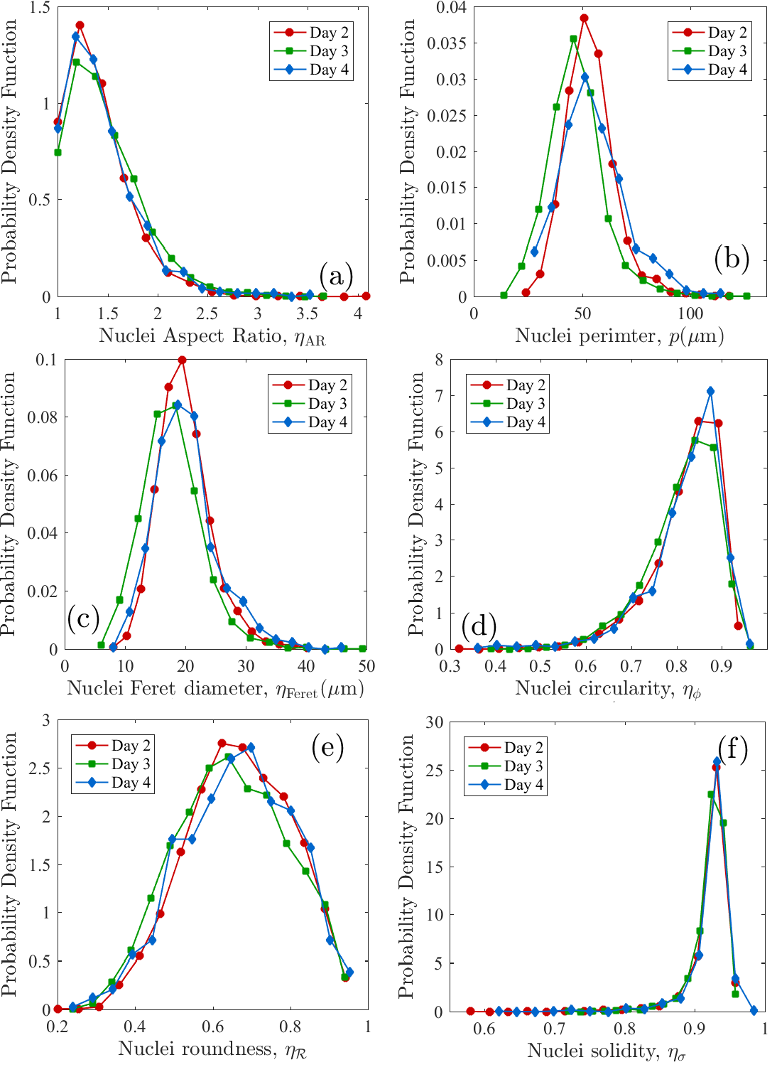
The Figure 10(a) shows the PDF for the nuclei aspect ratio following the colonies sampling day. Overall, the majority of cells have an aspect ratio of <2. Cells with elongated shapes are very few. This supports the applicability of the VD for an accurate tessellation of the colony.
Perimeter
The probability density function for nuclei perimeter is shown in Figure 10(b). It has a mean of m, and this value is similar for all sampling days.
Feret’s Diameter
The Feret’s diameter (), also know as maximum caliper measures the longest distance between any two points along the nucleus boundary. The probability density function (PDF) for the Feret’s diameter for the nuclei are shown in Figure 10(c), according to the sampling day. The PDFs have a mean m, with the maximum caliper being m.
Circularity
Circularity is a shape descriptor that indicates the degree of similarity with a circle, therefore as this quantity approaches 0, the shape is less circular. It is calculated using the equation , with and the nucleus area and perimeter respectively. The values for the PDF of the circularity are shown in Figure 10(d), with all three distributions centred around , therefore the nuclei shapes are highly circular.
Roundness
It is very similar to circularity but is insensitive to irregular borders along the perimeter. It is measured using the highest axis of the best fit ellipse. The equation for the roundness is . The results for the PDF for the roundness are shown in Figure 10(e).
Solidity
Describes the extent to which a shape is concave or convex. The solidity of a completely convex shape is 1, the farther the solidity deviates from 1, the concavity in the nucleus increases. It is calculated following the equation, , with representing the area of the convex hull that best encloses the nucleus boundary. The results are shown in Figure 10(f) and indicate that most of the nuclei have values close to 1.

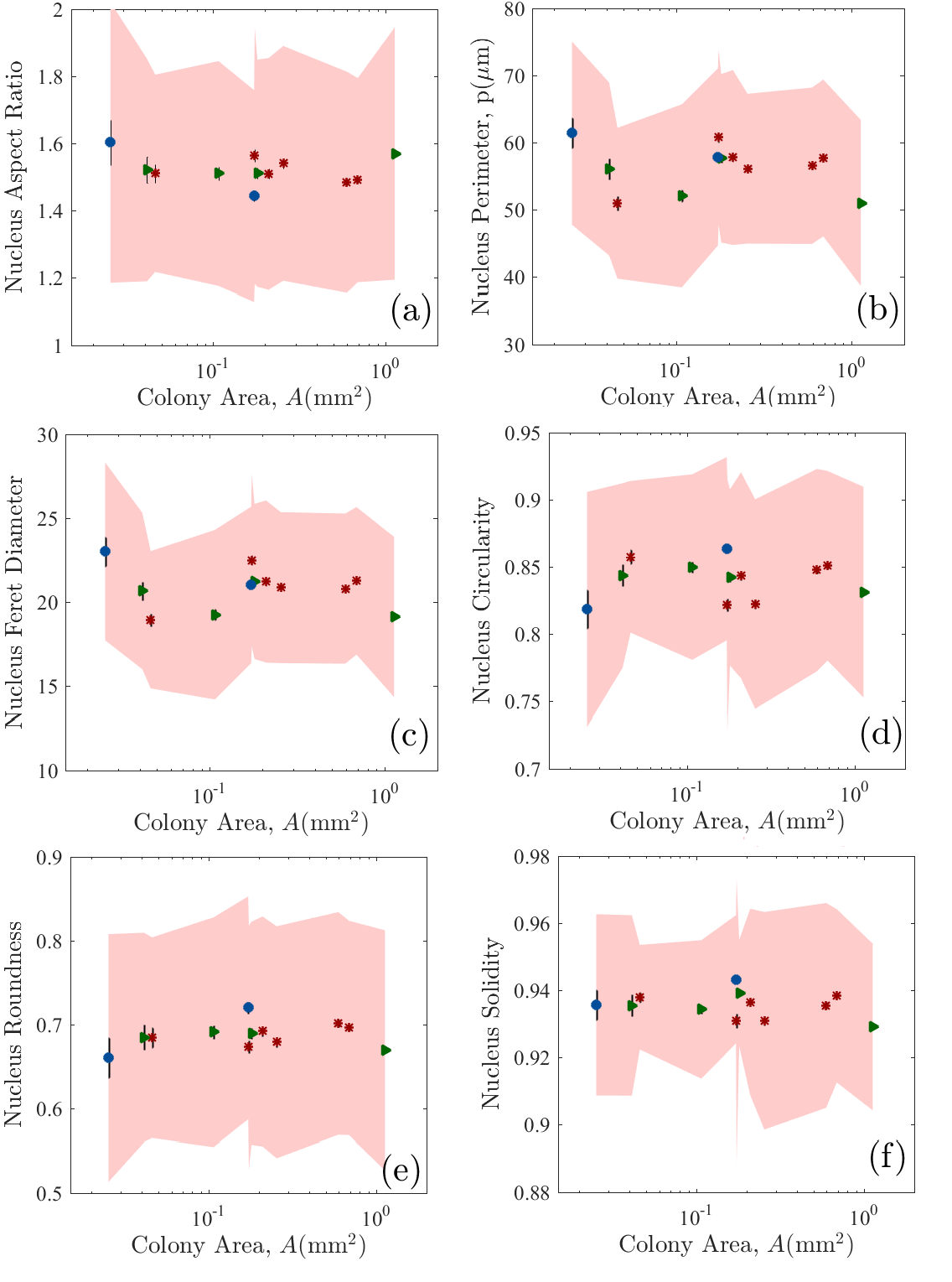
The averages for each shape descriptor are shown in Table 3 and the plots in Figure 11. The standard deviations range is the shaded region. The standard error of the mean is shown as a black line around the data point. Data points are presented with different symbols according to the day at which the image was taken (see legend panel f).
| () | Day | ||||
|---|---|---|---|---|---|
| 0.025 0.007† | 39 10 | 269 111 | 4.81 1.40 | 22.16 3.30 | 4 |
| 0.041 0.005 | 71 34 | 212 105 | 5.08 1.41 | 20.48 3.11 | 3 |
| 0.046 0.002 | 115 1.4 | 185 82 | 5.20 1.44 | 20.31 3.73 | 2 |
| 0.107 | 305 | 206 96 | 5.57 1.23 | 18.43 2.61 | 3 |
| 0.173 | 363 | 239 109 | 5.61 1.15 | 21.60 3.13 | 4 |
| 0.174 | 375 | 248 100 | 5.63 1.16 | 21.46 3.10 | 2 |
| 0.180 | 409 | 230 104 | 5.60 1.26 | 21.29 3.06 | 3 |
| 0.210 | 514 | 232 99 | 5.67 1.14 | 20.85 3.06 | 2 |
| 0.255 | 543 | 226 80 | 5.62 1.28 | 20.67 3.14 | 2 |
| 0.596 | 1026 | 222 92 | 5.76 1.12 | 20.37 3.15 | 2 |
| 0.690 | 1489 | 211 91 | 5.84 1.03 | 20.13 3.08 | 2 |
| 1982 | 179 87 | 5.91 0.96 | 18.56 3.11 | 3 |
| () | Day | ||||||
| 0.025 0.007† | 1.60 0.42 | 61.41 13.66 | 23.02 5.30 | 0.82 0.09 | 0.66 0.15 | 0.95 0.03 | 4 |
| 0.041 0.005 | 1.52 0.33 | 56.06 12.89 | 20.65 4.66 | 0.84 0.07 | 0.69 0.12 | 0.93 0.03 | 3 |
| 0.046 0.002 | 1.51 0.29 | 50.96 11.23 | 18.94 4.09 | 0.86 0.06 | 0.68 0.12 | 0.94 0.02 | 2 |
| 0.107 | 1.51 0.33 | 52.06 13.64 | 19.25 5.06 | 0.85 0.07 | 0.69 0.14 | 0.93 0.02 | 3 |
| 0.173 | 1.44 0.31 | 57.85 13.19 | 21.02 4.67 | 0.86 0.07 | 0.72 0.13 | 0.94 0.02 | 4 |
| 0.174 | 1.56 0.38 | 60.75 13.05 | 22.50 5.14 | 0.82 0.09 | 0.67 0.14 | 0.93 0.04 | 2 |
| 0.180 | 1.51 0.34 | 57.66 12.56 | 21.23 4.62 | 0.84 0.07 | 0.69 0.13 | 0.94 0.01 | 3 |
| 0.210 | 1.51 0.35 | 57.78 13.03 | 21.22 4.83 | 0.84 0.08 | 0.69 0.14 | 0.94 0.03 | 2 |
| 0.255 | 1.54 0.35 | 56.11 11.16 | 20.86 4.50 | 0.82 0.08 | 0.68 0.14 | 0.93 0.03 | 2 |
| 0.596 | 1.48 0.33 | 56.54 11.65 | 20.80 4.48 | 0.85 0.07 | 0.70 0.13 | 0.94 0.03 | 2 |
| 0.690 | 1.49 0.30 | 57.70 11.69 | 21.27 4.40 | 0.85 0.07 | 0.70 0.13 | 0.94 0.03 | 2 |
| 1.57 0.38 | 51.00 12.37 | 19.11 4.77 | 0.83 0.08 | 0.67 0.14 | 0.93 0.02 | 3 |
-
a
For the smallest colonies we obtained statistics from several samples, grouping them according to imaging day.
-
b
Only a portion of the colony was analysed.
The behaviour of the shape descriptor, although useful at the colony level for the detection of morphological changes between pluripotent and differentiated colonies, do not show a significant change at the nucleus level. The aspect ratio of the nuclei is in all cases, see Figure 11(a).
6.2 Edge detection
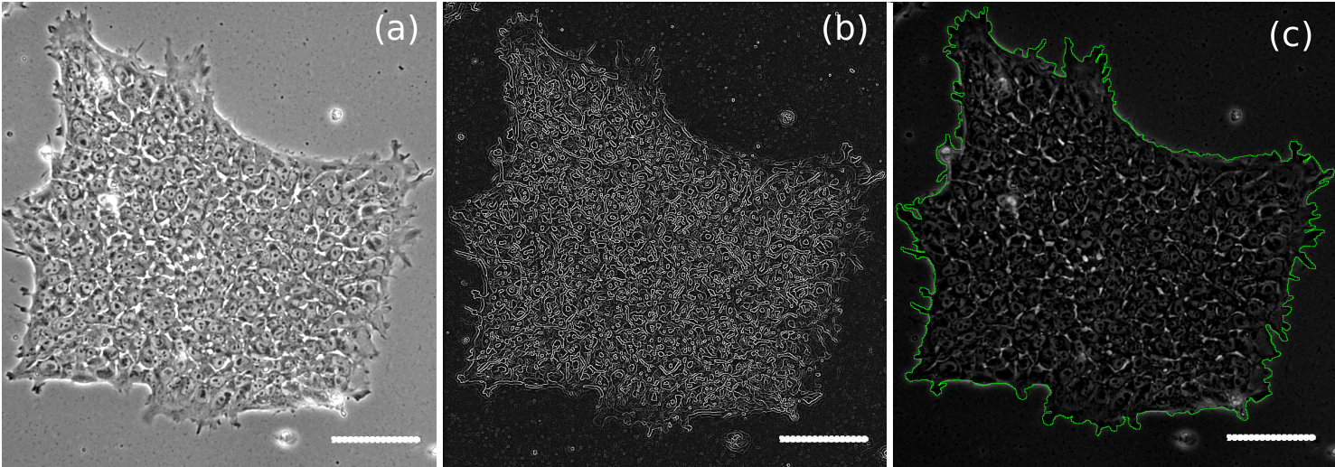
To detect the colony edges from the phase contrast images we use the canny Deriche algorithm included in ImageJ (Schneider et al., 2012) to aid in the identification of the border as shown in Figure 16(a) and (b). To obtain the boundary we traced manually the borders obtained in (b).
6.3 Colony reconstruction
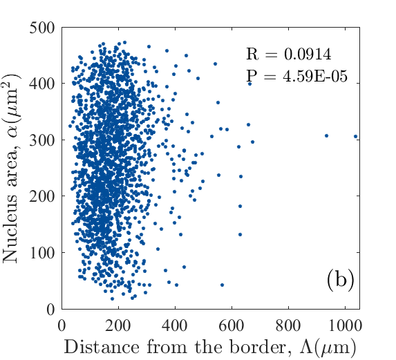
To measure the correlation between the cell nucleus size and its position within the colony we reconstructed the colony shown in Figure 2 using the Voronoi diagram (VD) bounded by the colony’s boundaries. In Figure 14(a) we show the reconstructed VD for the colony in Figure 2. Using the centroid position for each cell we calculated their (closest) distance from the boundary and plotted this as a function of the cell nucleus area , see Figure 14(b). Our results indicate that the cell sizes are not correlated to their position within the colony, with a correlation coefficient of .
Structural analysis through the radial distribution function
Radial distribution functions (RDF) are tools used widely in crystallography and soft condensed matter physics to characterise the structure of different materials (Rossler, 2004). The RDF, denoted with , determines the number of particles in a spherical shell of radius and thickness , i.e., . In other words, it describes the variation of the local cell density within a distance as viewed from the centring cell, relative to its bulk value. For ordered materials, such as crystals, the radial distribution function show an oscillating behaviour, where the peaks in are interpreted as the average inter-particle distances. The information contained in the RDF is a spatial average and has its limitations when the system is not isotropic.
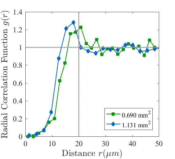
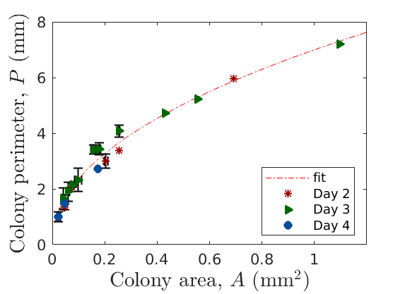
Figure 15 show results for the RDF for two colonies of different sizes with areas and , respectively. For both cases, the first peak is the best-defined one and corresponds to the distribution of the distance between the first nearest neighbours m and m respectively. The position of the second peak, gives the average distance or coordination, between second neighbours . We conclude that the colonies show a short-range order and the nearest coordination shells are visible in both cases. But, as we increase to account for the second nearest neighbours the second peak is washed out and broader than the first due to missing long-range order. The largest colony shows a second peak centred around m. Interestingly, the RDF shows that the cells in the largest colony () are more packed since the blue curve crosses the horizontal dotted line before the green curve. The results are shown in Figure 15 and indicate that is similar to the ones obtained for amorphous materials, therefore the structure of the material becomes blurred as the radius increases.
In biology, calculations of have been performed to study protein organisation, aggregation of particles on cell membranes, (Pearson et al., 1979; Heinrich et al., 1984). This tool has not been used previously in the literature to characterise dense aggregates of cells.
| Identifier | () | P () | Feret | MinFeret | L | Day | ||||
|---|---|---|---|---|---|---|---|---|---|---|
| DAY2_4x5 | 0.691 | 1489 | 5.960 | 0.245 | 1115.1 | 953.2 | 1.117 | 0.895 | 0.874 | 2 |
| DAY2_6x10 | 0.456 | 1026 | 2.747 | 0.759 | 931.6 | 691.4 | 1.365 | 0.733 | 0.962 | 2 |
| DAY2_7x10 | 0.209 | 514 | 3.180 | 0.260 | 644.8 | 513.1 | 1.233 | 0.811 | 0.840 | 2 |
| DAY2_8Ax10 | 0.047 | 114 | 1.428 | 0.292 | 305.6 | 252.3 | 1.125 | 0.889 | 0.810 | 2 |
| DAY2_8Bx10 | 0.045 | 116 | 1.283 | 0.341 | 348.5 | 190.7 | 1.938 | 0.516 | 0.845 | 2 |
| DAY2_9x10 | 0.071 | 206 | 1.979 | 0.228 | 405.0 | 275.3 | 1.428 | 0.700 | 0.827 | 2 |
| DAY2_10x10 | 0.254 | 543 | 3.390 | 0.278 | 702.4 | 573.3 | 1.132 | 0.883 | 0.883 | 2 |
| DAY2_11x10 | 0.199 | 375 | 2.825 | 0.314 | 607.3 | 481.8 | 1.238 | 0.808 | 0.872 | 2 |
| DAY3_1x10 | 0.164 | 457 | 3.249 | 0.196 | 553.2 | 452.5 | 1.165 | 0.858 | 0.842 | 3 |
| DAY3_2X10 | 0.056 | 151 | 1.770 | 0.226 | 344.5 | 279.6 | 1.224 | 0.817 | 0.816 | 3 |
| DAY3_3x10 | 0.255 | 862 | 3.930 | 0.208 | 716.1 | 555.3 | 1.183 | 0.846 | 0.845 | 3 |
| DAY3_4x10 | 0.107 | 305 | 2.616 | 0.197 | 460.5 | 356.8 | 1.298 | 0.770 | 0.873 | 3 |
| DAY3_5x10 | 0.080 | 198 | 2.187 | 0.211 | 444.4 | 341.4 | 1.192 | 0.839 | 0.763 | 3 |
| DAY3_6x5 | 1.097 | - | 7.205 | 0.263 | 1460.6 | 1040.9 | 1.420 | 0.703 | 0.888 | 3 |
| DAY3_7x10 | 0.252 | 703 | 4.230 | 0.177 | 685.4 | 529.9 | 1.213 | 0.824 | 0.848 | 3 |
| DAY3_10x10A | 0.056 | 83 | 1.535 | 0.299 | 334.2 | 275.7 | 1.255 | 0.797 | 0.839 | 3 |
| DAY3_10x10B | 0.038 | 74 | 1.922 | 0.129 | 363.6 | 215.7 | 1.848 | 0.541 | 0.651 | 3 |
| DAY3_11x10 | 0.088 | 229 | 2.023 | 0.270 | 410.0 | 341.6 | 1.100 | 0.909 | 0.837 | 3 |
| DAY3_12x10 | 0.069 | 166 | 1.983 | 0.219 | 394.8 | 289.6 | 1.376 | 0.727 | 0.803 | 3 |
| DAY3_14x10 | 0.182 | 440 | 3.664 | 0.171 | 710.0 | 391.8 | 1.960 | 0.510 | 0.844 | 3 |
| DAY3_16x10 | 0.159 | 359 | 3.410 | 0.172 | 594.0 | 462.6 | 1.223 | 0.818 | 0.778 | 3 |
| DAY3_17x5 | 0.430 | - | 4.718 | 0.243 | 974.8 | 614.9 | 1.607 | 0.622 | 0.876 | 3 |
| DAY3_18x10 | 0.175 | - | 3.320 | 0.200 | 629.9 | 461.7 | 1.379 | 0.725 | 0.830 | 3 |
| DAY3_19x10 | 0.076 | - | 2.095 | 0.217 | 433.9 | 292.2 | 1.579 | 0.633 | 0.775 | 3 |
| DAY3_20x10 | 0.180 | 409 | 3.222 | 0.218 | 643.6 | 439.3 | 1.609 | 0.622 | 0.833 | 3 |
| DAY3_21x10 | 0.147 | - | 3.593 | 0.143 | 629.5 | 380.3 | 1.831 | 0.546 | 0.792 | 3 |
| DAY3_23x5 | 0.555 | - | 5.232 | 0.255 | 1172.1 | 746.7 | 1.734 | 0.577 | 0.857 | 3 |
| DAY3_24x10 | 0.061 | - | 2.341 | 0.140 | 372.2 | 291.9 | 1.438 | 0.695 | 0.764 | 3 |
| DAY3_25x10 | 0.045 | 69 | 1.390 | 0.291 | 305.2 | 231.7 | 1.297 | 0.771 | 0.858 | 3 |
| DAY3_26x10 | 0.184 | - | 3.560 | 0.183 | 654.2 | 434.8 | 1.610 | 0.621 | 0.839 | 3 |
| DAY4_18x10 | 0.017 | 25 | 0.871 | 0.281 | 187.7 | 157.7 | 1.132 | 0.883 | 0.825 | 4 |
| DAY4_3x10 | 0.173 | 363 | 2.734 | 0.290 | 600.6 | 413.2 | 1.534 | 0.652 | 0.862 | 4 |
| DAY4_7x10 | 0.047 | 86 | 1.474 | 0.270 | 307.1 | 224.6 | 1.354 | 0.739 | 0.844 | 4 |
| DAY4_8x10 | 0.024 | 40 | 1.237 | 0.200 | 245.1 | 173.2 | 1.197 | 0.836 | 0.766 | 4 |
| DAY4_9x10 | 0.035 | 46 | 1.083 | 0.372 | 280.2 | 188.7 | 1.562 | 0.640 | 0.869 | 4 |
| DAY4_11x10 | 0.025 | 46 | 1.064 | 0.279 | 263.2 | 162.8 | 1.706 | 0.586 | 0.765 | 4 |
| DAY4_21x10 | 0.018 | 52 | 0.791 | 0.359 | 184.1 | 157.7 | 1.236 | 0.809 | 0.827 | 4 |
-
a
Cells were not counted, only features analysed.
