Modeling cell migration regulated by cell-ECM micromechanical coupling
Abstract
Cell migration in fibreous extracellular matrix (ECM) is crucial to many physiological and pathological processes such as tissue regeneration, immune response and cancer progression. During migration, individual cells can generate active pulling forces via actin filament contraction, which are transmitted to the ECM fibers through focal adhesion complexes, remodel the ECM, and eventually propagate to and can be sensed by other cells in the system. The microstructure and physical properties of the ECM can also significantly influence cell migration, e.g., via durotaxis and contact guidance. Here, we develop a computational model for cell migration regulated by cell-ECM micro-mechanical coupling. Our model explicitly takes into account a variety of cellular level processes including focal adhesion formation and disassembly, active traction force generation and cell locomotion due to actin filament contraction, transmission and propagation of tensile forces in the ECM, as well as the resulting ECM remodeling. We validate our model by accurately reproducing single-cell dynamics of MCF-10A breast cancer cells migrating on collagen gels and show that the durotaxis and contact guidance effects naturally arise as a consequence of the cell-ECM micro-mechanical interactions considered in the model. Moreover, our model predicts strongly correlated multi-cellular migration dynamics, which are resulted from the ECM-mediated mechanical coupling among the migrating cell and are subsequently verified in in vitro experiments using MCF-10A cells. Our computational model provides a robust tool to investigate emergent collective dynamics of multi-cellular systems in complex in vivo micro-environment and can be utilized to design in vitro micro-environments to guide collective behaviors and self-organization of cells.
I Introduction
Cell migration in fibreous extracellular matrix (ECM) is a complex dynamic process involving a series of intra-cellular and extra-cellular activities including the development of filopodia, formation of focal adhesion sites, locomotion due to actin filament contraction, and detachment of the rear end ref12 ; ref13 . Collective cell migration in a complex micro-environment is crucial to many physiological and pathological processes including tissue regeneration, immune response and cancer progression ref1 ; ref2 ; ref3 ; ref4 . Besides the well-established chemotaxis ref14 , the microstructure and physical properties of the ECM can also significantly influence cell migration via durotaxis ref15 ; ref16 ; Brown2009 , haptotaxis ref17 , and contact guidance ref18 ; ref19 ; Tranquillo1993 . For example, in durotaxis, a cell can sense and respond to the rigidity gradient in the local micro-environment, which in turn guides its migration Brown2009 .
A migrating cell also generates active pulling forces ref20 , which are transmitted to the ECM fibers via focal adhesion complexes ref21 ; ref22 ; ref23 . Such active forces remodel the local ECM, e.g., by re-orienting the collagen fibers, forming fiber bundles and increasing the local stiffness of ECM ref24 ; ref25 ; ref26 ; ref27 ; ref28 ; ref29 ; shaohua2019 . Recent studies have indicated that a delicate balance among the magnitude of the pulling forces, the cell-ECM adhesion strength, and the ECM rigidity is required to achieve an optimal mode of single cell migration ref30 . In a multi-cell system, the pulling forces generated by individual cells can give rise to a dynamically evolving force network (carried by the ECM fibers) in the system ref8 ; Frey07 ; ref5 ; ref6 ; ref7 ; ref8 ; ref9 ; ref10 ; ref11 . In other words, the active pulling forces generated by individual cells can propagate in the ECM and can be sensed by distant cells. This ECM-mediated mechanical coupling among the cells could further influence the migration of the individual cells, which in turn alters the ECM structure and properties, and thus the tensile force network. This feedback loop between the force network and cell migration could lead to a rich spectrum of collective migratory behaviors.
A variety of computational models have been developed to investigate the migration dynamics of both single cell and multi-cellular systems model01 ; model02 ; model03 as well as various sub-cellular processes involved in cell migration model04 ; model05 ; model06 ; model07 ; model08 ; model09 . For example, a migrating cell can be modelled as an “active particle” whose dynamics is mainly determined by an active self-propelling force, a random drift and various effective particle-particle and/or particle-environment interactions model10 ; model11 . A wide spectrum of collective dynamics have been observed and investigated in active-particle systems model11 . On the other hand, vertex-based models model12 and multi-state cellular Potts models model13 are usually employed to investigate the collective dynamics of densely packed sheets of cells, including the spontaneous cell sorting driven by differential adhesion and the epithelial to mesenchymal transition (EMT). Recently, cellular automaton models which explicitly consider the migration of invasive tumor cells following least-resistance paths have been devised to study the emergence of invasive dendritic structures composed of highly malignant tumor cells emanating from the primary tumor mass model14 ; model15 ; model16 ; model17 .
In the preponderance of existing cell migration models, the influence of the cell-ECM interactions and/or ECM-mediated indirect cell-cell interactions on collective migration dynamics either is not considered or is incorporated in an effective phenomenological manner. Recently, a computational model based on continuum mechanics has been developed that explicitly considers the micro-mechanical coupling of a migrating cell and the 2D substrate model18 . Durotaxis effects have been successfully reproduced from this model. Moreover, a novel model for investigating cell migration in model 2D ECM network guided by mechanical cues has been developed by considering coarse-grained cytoskeleton of a migrating cell as a part of the ECM network and the cells can hop between neighboring nodes of the network ref31 . Even in these novel models which explicitly take into account cell-ECM micro-mechanical couplings, a number of processes crucial to cell migratory behaviors such as focal adhesion formation and disassembly, actin filament contraction and the resulting continuous cell locomotion, the remodeling of ECM network and the influence of the complex microstructure and topology of ECM network have not been explicitly considered and incorporated into the models.
Here, we develop a computational model for cell migration regulated by cell-ECM micro-mechanical coupling, which could be employed to investigate collective migratory behaviors and emergent self-organizing multi-cellular patterns resulted from ECM-mediated mechanical signaling among the cells. Our model takes into account a variety of cellular level processes including focal adhesion formation and disassembly, active traction force generation and cell locomotion due to actin filament contraction, transmission and propagation of tensile forces in the ECM. We employ a node-bond (i.e., graph) representation to model the complex 3D ECM network microstructure, which is reconstructed based on confocal imaging data. In addition, we use a nonlinear mechanical model for the ECM networks, which incorporates buckling of collagen fibers upon compression and strain-hardening upon stretching. We validate our model by accurately reproducing single-cell dynamics of MCF-10A breast cancer cells migrating on collagen gels and show that the durotaxis and contact guidance effects naturally arise as a consequence of the cell-ECM micro-mechanical interactions considered in the model. Moreover, our model predicts strongly correlated multi-cellular migration dynamics, which are resulted from the ECM-mediated mechanical coupling among the migrating cell and are subsequently verified in in vitro experiments using MCF-10A cells.
The rest of the paper is organized as follows: In Sec. II, we describe the microstructural and mechanical model of the 3D ECM (mainly collagen I) networks. In Sec. III, we introduce our cell migration model and discuss the associated assumptions and limitations. In Sec. IV, we validate our model by producing single-cell migration dynamics of MCF-10A breast cancer cells on isotropic collagen network and investigate the cell migration dynamics on heterogeneous networks with stiffness gradient and aligned fibers. In Sec. V, we investigate collective multi-cellular dynamics resulted from ECM-mediated mechanical coupling among the migrating cells, and validate our results via in vitro experiments. In Sec. VI, we provide concluding remarks.
II Microstructure and mechanical model of 3D ECM network
II.1 Modeling ECM Network via Statistical Descriptors and Stochastic Reconstruction
In this section, we briefly describe the microstructural and micro-mechanical models for the ECM networks. The detailed descriptions of these models are provided in Refs. ref33 and ref11 , respectively. The 3D ECM, mainly composed of collagen type I gel, is modeled as a discrete network with a “graph” (i.e., node-bond) representation in a cubic simulation domain with linear size (), which is composed of nodes and bonds, depending on the collagen concentration. The average coordination number , i.e., the average number of bonds connected to each node, is given by . We mainly use fixed boundary (FB) conditions (i.e., the nodes within a certain distance from the boundaries of the simulation domain are fixed) in our simulations, but also confirm that using periodic boundary (PB) conditions does not affect the results for the large values used in our simulations.
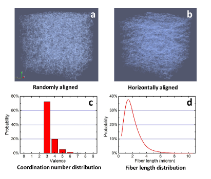
We employ a set of statistical descriptors for quantifying the network geometry and topology ref33 , which include the node density (corresponding to the collagen concentration), the fiber (or bond) length distribution function , the distribution of coordination number (i.e., the number of neighbors of a node) , and the average fiber orientation (measured as the average cosine value associated with the acute angle of a fiber made with respect to a prescribed direction). These statistical descriptors can be computed from 3D ECM network extracted from confocal images via skeletonization techniques ref25 . Fig. 1(c) and (d) respectively shows the coordination distribution and fiber length distribution for homogeneous collagen networks with a collagen concentration of , which will be used in our subsequent investigations. The average fiber length is and the average coordination number . Since the fibers are randomly oriented in homogeneous networks, the average fiber orientation metric . The node number density .
For a given set of network statistics (e.g., , , and ), we can generate realizations of the networks associated with the prescribed descriptors using stochastic reconstruction ref33 . In particular, we start from a randomly generated initial network with the prescribed node number density . From this initial network, the descriptors , , and are computed and compared to the corresponding prescribed descriptors. An energy functional is defined as the sum of the squared differences between the computed and corresponding prescribed the descriptors ref33 , i.e.,
| (1) |
Next, the initial network is perturbed by randomly displacing a node and/or removing/adding a bond to randomly selected pairs of nodes. A new energy for the new network is computed. If the new energy is lower than the old energy , the new network replaces the old one. Otherwise, the new network configuration replaces the old network with the probability , where is a virtual temperature, which possesses an initial large value and is gradually decreased. The network is continuously evolved in this way (more precisely, via simulated annealing method sim_annealing to allow even energy-increasing network during the initial stages) until , i.e., the computed descriptors match t he prescribed ones within a prescribed small tolerance. The detailed of this technique is provided in Ref. ref33 .
We note that one can either use experimentally obtained network statistics as the target descriptors in the reconstruction or can construct a set of feasible hypothetical statistical descriptors in order to control the geometry and topology of the constructed random network. Fig. 1(a) shows a reconstructed network based on the experimentally obtained statistics of the collagen gels, in which the fibers are randomly oriented. In order to investigate the effects of fiber alignment on cell migration dynamics, we also generate realizations of networks with horizontally aligned fibers (see Fig. 1(b)). This is achieved by setting with respect to the x-direction, and using the same , and of the homogeneous network. We note that the optimized of the reconstructed network is in fact a little smaller than unity (), due to additional topological and geometrical constraints specified by and . Nevertheless, the fiber alignment is already very significant in the reconstructed networks.
II.2 Micro-mechanical Model of ECM Networks
The ECM (collagen) fibers are highly non-linear, typically exhibiting buckling, strain-hardening and plastic behaviors ref25 ; MacKintosh05 ; Safran12 ; nat_method15 , which can significantly affect the propagation of the active forces in the system. The nonlinearity of the ECM fibers also induces a nontrivial coupling with the cell contractility, i.e., for small contraction, the fibers may be in the linear elastic regime, while for large contraction, the fibers may be in the strain-hardening or plastic regime ref11 . This in turns can affect the overall cell migration dynamics ref30 .
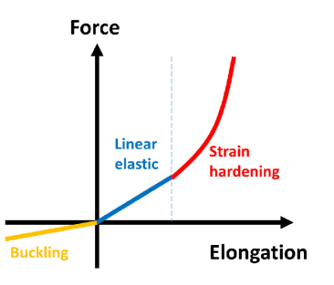
In this work, we will use a nonlinear micromechanical model for the ECM fiber, which is schematically illustrated in Fig. 2 ref11 ; nat_method15 . In particular, upon stretching, a fiber first enters a linear elastic regime, which is followed by a strong strain-hardening regime once the elongation is larger than a prescribed threshold. Upon compression, we consider the fiber immediately buckles and thus, possesses a much smaller compression modulus. The elongation stiffness of the fiber is thus given by
| (2) |
where and are respectively the Young’s modulus and cross-sectional area of the fiber bundle, and we use ref25 ; is elongation strain, and and are parameters for the strain-hardening model nat_method15 ; describes the effects of buckling ref11 . In addition, we consider the fiber segments as well as the cross links (nodes) can resist bending and employ a first-order bending approximation Frey07 , for which the bending energy is a function of transverse displacement of the two nodes of a fiber, i.e., , where the bending modulus ref25 , is the second moment of area, is the original length of the fiber segment, and . We also note that the effects of interstitial fluid, which quickly dissipates the kinetic energy generated due to cell contraction, are not explicitly considered.
Plasticity of the fibers is modeled as a time-dependent elongation of the fiber with a constant flow rate , i.e., , once the stretching force on the fiber is larger than a prescribed threshold . The flow rate can be calibrated based on experimental data available in literature ref29 . We note that this elongation due to plasticity effectively reduces the stiffness of the fiber, i.e., . In addition, we can easily construct a stiffness gradient in the ECM network, by introducing a position-dependent scaling factor, i.e., , where . It is clear that other forms of than the simple linear scaling could be employed to model more stiffness gradient. In the subsequent studies, we will use the simple constant gradient to investigate the durotaxis effects.
Once the cell contractions are applied (as described in Sec. III), an iteration procedure ref11 will be employed to find the force-balanced state of the network and obtain the forces on the fibers. The numerical procedure can be easily parallelized using OpenMP for large networks.
III Modeling cell migration regulated by cell-ECM micro-mechanical coupling
In this section, we present in detail the cell migration model, which is coupled with the ECM network model. We note that the current model is targeted for highly motile non-invasive cancer cells, such as the MCF-10A breast cancer cells, moving on 3D collagen gel (see Fig. 3 for illustration). In this case, the migrating cells are strongly coupled with the ECM via their micro-mechanical interactions without any ECM degradation, which is very challenging to accurately model. We will briefly discuss the generalization of the current model to incorporate ECM degradation in Sec. VI.
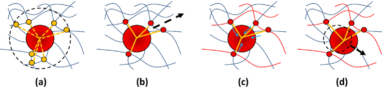
As illustrated in Fig. 3, our cell model consists of an elastic sphere representing the exclusion volume associated with cytoplasm and a set of cytoskeleton filaments connecting the cytoplasm sphere to the plasma membrane. As a starting point, we will not distinguish various types of actin assemblies and microtubules for simplicity, and only consider the contractility of the filaments. The plasma membrane is then modeled as the minimal hull associated with the end points of the filaments (see Fig. 3(A)). In the beginning of the simulation, a cell (i.e., a cytoplasm sphere and the associated cytoskeleton filaments) is introduced in the collagen network, and a random persistent direction is selected. The migration process is decomposed into cycles of successive events including (i) development of protrusion (due to active actin filaments polymerization) and formation of new adhesion sites, (ii) contraction of actin filaments and the resulting locomotion of the cell, and (iii) breaking of old adhesion sites. These events are modeled and simulated as described below:
(i) Protrusions are generated by the elongation (polymerization) of the actin filaments, which can be attached to the ECM fibers via focal adhesion. This process is modeled by adding new filaments connecting the center of mass of the cell (i.e., the center of the cytoplasm sphere) to a randomly selected node of the ECM network within (effective protrusion length) from the cell surface (see Fig. 3(a) and (b)), with the probability given by
| (3) |
where is the persistent direction of the cell, is vector connecting the cell center and the network node, is the largest stress on the fibers connected to the node, and are proportionality constants. This model implies that actin polymerization is more likely to occur in the polarized region of the cell Zallen2009 ; and that it is more likely to form an adhesion site on highly stressed fibers Munro2011 ; Matsumura2004 . Each adhesion site has a finite life span and breaks once is reached.
(ii) The contraction of an actin filament connecting the cytoplasm sphere and ECM network can generate a traction force () along the filament boey1998 ; boey1998II ; coughlin03 ; gordon2012 and a shrinkage of the filament length ( of the original length); see Fig. 3(c). This active force is transmitted to the ECM network through the “focal adhesion” node. Force boundary condition is imposed to this node (and other nodes connected to contracting filaments) and the deformed force-balanced network configuration is obtained as described in Sec. II.B. The length of the filament connecting the center cell and the displaced node is then computed. We then consider the contraction of this filament generates a displacement component for the cell enter, i.e.,
| (4) |
where is intrinsic contraction of the filament, and are respectively the distance between the cell center and the adhesion node before and after ECM deformation due to filament contraction, and is the unitary direction vector along the filament direction after ECM deformation.
(iii) Once the displacement components associated with all filaments are computed, the center of mass position of the cell is updated as follows (see Fig. 3(d)):
| (5) |
where the sum is taken for all filaments, and is the displacement component associated with the th filament. The persistent direction is updated as the direction of the cell displacement (i.e., ). We note that Eq. (4) and Eq. (5) imply that cell locomotion is due to actin filament contraction and depends on the stiffness of the local ECM.
(iv) All of the current adhesion sites are checked and those reach their life span are consider to break, leading to the detachment of the cell surface from the collagen fibers.
In the simulation, time is discretized such that a migration cycle is completed during the elapsing of one time step . The life time of focal adhesion sites , which is calibrated based on the experimental data (see Sec. IV for details). Once an entire migration cycle is completed, the position of cytoplasm sphere (and thus, the center of mass of the cell) is updated and the cell starts the next migration cycle, by repeating the steps (i) to (iv).
We also note that the cell-cell contact adhesion is not explicitly considered in this model, since our focus here is highly motile breast cancer cells with very weak cell-cell adhesion. In addition, we employ a minimal model for the contact inhibition effect for multi-cellular systems. In particular, we consider that if a pair of cells with radius () overlap, they feel a mutual repulsive force proportional to the linear overlap distance, i.e., , where is an effective elastic constant depending on the modulus of the cell, is the overlap distance, and is the cell center separation distance. In the subsequent sections, we will validate our model using single-cell migration experiments and employ the model to predict multi-cell migration dynamics.
IV Single-cell migration dynamics
In this section, we employ our model to investigate single cell migration dynamics and its regulation by the microstructure and mechanical properties of the micro-environment (i.e., the ECM network). We mainly focus on MCF-10A breast cancer cells in our simulations. The MCF-10A cell are highly motile non-metastatic cancer cells which exhibit strong micro-mechanical coupling of ECM networks when moving on collagen gels and do not degrade the collagen fibers qihui2019 . Therefore, this system provides an ideal system for testing our model. In the following discussions, we will directly use the experimental results to validate our model predictions. The experimental details are provided in Ref. qihui2019 .
IV.1 Migration dynamics of MCF-10A cells on isotropic collagen gel
We first employ our model to study the migration dynamics of individual MCF-10A breast cancer cells on isotropic collagen gels with randomly oriented fibers. It is well established that in the case, the overall cell dynamics can be captured by the active-particle model RMP , i.e.,
| (6) |
where is the particle center of mass, is an effective friction coefficient, is an effective constant self-propelling force, is the persistent direction which is subject to a random rotational diffusion and is a white-noise random vector RMP . The associated theoretical mean square displacement (MSD) is given by RMP
| (7) |
where is the diffusivity of the particle, is the persistent velocity and is the relaxation time for rotation diffusion of the persistent direction. It can be seen from Eq. (7) that for small , the particle exhibit ballistic dynamics with . At large , the system is diffusive, with .
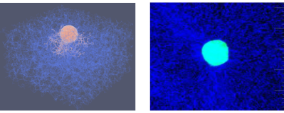
Figure 4 shows the 3D visualization of a single MCF-10A migrating on isotropic collagen gel with randomly oriented fibers (see the left panel). The 3D collagen network model is obtained via stochastic reconstruction based on the structural statistics extracted from confocal images, as described in Sec. IIA. The contraction of the actin filaments generates active tensile forces, which are transmitted to the collagen fibers and propagate in the ECM network. The fibers carrying large tensile forces are highlighted in red color. The right panel of the figure shows a confocal microscopy image of a migrating MCF-10A cell (bright blue) on isotropic collagen gel. It can be seen that the collagen fibers in the vicinity of the cell surface tend to orient perpendicularly to the cell surface, implying that the cell generates traction forces and pulls the fibers, consistent with the simulation results.
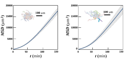
Figure 5 shows the mean square displacement (MSD) of a single MCF-10A cell migrating on isotropic collagen gel with randomly oriented fibers, respectively obtained from computer simulation (left panel) and in vitro experiment (right panel). The initial ballistic dynamics (i.e., ) can be clearly seen, which is followed by the diffusive dynamics (i.e., ). The cell diffusivity obtained from the simulations and experiments are respectively and , which agree well with one another. The insets of Fig. 5 show the trajectories of an ensemble cells respectively obtained from the simulations and experiments. It can be clearly seen that the cell migration is isotropic, as expected for a cell in a homogeneous micro-environment without any externally applied cues. These results clearly indicate the validity of our model.
IV.2 Migration dynamics of MCF-10A cells on collagen gel with aligned fibers
With our model validated by experiments, we now employ it to study cell migration in complex micro-environment, such as collagen gels with aligned fibers, which are difficult to fabricate experimentally. The 3D virtual ECM networks are stochastic constructed by maximizing the fiber orientation metric along the x-direction (see Sec. IIA for details). This leads to model networks with fibers mainly aligned along the x-direction (see Fig. 6).
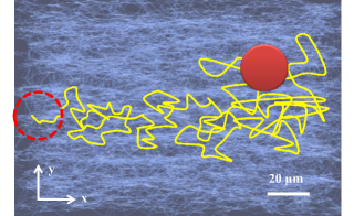
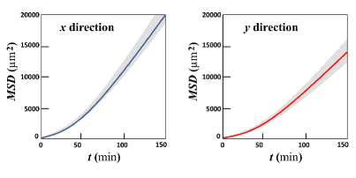
Figure 6 shows a typical trajectory of a MCF-10A cell migrating on 3D collagen gel with horizontally aligned fibers obtained from simulations. It can be clearly seen that the cell tends to migrate along the direction consistent with the fiber alignment direction (e.g., in this case, x-direction). This can also been seen quantitatively seen from the MSD analysis. Figure 7 shows the MSD of the migrating cell respectively along the x-direction (left panel) and y-direction (right panel). Anisotropy in the migration can be clearly observed, i.e., the cell moves much faster long the fiber alignment direction than the perpendicular direction.
We note that the phenomenon that cells tend to migrate along the fiber alignment direction is well known and termed as “contact guidance” ref18 ; ref19 . In our simulations, as the migrating cell pulls the ECM fibers, the large tensile forces are mainly carried by chains of aligned fibers, which are typically referred to as “force chains” ref11 ; ref33 . The high-stress fibers on the force chains are effective stiffer (e.g., due to strain hardening) and thus, can support large-magnitude locomotion steps along chain directions, and in this case, the fiber alignment direction.
IV.3 Migration dynamics of MCF-10A cells on collagen gel with a stiffness gradient
We now employ our model to study cell migration dynamics on collagen gels with a stiffness gradient. As described in Sec. IIA, the structural model of the 3D ECM is constructed based on the experimentally obtained statistics of a 2 mg/ml collagen gel with randomly oriented fibers. Once the 3D structural model is obtained, a linear stiffness distribution a long x-direction with a constant gradient is built. This is achieved by re-scaling the Young’s modulus of the fiber according to , where is the x-coordinate of the center of the fiber.
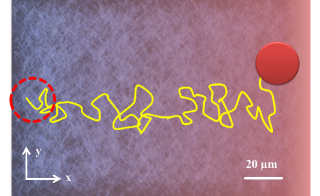
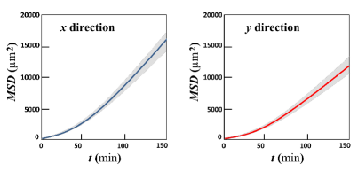
Figure 8 shows a typical trajectory of a MCF-10A cell migrating on 3D collagen gel with a stiffness gradient along the x-direction. Similar to the case of contact guidance, it can be clearly seen that the cell tends to migrate along the direction against the stiffness gradient along the positive x-direction. This can also been seen quantitatively seen from the MSD analysis. Figure 9 shows the MSD of the migrating cell respectively along the x-direction (left panel) and y-direction (right panel). Anisotropy in the migration can be clearly observed, i.e., the cell moves much faster long the stiffness gradient direction than the perpendicular direction. We note that an important distinction between migration anisotropy in this case and the contact guidance case is that here the cell migration is uni-directional, i.e., up the stiffness gradient; while in the contact guidance case, the migration is bi-directional, i.e., along the fiber alignment direction but the cells can go in both ways.
The phenomenon that cells migrate against stiffness gradient of the ECM is well known and termed as “durotaxis” ref15 ; ref16 ; Brown2009 . In our simulations, as the migrating cell pulls the ECM fibers, the stiffer fibers will possess smaller deformation, which in turn leads to larger locomotion components towards these fibers (c.f. Eq.(4)). The accumulated effect of many local migration steps is the overall biased migration up the stiffness gradient as observed in the experiments.
V Strongly correlated multi-cellular dynamics
In Sec. IV, we show that our computational model can capture the salient features of single-cell migration dynamics in both homogeneous and complex micro-environment. In this section, we employ the model to investigate multi-cellular migration dynamics. As mentioned in Sec. III, we do not explicitly model cell-cell adhesion here (due to the weak adhesion between the cancer cells) and use a minimal model for cell-cell repulsion due to contact inhibition (see Sec. III for details). In addition, in this study, we focus on relatively small system, containing cells.
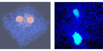
Figure 10(a) shows 3D visualization of a small portion (with a linear size ) of the simulation box which contains two closely spaced MCF-10A cells migrating on isotropic collagen gel with randomly oriented fibers. The active tensile forces generated by the cells (due to actin filament contraction) are transmitted to the collagen fibers. The collagen fibers carrying large tensile forces are highlighted in red. Figure 10(a) shows the confocal microscopy image of a pair of migrating MCF-10A cells (bright blue) on collagen gel. It can be clearly seen that the collagen fibers (dark blue) between the two cells form a mesoscopic scale structure, which is clearly distinguished from original homogeneous ECM network and is consistent with the meso-scale structure formed by the high-stress fibers in our simulations.
To quantify the correlations in the collective migration dynamics of multiple MCF-10A cells, we employ the velocity correlation function , i.e.,
| (8) |
where , , denote a pair of cells connected by the remodeled meso-scale ECM structures and denotes ensemble average over all different cell pairs. We note that in computing , we only consider a subsets of cell pairs, i.e., those between which the meso-scale structures are formed. This allows us to clearly obtain the effects of such meso-structure on the collective dynamics of the cells, if any. Due to cell’s mutual exclusion effects, for and is roughly the diameter of a cell. In addition, two cells separated by very large distances are not correlated, i.e., for large values. A positive indicates that the cells tend to move in the same direction, implying a net “flow” of cells in the system. On the other hand, a negative indicates that the cells move towards one another, implying the formation of aggregation or clusters.
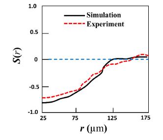
Figure 11 shows the velocity correlation function of MCF-10A cells migrating on isotropic collagen gel with randomly oriented fibers, respectively obtained from computer simulation (solid curve) and in vitro experiment (dashed curve). It can be seen that the simulation results agree very well with experimental data. Interestingly, the functions (beyond the trivial exclusion region) start from a very negative value (close to the minimal value -1) around , slowly increase to zero and then fluctuate around zero. This indicates the cells tend to move towards one another, facilitated by the mesoscopic structures of the remodeled ECM, which is also confirmed by time-lapse confocal data qihui2019 .
Our results indicate that strongly correlated cell migration dynamics is correlated with the meso-scale ECM structures due to cell remodeling. One possible reason is that the meso-structures are composed of many force chains (or a “force network”), which are in turn composed of fibers carrying large tensile forces. Therefore, the fibers in the meso-structures (at least in our simulations) are stiffer than the remaining stress-free fibers, which indicates that the meso-structures themselves are stiffer than the surrounding ECM. These stiffer meso-structures can then facilitate correlated cell migration via durotaxis, and also facilitate indirect mechanical coupling between the migrating cells.
VI Conclusions and discussion
In this paper, we develop a computational model for cell migration in complex micro-environment, which explicitly takes into account a variety of cellular level processes including focal adhesion formation and disassembly, active traction force generation and cell locomotion due to actin filament contraction, transmission and propagation of tensile forces in the ECM. We employ statistical descriptors obtainable from confocal imagining to quantify and control the 3D ECM network microstructure and use a nonlinear mechanical model for the ECM networks, which incorporates buckling of collagen fibers upon compression and strain-hardening upon stretching. We validate our model by accurately reproducing single-cell dynamics of MCF-10A breast cancer cells migrating on collagen gels and show that the durotaxis and contact guidance effects naturally arise as a consequence of the cell-ECM micro-mechanical interactions considered in the model. Moreover, our model predicts strongly correlated multi-cellular migration dynamics, which are resulted from the ECM-mediated mechanical coupling among the migrating cell and are subsequently verified in in vitro experiments using MCF-10A cells.
Although focusing on the non-metastatic MCF-10A breast cancer cells migrating on 3D collagen gels, our model can be generalize to investigate the migration of mesenchymal cells (e.g., invasive MDA-MB-231 breast cancer cells) in 3D ECM. The key modification is to explicitly model ECM degradation by the cells, which can be achieved using the following rule: A migrating cell degrades collagen fibers with a probability , with the distance from the fiber to the cell membrane. A degraded fiber is removed from the network in subsequent simulation steps. In addition, cell-cell adhesion can also be easily incorporated into the model to investigate a wide range of cell lines with different phenotypes. With proper modifications and generalizations, as well as efficient parallel implementation, it is expected that the model could be employed to investigate collective migratory behaviors and emergent self-organizing multi-cellular patterns resulted from ECM-mediated mechanical signaling among the cells.
Acknowledgements.
Y. Z., H. N. and Y. J. thank Arizona State University for the generous start-up funds and the University Graduate Fellowships. Q. F., X. W., and F. Y. thank the Chinese Academy of Sciences (CAS), the Key Research Program of Frontier Sciences of CAS (Grant No. QYZDB-SSW-SYS003). L. L. and R. L. thank the National Natural Science Foundation of China (Grants No. 11474345, No. 11674043, No. 11604030, and No. 11774394). B. S. thank the support from the Scialog Program sponsored jointly by Research Corporation for Science Advancement and the Gordon and Betty Moore Foundation. B. S. is partially supported by the Medical Research Foundation of Oregon and SciRIS-II award from Oregon State University and by the National Science Foundation Grant PHY-1400968.References
- (1) A. Aman, and T. Piotrowski, Cell migration during morphogenesis. Developmental Biology 341, 20-33 (2010)
- (2) P. Friedl, and D. Gilmour, Collective cell migration in morphogenesis, regeneration and cancer. Nature Reviews Molecular Cell Biology 10, 445 (2009).
- (3) A. Vaezi, C. Bauer, V. Vasioukhin, and E. Fuchs, Actin cable dynamics and Rho/Rock orchestrate a polarized cytoskeletal architecture in the early steps of assembling a stratified epithelium. Developmental Cell 3, 367-381 (2002).
- (4) S. Werner, T. Krieg, and H. Smola, Keratinocyte fibroblast interactions in wound healing. Journal of Investigative Dermatology 127, 998-1008 (2007).
- (5) A. J. Ridley, M.A. Schwartz, K. Burridge, R.A. Firtel, M.H. Ginsberg, G. Borisy, J.T. Parsons, and A.R. Horwitz, Cell migration: integrating signals from front to back. Science 302, 1704-1709 (2003).
- (6) P. Friedl, and E.-B. Brocker, The biology of cell locomotion within three-dimensional extracellular matrix. Cellular and Molecular Life Sciences CMLS 57, 41-64 (2000).
- (7) H. Szurmant, and G.W. Ordal, Diversity in chemotaxis mechanisms among the bacteria and archaea. Microbiology and Molecular Biology Reviews 68, 301-319 (2004).
- (8) S. V. Plotnikov, A.M. Pasapera, B. Sabass, and C.M. Waterman, Force fluctuations within focal adhesions mediate ECM-rigidity sensing to guide directed cell migration. Cell 151, 1513-1527 (2012).
- (9) R. Sunyer, V. Conte, J. Escribano, A. Elosegui-Artola, A. Labernadie, L. Valon, D. Navajas, J.M. Garc a-Aznar, J.J. Munoz, and P. Roca-Cusachs, Collective cell durotaxis emerges from long-range intercellular force transmission. Science 353, 1157-1161 (2016).
- (10) E. Hadjipanayi, V. Mudera, and R. A. Brown, Guiding cell migration in 3D: a collagen matrix with graded directional stiffness. Cell Motil. Cytoskeleton 66 121-8 (2009).
- (11) S. B. Carter, Haptotaxis and the mechanism of cell motility. Nature 213, 256 (1967).
- (12) P. P. Provenzano, D.R. Inman, K.W. Eliceiri, S.M. Trier, and P.J. Keely, Contact guidance mediated three-dimensional cell migration is regulated by Rho/ROCK-dependent matrix reorganization. Biophysical Journal 95, 5374-5384 (2008).
- (13) J. H. Wang, and E.S. Grood, The strain magnitude and contact guidance determine orientation response of fibroblasts to cyclic substrate strains. Connective Tissue Research 41, 29-36 (2000).
- (14) S. Guido and R. T. Tranquillo, A methodology for the systematic and quantitative study of cell contact guidance in oriented collagen gels. Correlation of fibroblast orientation and gel birefringence. J. Cell Sci. 105, 317-31 (1993).
- (15) S. Wang, and P.G. Wolynes, Active contractility in actomyosin networks. Proceedings of the National Academy of Sciences 109, 6446-6451 (2012).
- (16) T. Lecuit, P.-F. Lenne, and E. Munro, Force generation, transmission, and integration during cell and tissue morphogenesis. Annual Review of Cell and Developmental Biology 27, 157-184 (2011).
- (17) M. A. Schwartz, Integrins and extracellular matrix in mechanotransduction. Cold Spring Harbor perspectives in biology, a005066 (2010).
- (18) G. Totsukawa, Y. Wu, Y. Sasaki, D.J. Hartshorne, Y. Yamakita, S. Yamashiro, and F. Matsumura, Distinct roles of MLCK and ROCK in the regulation of membrane protrusions and focal adhesion dynamics during cell migration of fibroblasts. The Journal of Cell Biology 164, 427-439 (2004).
- (19) C. A. Jones, M. Cibula, J. Feng, E.A. Krnacik, D.H. McIntyre, H. Levine, and B. Sun, Micromechanics of cellularized biopolymer networks. Proceedings of the National Academy of Sciences 112, E5117-E5122 (2015).
- (20) S. B. Lindstrom, D.A. Vader, A. Kulachenko, and D.A. Weitz, Biopolymer network geometries: characterization, regeneration, and elastic properties. Physical Review E 82, 051905 (2010).
- (21) H. Mohammadi, P.D. Arora, C.A. Simmons, P.A. Janmey, and C.A. McCulloch, Inelastic behaviour of collagen networks in cell matrix interactions and mechanosensation. Journal of The Royal Society Interface 12, 20141074 (2015).
- (22) S. Nam, K.H. Hu, M.J. Butte, and O. Chaudhuri, Strain-enhanced stress relaxation impacts nonlinear elasticity in collagen gels. Proceedings of the National Academy of Sciences, 201523906 (2016).
- (23) Nam, S., J. Lee, D.G. Brownfield, and O. Chaudhuri, Viscoplasticity enables mechanical remodeling of matrix by cells. Biophysical journal, 2016. 111(10): p. 2296-2308.
- (24) J. Kim, J. Feng, C.A. Jones, X. Mao, L.M. Sander, H. Levine, and B. Sun, Stress-induced plasticity of dynamic collagen networks. Nature Communications 8, 842 (2017).
- (25) S. Chen, W. Xu, J. Kim, H. Nan, Y. Zheng, B. Sun, and Y. Jiao, Novel inverse finite-element formulation for reconstruction of relative local stiffness in heterogeneous extra-cellular matrix and traction forces on active cells. Physical Biology 16, 036002 (2019).
- (26) A. D. Doyle, N. Carvajal, A. Jin, K. Matsumoto, and K.M. Yamada, Local 3D matrix microenvironment regulates cell migration through spatiotemporal dynamics of contractility-dependent adhesions. Nature Communications 6, 8720 (2015).
- (27) C. Heussinger and E. Frey, Force distributions and force chains in random stiff fiber networks. Eur. Phys. J. E 24 47 53 (2007).
- (28) F. Grinnell, and W.M. Petroll, Cell motility and mechanics in three-dimensional collagen matrices. Annual Review of Cell and Developmental Biology 26, 335-361 (2010).
- (29) Y. L. Han, P. Ronceray, G. Xu, A. Malandrino, R.D. Kamm, M. Lenz, C.P. Broedersz, and M. Guo, Cell contraction induces long-ranged stress stiffening in the extracellular matrix. Proceedings of the National Academy of Sciences 115, 4075-4080 (2018).
- (30) X. Ma, M.E. Schickel, M.D. Stevenson, A.L. Sarang-Sieminski, K.J. Gooch, S.N. Ghadiali, and R.T. Hart, Fibers in the extracellular matrix enable long-range stress transmission between cells. Biophysical Journal 104, 1410-1418 (2013).
- (31) P. Ronceray, C.P. Broedersz, and M. Lenz, Fiber networks amplify active stress. Proceedings of the National Academy of Sciences 113, 2827-2832 (2016).
- (32) H. Wang, A. Abhilash, C.S. Chen, R.G. Wells, and V.B. Shenoy, Long-range force transmission in fibrous matrices enabled by tension-driven alignment of fibers. Biophysical Journal 107, 2592-2603 (2014).
- (33) F. Beroz, L.M. Jawerth, S. Munster, D.A. Weitz, C.P. Broedersz, and N.S. Wingreen, Physical limits to biomechanical sensing in disordered fibre networks. Nature Communications 8, 16096 (2017).
- (34) L. Liang, C. Jones, S. Chen, B. Sun, and Y. Jiao, Heterogeneous force network in 3D cellularized collagen networks. Physical Biology 13, 066001 (2016).
- (35) M. H. Zaman, R. D. Kamm, P. Matsudaria, and D. A. Lauffenburger. Computational model for cell migration in three-dimensional matrices. Biophys. J. 89, 1389 (2005).
- (36) A. Vaziri and A. Gopinath. Cell and biomolecular mechanics in silico. Nature Materials 7, 15 (2008).
- (37) P. Masuzzo, M. Van Troys, C. Ampe, and L. Martens, Taking aim at moving targets in computational cell migration. Trends in cell biology 26, 88 (2016).
- (38) F. Ziebert, S. Swaminathan, and I. S. Aranson. Modeling for self-polarization and motility of keratocyte fragments. J. R. Soc. Interface 9, 1084 (2011).
- (39) D. Shao, W. J. Rappel, and H. Levine. Computational model for cell morphodynamics. Phys. Rev. Lett. 105, 108104 (2010).
- (40) D. Shao, H. Levine, and W. J. Rappel. Coupling actin flow, adhesion, and morphology in a computational cell motility model. Proc. Natl. Acad. Sci. USA 109, 6851 (2012).
- (41) U. Z. George, A. Stephanou, and A. Madzvamuse. Mathematical modeling and numerical simulations of actin dynamics in the eukaryotic cell. J. Math. Biol. 66, 547 (2013).
- (42) T. C. Bidone, W. Jung, D. Maruri, C. Borau, R. D. Kamm, and T. Kim, Morphological transformation and force generation of active cytoskeletal networks, PLoS Comput. Biol. 13, e1005277 (2017).
- (43) M. C. Kim, J. Whisler, Y. R., Silberberg, et al. Cell invasion dynamics into a three dimensional extracellular matrix fibre network. PLoS Comput. Biol. 11, e1004535 (2015).
- (44) T. Vicsek, A. Czirok, E. Ben-Jacob, I. Cohen, and O. Shochet, Novel type of phase transition in a system of self-driven particles. Physical review letters 75, 1226 (1995).
- (45) C. Bechinger, R. Di Leonardo, H. Lowen, C. Reichhardt, G. Volpe, Active particles in complex and crowded environments. Reviews of Modern Physics 88, 045006 (2016).
- (46) D. Bi, J. Lopez, J. Schwarz, M. L. Manning, A density-independent rigidity transition in biological tissues, Nature Physics 11, 1074 (2015).
- (47) F. Graner, and J. A. Glazier, Simulation of biological cell sorting using a two-dimensional extended Potts model. Physical Review Letters 69, 2013 (1992).
- (48) Y. Jiao and S. Torquato, Emergent Properties from a Cellular Automaton Model for Invasive Tumor Growth in Heterogeneous Environment, PLoS Computational Biology 7, 1002314 (2011).
- (49) Y. Jiao and S. Torquato, Diversity of Dynamics and Morphologies of Invasive Solid Tumors, AIP Advances 2, 011003 (2012)
- (50) Y. Jiao and S. Torquato, Evolution and Morphology of Microenvironment-Enhanced Malignancy of Three-Dimensional Invasive Solid Tumors, Physical Review E 87, 052707 (2013)
- (51) H. Xie, Y. Jiao, Q. Fan, et. al., Modeling Three-dimensional Invasive Solid Tumor Growth in Heterogeneous Microenvironment under Chemotherapy, PLoS One 13, e0206292 (2018)
- (52) H. Abdel-Rahman, B. Thomas, and T. Kim, A mechanical model for durotactic cell migration, ACS Biomater Sci Eng (2019)
- (53) M. Dietrich, H. Le Roy, D. B. Bruckner, H. Engelke, R. Zantl, J. O. Radler and C. P. Broedersz, Guiding 3D cell migration in deformed synthetic hydrogel microstructures, Soft Matter 14, 2816 (2018).
- (54) Nan, H., L. Liang, G. Chen, L. Liu, R. Liu, and Y. Jiao, Realizations of highly heterogeneous collagen networks via stochastic reconstruction for micromechanical analysis of tumor cell invasion. Physical Review E 97, 033311 (2018).
- (55) S. Kirkpatrick, C. D. Gelatt, and M. P. Vecchi, Optimization by simulated annealing. Science 220, 671 (1983).
- (56) D. A. Head, A. J. Levine, and F. C. MacKintosh, Mechanical response of semiflexible networks to localized perturbations. Phys. Rev. E 72 061914 (2005).
- (57) Y. Shokef and S. A. Safran, Scaling laws for the response of nonlinear elastic media with implications for cell mechanics. Phys. Rev. Lett. 108, 178103 (2012).
- (58) J. Steinwachs, C. Metzner, K. Skodzek, et. al. Three-dimensional force microscopy of cells in biopolymer networks. Nat. Method 13 171-6 (2016).
- (59) R. Fernandez-Gonzalez, M. S. Simoes, J. C. Roper, S. Eaton, and J. A. Zallen. Myosin ii dynamics are regulated by tension in intercalating cells. Dev. Cell 17, 736 743 (2009).
- (60) T. Lecuit, P. Lenne, and E. Munro. Force generation, transmission and integration during cell and tissue morphogenesis. Annu. Rev. Cell Dev. Biol. 27, 157 184 (2011).
- (61) G. Totsukawa, Y. Wu, Y. Sasaki, D. J. Hartshorne, Y. Yamakita, S. Yamashiro, and F. Matsumura. Distinct roles of mlck and rock in the regulation of membrane protrusions and focal adhesion dynamics during cell migration of fibroblasts. J. Cell Biol. 164, 427 439 (2004).
- (62) S. K. Boey, D. H. Boal, and D. Discher. Simulations of the erythrocyte cytoskeleton at large deformation. i. microscopic models. Biophys. J. 75, 1573 1583 (1998).
- (63) D. Discher, D. H. Boal, and S. K. Boey. Simulations of the erythrocyte cytoskeleton at large deformation. ii. micropipette aspiration. Biophys. J. 75, 1584 1597 (1998).
- (64) M. F. Coughlin and D. Stamenovic. A prestressed cable network model for the adherent cell cytoskeleton. Biophys. J. 84, 1328 1336 (2003).
- (65) D. Gordon, A. Bernheim-Groswasser, C. Keasar, and O. Farago. Hierarchical self-organization of cytoskeletal active networks. Phys. Biol. 9, 026005 (2012).
- (66) Q. Fan, Y. Zheng, H. Nan, Y. Jiao, and F. Ye, Strongly correlated cell dynamics induced by ECM-mediated long-range mechanical coupling, submitted.
- (67) C. Bechinger, R. D. Leonardo, H. Lowen, C. Reichhardt, G. Volpe and G. Volpe, Active particles in complex and crowded microenvironments, Rev. Mod. Phys. 88, 045006 (2016).