Efficient video indexing for monitoring disease activity and progression in the upper gastrointestinal tract
Abstract
Endoscopy is a routine imaging technique used for both diagnosis and minimally invasive surgical treatment. While the endoscopy video contains a wealth of information, tools to capture this information for the purpose of clinical reporting are rather poor. In date, endoscopists do not have any access to tools that enable them to browse the video data in an efficient and user friendly manner. Fast and reliable video retrieval methods could for example, allow them to review data from previous exams and therefore improve their ability to monitor disease progression. Deep learning provides new avenues of compressing and indexing video in an extremely efficient manner. In this study, we propose to use an autoencoder for efficient video compression and fast retrieval of video images. To boost the accuracy of video image retrieval and to address data variability like multi-modality and view-point changes, we propose the integration of a Siamese network. We demonstrate that our approach is competitive in retrieving images from 3 large scale videos of 3 different patients obtained against the query samples of their previous diagnosis. Quantitative validation shows that the combined approach yield an overall improvement of 5% and 8% over classical and variational autoencoders, respectively.
Index Terms— Endoscopy, deep learning, autoencoders, Siamese network, image retrieval
1 Introduction
Due to the lack of efficient tools for indexing and retrieval, the wealth of information contained in endoscopy video is only rarely used for improving clinical reporting and diagnosis. Today only manually selected still frames (low quality screenshots) are included into clinical reports. In addition, it is not feasible for endoscopists to look into long videos. Computer assisted methods for extracting clinically relavant video frames from a larger video stream do not exist. Compressing and indexing the entire video in an extremely efficient manner would open up the possibility to use corresponding data from previous exams to enable the monitoring of disease progression and response to therapy. Content-based image retrieval (CBIR) methods allow to locate images of interest (query) in target databases. Such techniques have been already applied on large scale medical databases [1, 2, 3].
Oesophageal carcinoma is the sixth leading cause of mortality and the eight most common cancer worldwide. The overall 5-year survival of patients with
oesophageal carcinoma ranges from 15% to 25%; while earlier diagnosis can result in improved survival rate [4]. Endoscopy is a routine clinical procedure for both diagnosis and early treatment of pre-cancerous malignancies seen in oesophagus commonly referred as “Barrett’s Oesophagus” (BE). Patients with BE have a 30–125-fold increased lifetime risk of developing oesophageal cancer. Barrett’s is defined as the substitution of the normal stratified squamous epithelium of the oesophagus with a columnar cell lining and can be visualized using an endoscope. It is therefore required that patients with BE must undergo periodic endoscopies during which biopsies are taken to examine the cancer risk. The goal of this work is to develop an approach that can effectively support monitoring of these patients utilizing information content of video endoscopy at an optimal compression, speed and retrieval accuracy which are important factors for clinical usability.
2 Related work
Existing CBIR approaches are typically not suitable for clinical use as they utilise a very low resolution representation for the video data. Important diagnostically relevant detail is often lost. Such system use derived features such as texture, colour, shape as well as local spatial properties to learn a low-dimensional representation. Often binary coding or hashing are used. Query images are then compared with low-dimensional representation of target images for fast image retrieval. The efficiency of CBIR directly depends on the used feature extraction and representation. Liu et al. [5] used colour difference histogram utilizing colours and edge orientations for better feature representation. Murala et al. [1] employed local binary patterns (LBP) to extract features from low-level wavelet sub-bands in CT and MRI data. Scale invariant feature transform (SIFT) were used in [2] to derive representation for bag-of-visual words. Ye et al. [3] used LBP based 496-dimensional image histogram descriptor and a hashing technique based on random forest for real time biopsy retargeting utilizing video endoscopy. All of these previous approaches are based on hand-crafted features and do not incorporate high-level semantic (semantic gap).
Recently, approaches that make use of advances in deep learning have demonstrated considerable success in learning both low- and high-level semantically relevant features. Ahmad et al. [6] suggested using a convolutional neural network (CNN) based salient features to retrieve endoscopic images. Convolutional kernels from the first layer of a pre-trained AlexNet model was used along with a pooling strategy for achieving compact 96 bin histogram. We argue that variations in endoscopy data can not be captured using such pre-trained models as the features in endocopic images are very different from natural images (used by such pre-trained networks). Masci and collegues [7] demontrated that unsupervised convolution autoencoders can be used to extract salient features. Krizhevsky and Hinton [8] revealed that deep autoencoders can be applied for extremely fast image retrieval task that is independent of database sizes due to high data compression capability of autoencoders. Stacked autoencoders were used by Sharma and colleagues [9] on medical images (x-ray data).
Two challenges need to be addressed before image automated retrieval from endoscopy video can be applied in the clinical setting: 1) Efficient compression - the number of images per video is extremely large (nearly 15-40 thousands) which demands for large storage (nearly 1.5-4 GB per video) and 2) preservation of diagnostically relevant features - while it is necessary to preserve a high level of anatomical detail, various artefacts need to be discarded. We argue that autoencoders are extremely well suited to address the first problem. However, in the presence of challenges posed by data variabilities, autoencoders can fail to accurately retrieve images in restricted search space. In this paper, we propose to use autoencoders (AE) and Siamese network working side-by-side for achieving better compression, and fast and accurate image retrieval. We demonstrated that combining classical AE with Siamese network or variational autoencoder (VAE, [10]) with Siamese results in improved accuracy. We observed that VAE compresses data nearly 70 folds more than classical AE at an improved retrieval speed but with a compromise in accuracy. However, when combined with Siamese network, it yields very competitive retrieval results. We have compared retrieval performances with and without Siamese network for both autoencoders on 3 different BE patient videos. The rest of the paper is organized as follows: Section 3 briefly describes both classical and variational autoencoders, Siamese networks and our combined approach for image retrieval task. Section 4 evaluates our combined approaches utilizing oesophageal endoscopic video data and Section 5 concludes the paper.
3 Method
| Method | Training | Compression | Testing(s) | |||||
|---|---|---|---|---|---|---|---|---|
| Samples | Epochs | Time(s) | Data | Comp. | Time(s) | Load | Execute | |
| AE | 33k/15k | 500 | 18/epoch | 15k | 160 MB | 1.63 | 3 | 0.16 |
| VAE | 33k/15k | 500 | 21/epoch | 15k | 1.7 MB | 1.21 | 2 | 0.18 |
| Siamese | 900/100 | 1000 | 48/epoch | - | - | - | 1.05 | 1.35 |
After providing a brief description of autoencoders and Siamese networks we motivate on why these should be combined and present our approach for efficient retrieval of video endoscopy.
3.1 Autoencoder
An autoencoder is an unsupervised machine learning algorithm that is capable of learning efficient and compressed data representation referred as “coding“ or “latent-space representation“, say . A reverse process “decoding“ is performed to achieve outputs similar to the input data. Decoder tries to reconstruct using fewer number of bits from the bottleneck (latent-space). A latent-space representation is learned when the dissimilarity between decoder output and input data is minimized.
Here, a convolutional autoencoder (CAE, [7]) that can be trained in an end-to-end fashion is used. Our architecture consists of 3 convolution filter layers with ‘relu‘ activation function and a fully connected last layer (32 dense connections). A subsequent downsampling with stride 2 is performed at each layer for encoder and a similar architecture with upsampling with stride 2 is performed for decoder. The decoder unflattens the encoder output first and then upscales it using similar size (mirrored) convolution filters. Cross-entropy has been used as a loss function with an Adadelta optimizer and a relu activation. The image size used for training is . There are in total of 4,089,283 trainable parameters. We have trained our CAE for 500 epochs utilizing 33,000 samples for training and 15,000 for validation.
3.2 Variational autoencoder
In contrast to classical autoencoders where the information regarding input data distribution is not known, variational autoencoders [10] assert the latent-space representation to be drawn from a unit normal distribution, . Thus, such autoencoders are effective generative models that can produce samples from the learned unit normal distribution. For our image retrieval task, the encoded target embedding might not exactly match the encoded query but VAEs have tremendous strength to learn more meaningful latent representations yielding in an effective search space.
We have used the same architecture (refer Sec.3.1) for our encoder-decoder network in VAE. However, the final layer of VAE encoding consists of mean and standard deviation encoding (vectors) for ’n’ ( for our case) latent embeddings. The actual coding is then randomly sampled from a unit normal distribution for decoding. Negative of cross-entropy loss is minimized using an Adam optimizer. In order to push the autoencoder to learn unit normal distribution, a second loss “latent loss“ is used which is computed as KL-divergence between the target normal distribution and the actual coding. The total trainable parameters are 1,362,480 which is lot less than classical autoencoder as only mean and variances in the data are learnt. In our retrieval task, we compare only mean embeddings between the encoded target dataset and query images. This reduces both the computational complexity and embedding file size (see Tab.1). We have used a 10-dimensional latent variable space and trained our VAE for 500 epochs utilizing 33,000 samples for training and 15,000 for validation.
3.3 Siamese network
A Siamese network [11] learns to differentiate between two input images and consists of two identical neural networks, say and , for doing this. The dissimilarities are computed as a contrastive loss function:
| (1) |
where is the Euclidean distance between outputs of sister networks, is the class label and is the margin value. Both sister networks and have exact same weights.
We have trained a Siamese network for dealing with multi-modality and varying view-points in our oesophageal endoscopy dataset. For this we created a database consisting of 100 sets of images with 10 images each (in total 1000) that included WL, NBI and 8 different viewing angles. Paired multi-modality images were generated by using a trained domain adaptive network (refere ‘cycleGAN’ [12]). CycleGAN was trained using 300 pairs of WL and NBI images. For addressing view-point changes in endoscopy, simulated images using different rotation angles () were generated. An Adam optimizer was used to minimize contrastive loss (see Eq. 1). The network was trained for 1000 epochs and 100 iterations with learning parameter of 0.005.
3.4 Combined approach
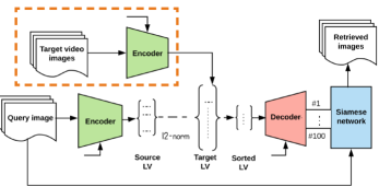
An overview of the proposed combined approach is presented in Fig. 1. First, both the autoencoder and the Siamese network are trained separately (see Section 3.1-3.3). Then, the trained autoencoder is used to compress the target video into a low dimensional latent variable space vector (target LV). Offline batch processing can be used to encode all target videos needed for patient follow-up. Thanks to the very high compression ratios this method achieves, only a modest amount of storage is required to hold the compressed videos. Given a new query image, the trained autoencoder is used to project it into the latent space. The resulting latent variable space vector (source LV) is then compared with the target LV using an metric. 100 LVs are sorted based on their similarity scores and are processed to either a decoder or an image list. The trained Siamese network is then used to penalise any images which don’t satisfy the similarity requirement. The distance output of the Siamese network is used to produce the final ranking of the candidate images. Finally, the -best retrieved images (in our case ) are being presented.
4 Experiments
10 oesophageal endoscopy videos were used in this study. Each video consists of more than 15,000 frames. 4 different patient video images were combined randomly and used in different proportions for training purposes of our networks (see Table 1) and 6 videos of 3 different patients were used for our frame retrieval test (3 for query and 3 for retrieval).
| Method | Patient | Average | ||||||||||
|---|---|---|---|---|---|---|---|---|---|---|---|---|
| #1 | #2 | #3 | ||||||||||
| TP | FP | P | TP | FP | P | TP | FP | P | TP | FP | P | |
| VAE | 356 | 134 | 0.73 | 351 | 139 | 0.71 | 437 | 53 | 0.89 | 381.3 | 108 | 0.77 |
| AE | 417 | 73 | 0.85 | 393 | 97 | 0.80 | 456 | 34 | 0.93 | 422 | 68 | 0.86 |
| VAE-Siamese | 399 | 91 | 0.81 | 410 | 80 | 0.83 | 439 | 51 | 0.89 | 416 | 74 | 0.85 |
| AE -Siamese | 437 | 53 | 0.89 | 437 | 53 | 0.89 | 473 | 17 | 0.96 | 449 | 41 | 0.91 |
4.1 Quantitative results

Query #1

Query #2


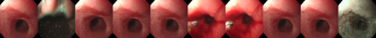


Table 2 shows image retrieval performance of both the sole application of autoencoders and our combined approaches. It can be seen that Siamese network improves the average retrieval precision by 8% and 5%, respectively, for VAE and AE. AE-Siamese yields the best results for all 3 patient cases (89%, 89% and 96%) and on average 6% higher precision than VAE-Siamese. However, from Tab. 1 it can be observed that VAE has the best compression performance, nearly 70 folds more than AE, and a very reasonable average precision of 85% (see Tab. 2).
4.2 Qualitative results
Fig. 2 presents visual analysis of image retrieval on two query video images in our dataset. These query images were searched in a same patient video archived 6 months earlier and consisted of nearly 21,462 image frames. For query #1 (top block), VAE (1st row) has more mismatches than AE (2nd row) and the retrieved images in both cases are not ordered even if they are matched. We can observe that VAE-Siamese (3rd row) has improved matches while AE-Siamese (4th row) has also been ordered better. Similarly, for query #2 (bottom block), AE (2nd row) has better matches than VAE (1st row). Infact, AE has perfect match for same modality. However, utilizing Siamese network on top, both VAE and AE (3rd and 4th rows, respectively) are able to capture more variabilities for the same site and includes NBI multi-modality cases.
5 Conclusion
While autoencoders allow for better compression of large-scale endoscopy data eliminating the need for large archival spaces, our experiments demonstrated that a Siamese network on top can be used to provide a very effective similarity score for improved retrieval accuracy of clinically significant frames. Our resulting system can thus achieve high compression and maintain a feature representation that keeps diagnostically relevant detail in the context of monitoring Barrett’s oesophagus. Our future work will include combining of text information on the report with image data for obtaining more accurate and meaningful image retrieval of oesophageal endoscopic videos.
Acknowledgement
SA is supported by the NIHR Oxford BRC. JR is funded by EPSRC EP/M013774/1 Seebibyte.
References
- [1] Subrahmanyam Murala, R. P. Maheshwari, and R. Balasubramanian, “Directional binary wavelet patterns for biomedical image indexing and retrieval,” Journal of Medical Systems, vol. 36, no. 5, pp. 2865–2879, Oct 2012.
- [2] Antonio Foncubierta-Rodríguez, Alba García Seco de Herrera, and Henning Müller, “Medical image retrieval using bag of meaningful visual words: Unsupervised visual vocabulary pruning with plsa,” in Proceedings of the 1st ACM International Workshop on Multimedia Indexing and Information Retrieval for Healthcare, 2013, pp. 75–82.
- [3] Menglong Ye, Edward Johns, Benjamin Walter, Alexander Meining, and Guang-Zhong Yang, “An image retrieval framework for real-time endoscopic image retargeting,” International Journal of Computer Assisted Radiology and Surgery, vol. 12, no. 8, pp. 1281–1292, Aug 2017.
- [4] Arjun Pennathur, Michael K Gibson, Blair A Jobe, and James D Luketich, “Oesophageal carcinoma,” Lancet, vol. 381, no. 9864, pp. 400–12, Feb 2013.
- [5] Guang-Hai Liu and Jing-Yu Yang, “Content-based image retrieval using color difference histogram,” Pattern Recognition, vol. 46, no. 1, pp. 188 – 198, 2013.
- [6] Jamil Ahmad, Khan Muhammad, Mi Young Lee, and Sung Wook Baik, “Endoscopic image classification and retrieval using clustered convolutional features,” Journal of Medical Systems, vol. 41, no. 12, pp. 196, Oct 2017.
- [7] Jonathan Masci, Ueli Meier, Dan Cireşan, and Jürgen Schmidhuber, “Stacked convolutional auto-encoders for hierarchical feature extraction,” in Proceedings of the 21th International Conference on Artificial Neural Networks (ICANN). 2011, pp. 52–59, Springer-Verlag.
- [8] Alex Krizhevsky and Geoffrey E. Hinton, “Using very deep autoencoders for content-based image retrieval,” in 19th European Symposium on Artificial Neural Networks (ESANN), April 2011.
- [9] S. Sharma, I. Umar, L. Ospina, D. Wong, and H. R. Tizhoosh, “Stacked autoencoders for medical image search,” in International Symposium on Visual Computing (ISVC). 2016, pp. 45–54, LNCS, Springer.
- [10] Diederik P. Kingma and Max Welling, “Auto-encoding variational bayes.,” CoRR, 2013.
- [11] Gregory Koch, Richard Zemel, and Ruslan Salakhutdinov, “Siamese neural networks for one-shot image recognition,” in ICML Deep Learning workshop, 2015.
- [12] J. Zhu, T. Park, P. Isola, and A. A. Efros, “Unpaired image-to-image translation using cycle-consistent adversarial networks,” in IEEE International Conference on Computer Vision (ICCV), Oct 2017, pp. 2242–2251.