Permanent address: ]Institute of Atomic and Molecular Physics, Jilin University, Changchun 130012, ChinaPresent address: ]Department of Chemistry, University of Basel, Klingelbergstrasse 80, 4056 Basel, SwitzerlandPresent address: ]Institute for Molecules and Materials, Radboud University, Heijendaalseweg 135, 6525 AJ Nijmegen, The Netherlands
Pure Molecular Beam of Water Dimer
Abstract
Spatial separation of water dimers from water monomers and larger water-clusters through the electric deflector is presented. A beam of water dimers with purity and a rotational temperature of K was obtained. Following strong-field ionization using a 35 fs laser pulse with a wavelength centered around 800 nm and a peak intensity of we observed proton transfer and of ionized water dimers broke apart into hydronium ions and neutral OH.
I Introduction
Hydrogen bonding between water molecules plays an important role in aqueous systems, e. g., for biomolecules that are surrounded by solvents. It is responsible for the unique properties of water, such as its high boiling point Jeffrey (1997). While hydrogen bonds have been studied extensively in many different molecular systems Liu et al. (1996); Nauta and Miller (2000); Dunning et al. (2015); Berden et al. (1996); Korter et al. (1998); Sobolewski and Domcke (2001); Ren et al. (2018), one of the most important models remains the water dimer, somehow the smallest drop of water. Numerous studies have been conducted on this benchmark system and its structure with a single hydrogen bond is well known Odutola and Dyke (1980); Yu and van Gunsteren (2004); Dyke et al. (1977); Dyke and Muenter (1972).
Water molecules and water-clusters have been studied using various techniques to describe dynamics such as proton motion Marchenko et al. (2018) or chemical processes, e. g., reactive collisions Kilaj et al. (2018). For investigations of ultrafast molecular dynamics, such as energy and charge transfer across hydrogen bonds in molecular systems, photoion-photoion coincidence measurements at free-electron lasers are developing as a powerful tool Boll et al. (2016); Kierspel (2016); Ren et al. (2018) and this approach was also used to study hydrogen bonding in the water dimer at a synchrotron Jahnke et al. (2010). Other spectroscopic techniques utilizing synchrotron facilities Winter et al. (2007); Smith et al. (2004) or table-top laser-systems Keutsch and Saykally (2001); Berden et al. (1996); Korter et al. (1998); Zwier (1996) further improved the knowledge about hydrogen bonding in water and water-clusters.
Most of these experiments investigating the dynamics of hydrogen-bonded systems would benefit from samples of identical molecules in a well-defined initial state. The widely used supersonic expansion technique provides cold molecular beams down to rotational temperatures of K Scoles (1988); Even et al. (2000); Johny et al. (2019). However, cluster expansions do not produce single-species beams, but a mixture of various cluster stoichiometries. Hence, only low concentrations of specific species can be achieved. In the case of water molecules, supersonic expansion produces a cold beam of various water clusters Liu et al. (1996) with a water dimer concentration of only a few percent Jahnke et al. (2010); Paul et al. (1997). This leads to small experimental event rates and requires long measurement times, e. g., in coincidence detection schemes Jahnke et al. (2010); Kierspel (2016). These experiments with a mixture of molecules in a molecular beam are only feasible if it can be disentangled which molecule was actually measured. Therefore, these mixtures severely limit the applicable techniques. A pure beam of water dimers would significantly speed up the measurements, when unwanted backgrounds from carrier gas and larger water-clusters are avoided, or simply enable such experiments.
The electrostatic deflector is an established method to spatially separate the molecules of interest from the carrier gas and to separate different species within a cold molecular beam Chang et al. (2015). This includes the separation of molecular conformers Filsinger et al. (2008, 2009a); Kierspel et al. (2014); Teschmit et al. (2018), individual quantum states of small molecules Nielsen et al. (2011); Horke et al. (2014), as well as specific molecular clusters Trippel et al. (2012); You et al. (2018); Johny et al. (2019). The deflector was previously utilized in investigations of water, e. g., to determine the rotational temperatures of “warm” molecular beams of water Moro et al. (2007), to separate its para and ortho species Horke et al. (2014), and to measure the dipole moment of small water-clusters Moro et al. (2006). Alternatively, separation by the cluster species’ distinct collision cross sections, i. e., by the transverse momentum changes due to scattering with a perpendicular rare-gas beam, was demonstrated Buck and Huisken (2000); this method is especially amenable to larger cluster sizes Pradzynski et al. (2012). Such spatially separated single-species samples enable, for instance, advanced imaging applications of water-clusters using non-species-specific techniques, as well as the study of size-specific effects and the transition from single-molecule to bulk behavior.
II Experimental Methods
Here, the electrostatic deflector was used to spatially separate water dimers from water monomers as well as larger water-clusters in a molecular beam formed by supersonic expansion. The experimental setup was described previously Chang et al. (2015); Trippel et al. (2018). Briefly, liquid water was placed in the reservoir of an Even-Lavie valve Even et al. (2000), heated to , seeded in 100 bar of helium, and expanded into vacuum with a nominal driving-pulse duration of 19.5 µs and at a repetition rate of 250 Hz. The produced molecular beam was doubly skimmed, 6.5 cm ( mm) and 30.2 cm ( mm) downstream from the nozzle, directed through the electrostatic deflector Kienitz et al. (2017) of 154 mm length and with a nominal field strength of 50 with an applied voltage of 8 kV across the deflector, before passing through a third skimmer ( mm). The deflector was placed 4.4 cm behind the tip of the second skimmer. In the center of a time-of-flight (TOF) mass spectrometer, 134.5 cm downstream from the nozzle, molecules were strong-field ionized by a 35 fs short laser pulse with a central wavelength around 800 nm and a pulse energy of 170 µJ. Focusing to 65 µm yielded a peak intensity of . The generated ions were accelerated toward a microchannel-plate detector combined with a phosphor screen and the generated signal was recorded with a digitizer. The valve, skimmers, and deflector were placed on motorized translation stages, which allowed movement of the molecular beam through the ionization laser focus and the recording of vertical molecular-beam-density profiles without moving the laser focus, resulting in fixed imaging conditions Küpper et al. (2014); Stern et al. (2014); Filsinger et al. (2009b).
While the employed strong-field ionization is a general, non-species specific ionization technique, it can also lead to fragmentation of molecules, such that recorded mass spectra (MS) do not directly reflect the composition of the molecular beam. In combination with the species-specific deflection process, however, this can be disentangled and, thus, even allows for the investigation of strong-field-induced fragmentation processes of a single species.
III Results and Discussion
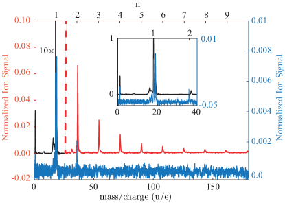
TOF-MS of the direct and the deflected beams are shown in Fig. 1 . The spectrum of the undeflected beam shows water-cluster ions up to and protonated water-cluster ions up to . Even larger clusters were likely formed in the supersonic expansion, but were not observed in the recorded TOF interval. We point out that all clusters that reach the interaction region are neutral clusters of the type , and protonated clusters must result from the interactions with the femtosecond laser, i. e., due to fragmentation during or after the strong-field-ionization process.
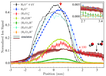
Vertical molecular-beam-density profiles for water ions , water dimer ions , and protonated water-cluster ions H+ up to , with a potential difference of 8 kV applied across the deflector, are shown in Fig. 2 . For comparison, a field-free vertical profile for the water ion with 0 kV across the deflector is also shown. The vertical molecular-beam-density profiles have been normalized to the area of the field-free spatial profile of the water ion. For visibility the water dimer profile has been scaled by a factor of 100 after normalization. While the field-free molecular beam profile is centered around 0 mm, application of a voltage of 8 kV to the deflector shifted the peak of water ions, water dimer ions, and protonated water-cluster ions by +0.5 mm, as indicated by the red arrow in Fig. 2 . In addition, water dimer ions showed a broadening and an increase of signal at around +3 mm, indicated by a black arrow in Fig. 2 .
In the inset of Fig. 2 the region around mm is shown enlarged with a magnification factor of 5 applied to and H+ with . The corresponding TOF-MS in the deflected part of the beam at a position of +3 mm is highlighted in Fig. 1 by the blue line. Not just water dimer ions, but also hydronium ions, , and water ions, , showed an increased signal in the deflected beam. The shape of the vertical beam profiles for these ions matched the water dimer profile in the region of 2.8–3.5 mm, indicating that they originated from the same parent molecule.
The water dimer ion was the largest non-protonated cluster measured in this setup. To verify that the water dimer ion was originating from the water dimer, the deflection behaviour of water-clusters inside the electrostatic deflector was simulated. Therefore, the Stark energies and effective dipole moments of water monomers and water-clusters were calculated with the freely available CMIstark software package Chang et al. (2014) using rotational constants, dipole moments, and centrifugal distortion constants from the literature DeLucia et al. (1974); Coudert and Hougen (1990); Shostak et al. (1991); Malomuzh et al. (2014); Dyke et al. (1977), see Suppl. Inf. Table I; contributions of the polarizability to the Stark effect could safely be ignored Maroulis (2000); Kienitz et al. (2017). The rotational constants of the water dimer are significantly smaller than for the water monomer, leading to a larger effective dipole moment for the water dimer than for water and a larger acceleration in the electric field in the deflector, see Fig. 3 and Table I of the Suppl. Inf. for further information.
The simulated vertical molecular-beam-density profiles of the water monomer and the water dimer are shown in Fig. 2 . The deviations between the measured and simulated undeflected vertical beam profiles are ascribed to imperfect alignment of experimental setup, which was not taken into account in the simulations. Due to the rotational-state dependence of the Stark effect, the deflection of a molecular beam in an electrostatic field depends on the rotational temperature of the molecular ensemble Chang et al. (2015) and the best fit for the profiles of the water monomer and the water dimer at a deflector voltage of 8 kV was obtained assuming a Boltzmann population distribution of rotational states corresponding to K.
Not only deflection of water-clusters measured as a mass of 36 amu, but also of water-clusters detected as protonated-clusters have been measured, for instance, for , as indicated by the red arrow and symbols in Fig. 2 . Trajectory simulations for water-clusters with using a rotational temperature of K were performed to understand the origin of this deflection behavior. For the water hexamer three and for the water heptamer two conformers have been simulated, assuming an equal population of the conformers. These showed that, based on the different effective dipole moments, a different deflection is expected for different water-clusters, see Fig. 5 of the Suppl. Inf.. Since the detected protonated water-clusters arose from the strong-field fragmentation of larger neutral clusters in the interaction region, the measured vertical protonated-cluster density profiles are a superposition of several neutral water-cluster density profiles. Thus, it is not possible to compare the individual simulated molecular-beam-density profiles of neutral clusters directly with the measured protonated water-cluster density profiles. Instead, at each position of the deflection profile the signal from all water-clusters has been summed up, both for the simulated and the measured molecular-beam-density profiles. The result yields a comparable amount of deflection for simulated and measured molecular-beam-density profiles, see Suppl. Inf. Fig. 6. The shift of 0.5 mm can, therefore, originate from the superimposed molecular-beam-density profiles from different larger clusters due to fragmentation into smaller water-clusters. The same shift is visible for and , which indicates that water-clusters are also fragmenting into and . Nevertheless, the simulation for water-clusters shows that the water dimer deflected the most, reaching a position of +3 mm and above, see Suppl. Inf. Fig. 4 and Fig. 5. Of all the other clusters considered, only the water hexamer in its prism and book forms reaches to a position up to 3.2 mm with the falling edge of the profile. In our experiments the water hexamer and higher order clusters have only been measured as fragments, such that the concentration and size distribution of neutral clusters in the molecular beam is unknown. However, the measured fragment distributions strongly suggest that significantly larger clusters are not present, since the ion signals decay exponentially and it is known that clusters primarily fragment through loss of single water molecules Angel and Stace (2001); Belau et al. (2007); Liu et al. (2011).
The TOF-MS in the deflected part of the beam, shown in Fig. 1 , contains peaks corresponding to H+, O+, OH+, , and , in addition to the water dimer ion. As mentioned before the short-pulse ionization can lead to fragmentation. For the water dimer, two fragmentation channels were reported for electron-impact ionization with 70 eV electrons Angel and Stace (2001): either an ion and a neutral OH are formed or a ion and a neutral water monomer . Using a size-selection method and infrared spectroscopy, has been reported as a fragment of the water dimer Buck and Huisken (2000). Comparison of the vertical molecular-beam-density profiles of the deflected molecules allowed further investigation of the fragmentation channels of the water dimer. The measured vertical molecular-beam-density profiles of these molecules showed a similar deflection behavior in the region of 2.8 to 3.5 mm as the water dimer, see Fig. 2 and Suppl. Inf. Fig. 1. The observed constant ratio of those fragments over this spatial region indicates that all these fragments originated from the water dimer.
Comparing the intensity of the fragments of the water dimer, and and (, in the deflected beam, the fragmentation ratios of the intact water dimer were estimated. These showed that of the water dimer fragmented into one ionized water molecule, while of the water dimer underwent most likely proton transfer and formed a hydronium ion. Only of the water dimer present in the molecular beam stayed intact after ionization.
The actual number of water dimer molecules per shot in the deflected molecular beam was estimated to within the laser focus using the known fragmentation ratios of and , while the fragmentation channels of H+, O+, OH+ have not been included. Taking the known fragmentation channels into account, the fraction of the water dimer within the molecular beam was evaluated. Comparing the ratios between the water dimer and all other species visible in the TOF, a water dimer fraction of in the center of the undeflected beam and of in the deflected beam, at a position of +3 mm, was achieved. Thus, using the electrostatic deflector the fraction of the water dimer within the interaction region could be increased by nearly a factor of 24.
IV Conclusions
In summary, a high-purity beam of water dimers was created using the electrostatic deflector, which spatially separated water dimers from other species present in the molecular beam. The resulting water dimer sample had a purity of . The fragmentation products and ratios of the water dimer following strong-field ionization using a 35 fs laser pulse with a wavelength centered around 800 nm and peak intensity of were studied, with of the water dimer found to form a hydronium ion and fragmenting into one water cation and one neutral water monomer, while of the water dimer stayed intact. The deflection profiles could be simulated using a rigid-rotor model and an initial rotational temperature of 1.5(5) K.
The produced clean samples of water dimers are well suited for non-species-specific experiments, e. g., reactive-collisions, diffractive imaging, or ultrafast spectroscopies Kilaj et al. (2018); Hensley et al. (2012); Küpper et al. (2014). Even for experiments that can distinguish different species, for example photoion-photoion coincidence measurements Ren et al. (2018); Kierspel et al. (2018), the produced clean beams will enable significantly faster measurements of this important hydrogen-bonded model system, e. g., because unwanted backgrounds are avoided. Furthermore, the electrostatic separation technique can be used to separate different conformers Chang et al. (2015), which could be highly interesting in the purification and studies of larger water-clusters that exhibit multiple conformers Gregory et al. (1997).
Acknowledgments
This work has been supported by the Clusters of Excellence “Center for Ultrafast Imaging” (CUI, EXC 1074, ID 194651731) and “Advanced Imaging of Matter” (AIM, EXC 2056, ID 390715994) of the Deutsche Forschungsgemeinschaft (DFG), by the European Union’s Horizon 2020 research and innovation program under the Marie Skłodowska-Curie Grant Agreement 641789 “Molecular Electron Dynamics investigated by Intense Fields and Attosecond Pulses” (MEDEA), by the European Research Council under the European Union’s Seventh Framework Program (FP7/2007-2013) through the Consolidator Grant COMOTION (ERC-Küpper-614507), and by the Helmholtz Gemeinschaft through the “Impuls- und Vernetzungsfond”. L.H. acknowledges a fellowship within the framework of the Helmholtz-OCPC postdoctoral exchange program and J.O. gratefully acknowledges a fellowship by the Alexander von Humboldt Foundation.
Supporting Information Description
Supporting Information Available: Description of the fragmentation correction method and the trajectory simulations
References
- Jeffrey (1997) G. A. Jeffrey, An Introduction to Hydrogen Bonding (Oxford University Press, 1997).
- Liu et al. (1996) K. Liu, J.D. Cruzan, and R.J. Saykally, “Water clusters,” Science 271, 929–933 (1996).
- Nauta and Miller (2000) K. Nauta and R. E. Miller, “Formation of cyclic water hexamer in liquid helium: The smallest piece of ice,” Science 287, 293 (2000).
- Dunning et al. (2015) G. T. Dunning, D. R. Glowacki, T. J. Preston, S. J. Greaves, G. M. Greetham, I. P. Clark, M. Towrie, J. N. Harvey, and A. J. Orr-Ewing, “Vibrational relaxation and microsolvation of DF after F-atom reactions in polar solvents,” Science 347, 530–533 (2015).
- Berden et al. (1996) G. Berden, W. L. Meerts, M. Schmitt, and K. Kleinermanns, “High resolution UV spectroscopy of phenol and the hydrogen bonded phenol-water cluster,” J. Chem. Phys. 104, 972 (1996).
- Korter et al. (1998) Timothy M. Korter, David W. Pratt, and Jochen Küpper, “Indole-H2O in the gas phase. Structures, barriers to internal motion, and transition moment orientation. Solvent reorganization in the electronically excited state,” J. Phys. Chem. A 102, 7211–7216 (1998).
- Sobolewski and Domcke (2001) A. L. Sobolewski and W. Domcke, “Photoinduced electron and proton transfer in phenol and its clusters with water and ammonia,” J. Phys. Chem. A 105, 9275–9283 (2001).
- Ren et al. (2018) X. Ren, E. Wang, A. D. Skitnevskaya, A. B. Trofimov, G. Kirill, and A. Dorn, “Experimental evidence for ultrafast intermolecular relaxation processes in hydrated biomolecules,” Nat. Phys. 79, 1745 (2018).
- Odutola and Dyke (1980) J. A. Odutola and T. R. Dyke, “Partially deuterated water dimers: Microwave spectra and structure,” J. Chem. Phys. 72, 5062–5070 (1980).
- Yu and van Gunsteren (2004) H. Yu and W. F. van Gunsteren, “Charge-on-spring polarizable water models revisited: From water clusters to liquid water to ice,” J. Chem. Phys. 121, 9549–9564 (2004).
- Dyke et al. (1977) T. R. Dyke, K. M. Mack, and J. S. Muenter, “The structure of water dimer from molecular beam electric resonance spectroscopy,” J. Chem. Phys. 66, 498–510 (1977).
- Dyke and Muenter (1972) T. R. Dyke and J. S. Muenter, “Molecular-beam electric deflection studies of water polymers,” J. Chem. Phys. 57, 5011 (1972).
- Marchenko et al. (2018) T. Marchenko, L. Inhester, G. Goldsztejn, O. Travnikova, L. Journel, R. Guillemin, I. Ismail, D. Koulentianos, D. Céolin, R. Püttner, M. N. Piancastelli, and M. Simon, “Ultrafast nuclear dynamics in the doubly-core-ionized water molecule observed via auger spectroscopy,” Phys. Rev. A 98, 063403 (2018).
- Kilaj et al. (2018) Ardita Kilaj, Hong Gao, Daniel Rösch, Uxia Rivero, Jochen Küpper, and Stefan Willitsch, “Observation of different reactivities of para- and ortho-water towards trapped diazenylium ions,” Nat. Commun. 9, 2096 (2018).
- Boll et al. (2016) Rebecca Boll, Benjamin Erk, Ryan Coffee, Sebastian Trippel, Thomas Kierspel, Cédric Bomme, John D. Bozek, Mitchell Burkett, Sebastian Carron, Ken R. Ferguson, Lutz Foucar, Jochen Küpper, Tatiana Marchenko, Catalin Miron, Minna Patanen, Timur Osipov, Sebastian Schorb, Marc Simon, Michelle Swiggers, Simone Techert, Kiyoshi Ueda, Christoph Bostedt, Daniel Rolles, and Artem Rudenko, “Charge transfer in dissociating iodomethane and fluoromethane molecules ionized by intense femtosecond x-ray pulses,” Struct. Dyn. 3, 043207 (2016).
- Kierspel (2016) Thomas Kierspel, Imaging structure and dynamics using controlled molecules, Dissertation, Universität Hamburg, Hamburg, Germany (2016).
- Jahnke et al. (2010) T. Jahnke, H. Sann, T. Havermeier, K. Kreidi, C. Stuck, M. Meckel, M. Schöffler, N. Neumann, R. Wallauer, S. Voss, A. Czasch, O. Jagutzki, A. Malakzadeh, F. Afaneh, Th. Weber, H. Schmidt-Böcking, and R. Dörner, “Ultrafast energy transfer between water molecules,” Nat. Phys. 6, 139–142 (2010).
- Winter et al. (2007) Bernd Winter, Emad F. Aziz, Uwe Hergenhahn, Manfred Faubel, and Ingolf V. Hertel, “Hydrogen bonds in liquid water studied by photoelectron spectroscopy,” J. Chem. Phys. 126, 124504 (2007).
- Smith et al. (2004) Jared D. Smith, Christopher D. Cappa, Kevin R. Wilson, Benjamin M. Messer, Ronald C. Cohen, and Richard J. Saykally, “Energetics of hydrogen bond network rearrangements in liquid water,” Science 306, 851–853 (2004).
- Keutsch and Saykally (2001) Frank N. Keutsch and Richard J. Saykally, “Inaugural article: Water clusters: Untangling the mysteries of the liquid, one molecule at a time,” PNAS 98, 10533–10540 (2001).
- Zwier (1996) T. S. Zwier, “The spectroscopy of solvation in hydrogen-bonded aromatic clusters,” Annu. Rev. Phys. Chem. 47, 205–241 (1996).
- Scoles (1988) G. Scoles, ed., Atomic and molecular beam methods, Vol. 1 (Oxford University Press, New York, NY, USA, 1988).
- Even et al. (2000) U. Even, J. Jortner, D. Noy, N. Lavie, and N. Cossart-Magos, “Cooling of large molecules below 1 K and He clusters formation,” J. Chem. Phys. 112, 8068–8071 (2000).
- Johny et al. (2019) Melby Johny, Jolijn Onvlee, Thomas Kierspel, Helen Bieker, Sebastian Trippel, and Jochen Küpper, “Spatial separation of pyrrole and pyrrole-water clusters,” Chem. Phys. Lett. 721, 149–152 (2019), arXiv:1901.05267 [physics] .
- Paul et al. (1997) J. B. Paul, C. P. Collier, R. J. Saykally, J. J. Scherer, and A. O’Keefe, “Direct measurement of water cluster concentrations by infrared cavity ringdown laser absorption spectroscopy,” J. Phys. Chem. A 101, 5211–5214 (1997).
- Chang et al. (2015) Yuan-Pin Chang, Daniel A. Horke, Sebastian Trippel, and Jochen Küpper, “Spatially-controlled complex molecules and their applications,” Int. Rev. Phys. Chem. 34, 557–590 (2015), arXiv:1505.05632 [physics] .
- Filsinger et al. (2008) Frank Filsinger, Undine Erlekam, Gert von Helden, Jochen Küpper, and Gerard Meijer, “Selector for structural isomers of neutral molecules,” Phys. Rev. Lett. 100, 133003 (2008), arXiv:0802.2795 [physics] .
- Filsinger et al. (2009a) Frank Filsinger, Jochen Küpper, Gerard Meijer, Jonas L. Hansen, Jochen Maurer, Jens H. Nielsen, Lotte Holmegaard, and Henrik Stapelfeldt, “Pure samples of individual conformers: the separation of stereo-isomers of complex molecules using electric fields,” Angew. Chem. Int. Ed. 48, 6900–6902 (2009a).
- Kierspel et al. (2014) Thomas Kierspel, Daniel A. Horke, Yuan-Pin Chang, and Jochen Küpper, “Spatially separated polar samples of the cis and trans conformers of 3-fluorophenol,” Chem. Phys. Lett. 591, 130–132 (2014), arXiv:1312.4417 [physics] .
- Teschmit et al. (2018) Nicole Teschmit, Daniel A. Horke, and Jochen Küpper, “Spatially separating the conformers of the dipeptide Ac-Phe-Cys-NH2,” Angew. Chem. Int. Ed. 57, 13775–13779 (2018), arXiv:1805.12396 [physics] .
- Nielsen et al. (2011) Jens H. Nielsen, Paw Simesen, Christer Z. Bisgaard, Henrik Stapelfeldt, Frank Filsinger, Bretislav Friedrich, Gerard Meijer, and Jochen Küpper, “Stark-selected beam of ground-state OCS molecules characterized by revivals of impulsive alignment,” Phys. Chem. Chem. Phys. 13, 18971–18975 (2011), arXiv:1105.2413 [physics] .
- Horke et al. (2014) Daniel A. Horke, Yuan-Pin Chang, Karol Długołęcki, and Jochen Küpper, “Separating para and ortho water,” Angew. Chem. Int. Ed. 53, 11965–11968 (2014), arXiv:1407.2056 [physics] .
- Trippel et al. (2012) Sebastian Trippel, Yuan-Pin Chang, Stephan Stern, Terry Mullins, Lotte Holmegaard, and Jochen Küpper, “Spatial separation of state- and size-selected neutral clusters,” Phys. Rev. A 86, 033202 (2012), arXiv:1208.4935 [physics] .
- You et al. (2018) Hyun Sik You, Junggil Kim, Songhee Han, Doo-Sik Ahn, Jean Sun Lim, and Sang Kyu Kim, “Spatial isolation of conformational isomers of hydroquinone and its water cluster using the stark deflector,” J. Phys. Chem. A 122, 1194 (2018).
- Moro et al. (2007) Ramiro Moro, Jaap Bulthuis, Jonathon Heinrich, and Vitaly V. Kresin, “Electrostatic deflection of the water molecule: A fundamental asymmetric rotor,” Phys. Rev. A 75, 013415 (2007).
- Moro et al. (2006) Ramiro Moro, Roman Rabinovitch, Chunlei Xia, and Vitaly V. Kresin, “Electric dipole moments of water clusters from a beam deflection measurement,” Phys. Rev. Lett. 97, 123401 (2006).
- Buck and Huisken (2000) Udo Buck and Friedrich Huisken, “Infrared spectroscopy of size-selected water and methanol clusters,” Chem. Rev. 100, 3863–3890 (2000).
- Pradzynski et al. (2012) Christoph C. Pradzynski, Richard M. Forck, Thomas Zeuch, Petr Slavíček, and Udo Buck, “A fully size-resolved perspective on the crystallization of water clusters,” Science 337, 1529–1532 (2012).
- Trippel et al. (2018) Sebastian Trippel, Melby Johny, Thomas Kierspel, Jolijn Onvlee, Helen Bieker, Hong Ye, Terry Mullins, Lars Gumprecht, Karol Długołęcki, and Jochen Küpper, “Knife edge skimming for improved separation of molecular species by the deflector,” Rev. Sci. Instrum. 89, 096110 (2018), arXiv:1802.04053 [physics] .
- Kienitz et al. (2017) Jens S. Kienitz, Karol Długołęcki, Sebastian Trippel, and Jochen Küpper, “Improved spatial separation of neutral molecules,” J. Chem. Phys. 147, 024304 (2017), arXiv:1704.08912 [physics] .
- Küpper et al. (2014) Jochen Küpper, Stephan Stern, Lotte Holmegaard, Frank Filsinger, Arnaud Rouzée, Artem Rudenko, Per Johnsson, Andrew V. Martin, Marcus Adolph, Andrew Aquila, Saša Bajt, Anton Barty, Christoph Bostedt, John Bozek, Carl Caleman, Ryan Coffee, Nicola Coppola, Tjark Delmas, Sascha Epp, Benjamin Erk, Lutz Foucar, Tais Gorkhover, Lars Gumprecht, Andreas Hartmann, Robert Hartmann, Günter Hauser, Peter Holl, Andre Hömke, Nils Kimmel, Faton Krasniqi, Kai-Uwe Kühnel, Jochen Maurer, Marc Messerschmidt, Robert Moshammer, Christian Reich, Benedikt Rudek, Robin Santra, Ilme Schlichting, Carlo Schmidt, Sebastian Schorb, Joachim Schulz, Heike Soltau, John C. H. Spence, Dmitri Starodub, Lothar Strüder, Jan Thøgersen, Marc J. J. Vrakking, Georg Weidenspointner, Thomas A. White, Cornelia Wunderer, Gerard Meijer, Joachim Ullrich, Henrik Stapelfeldt, Daniel Rolles, and Henry N. Chapman, “X-ray diffraction from isolated and strongly aligned gas-phase molecules with a free-electron laser,” Phys. Rev. Lett. 112, 083002 (2014), arXiv:1307.4577 [physics] .
- Stern et al. (2014) Stephan Stern, Lotte Holmegaard, Frank Filsinger, Arnaud Rouzée, Artem Rudenko, Per Johnsson, Andrew V. Martin, Anton Barty, Christoph Bostedt, John D. Bozek, Ryan N. Coffee, Sascha Epp, Benjamin Erk, Lutz Foucar, Robert Hartmann, Nils Kimmel, Kai-Uwe Kühnel, Jochen Maurer, Marc Messerschmidt, Benedikt Rudek, Dmitri G. Starodub, Jan Thøgersen, Georg Weidenspointner, Thomas A. White, Henrik Stapelfeldt, Daniel Rolles, Henry N. Chapman, and Jochen Küpper, “Toward atomic resolution diffractive imaging of isolated molecules with x-ray free-electron lasers,” Faraday Disc. 171, 393 (2014), arXiv:1403.2553 [physics] .
- Filsinger et al. (2009b) Frank Filsinger, Jochen Küpper, Gerard Meijer, Lotte Holmegaard, Jens H. Nielsen, Iftach Nevo, Jonas L. Hansen, and Henrik Stapelfeldt, “Quantum-state selection, alignment, and orientation of large molecules using static electric and laser fields,” J. Chem. Phys. 131, 064309 (2009b), arXiv:0903.5413 [physics] .
- Chang et al. (2014) Y.-P. Chang, F. Filsinger, B. Sartakov, and J. Küpper, “CMIstark: Python package for the stark-effect calculation and symmetry classification of linear, symmetric and asymmetric top wavefunctions in dc electric fields,” Comp. Phys. Comm. 185, 339–349 (2014), arXiv:1308.4076 [physics] .
- DeLucia et al. (1974) Frank C. DeLucia, Paul Helminger, and William H. Kirchhoff, “Microwave spectra of molecules of astrophysical interest V. Water vapor,” J. Phys. Chem. Ref. Data 3, 211–219 (1974).
- Coudert and Hougen (1990) L. H. Coudert and J. T. Hougen, “Analysis of the microwave and far infrared spectrum of the water dimer,” Journal Of Molecular Spectroscopy 139, 259–277 (1990).
- Shostak et al. (1991) Shelley L Shostak, William L Ebenstein, and John S Muenter, “The dipole moment of water. I. dipole moments and hyperfine properties of H2O and HDO in the ground and excited vibrational states,” J. Chem. Phys. 94, 5875 (1991).
- Malomuzh et al. (2014) N. P. Malomuzh, V. N. Makhlaichuk, and S. V. Khrapatyi, “Water dimer dipole moment,” Russian Journal of Physical Chemistry A 88, 1431–1435 (2014).
- Maroulis (2000) George Maroulis, “Static hyperpolarizability of the water dimer and the interaction hyperpolarizability of two water molecules,” J. Chem. Phys. 113, 1813–1820 (2000).
- Angel and Stace (2001) L. Angel and A. J. Stace, “Dissociation patterns of (H2O) cluster ions, for n=2–6,” Chem. Phys. Lett. 345, 277–281 (2001).
- Belau et al. (2007) Leonid Belau, Kevin R Wilson, Stephen R Leone, and Musahid Ahmed, “Vacuum ultraviolet (VUV) photoionization of small water clusters,” J. Phys. Chem. A 111, 10075–10083 (2007).
- Liu et al. (2011) Xiaojie Liu, Wen-Cai Lu, C.Z. Wang, and K.M. Ho, “Energetic and fragmentation stability of water clusters (h2o)n, n=2–30,” Chem. Phys. Lett. 508, 270 – 275 (2011).
- Hensley et al. (2012) Christopher J. Hensley, Jie Yang, and Martin Centurion, “Imaging of isolated molecules with ultrafast electron pulses,” Phys. Rev. Lett. 109, 133202 (2012).
- Kierspel et al. (2018) Thomas Kierspel, Cédric Bomme, Michele Di Fraia, Joss Wiese, Denis Anielski, Sadia Bari, Rebecca Boll, Benjamin Erk, Jens S. Kienitz, Nele L. M. Müller, Daniel Rolles, Jens Viefhaus, Sebastian Trippel, and Jochen Küpper, “Photophysics of indole upon x-ray absorption,” Phys. Chem. Chem. Phys. 20, 20205 (2018), arXiv:1802.02964 [physics] .
- Gregory et al. (1997) J. K. Gregory, D. C. Clary, K. Liu, M. G. Brown, and R. J. Saykally, “The water dipole moment in water clusters,” Science 275, 814–817 (1997).