Beta-delayed-neutron studies of 135,136Sb and 140I performed with trapped ions
Abstract
Beta-delayed-neutron (n) spectroscopy was performed using the Beta-decay Paul Trap and an array of radiation detectors. The n branching ratios and energy spectra for 135,136Sb and 140I were obtained by measuring the time of flight of recoil ions emerging from the trapped ion cloud. These nuclei are located at the edge of an isotopic region identified as having n branching ratios that impact the r-process abundance pattern around the peak. For 135,136Sb and 140I, n branching ratios of 14.6(11), 17.6(28), and 7.6(28) were determined, respectively. The n energy spectra obtained for 135Sb and 140I are compared with results from direct neutron measurements, and the n energy spectrum for 136Sb has been measured for the first time.
pacs:
I Introduction
Beta-delayed-neutron (n) emission is a process that can occur for neutron-rich nuclei sufficiently far from stability. In this process, a precursor nucleus undergoes decay to a highly excited state in the daughter nucleus above the neutron-separation energy that emits a neutron. The properties of n-emitting nuclei are important in various areas of basic and applied sciences, including nuclear astrophysics, nuclear energy, and nuclear structure.
The astrophysical rapid neutron-capture process (r process) is believed to be responsible for the production of roughly half of the elements heavier than iron Burbidge et al. (1957); Cameron (1957). In the r process, neutron-rich nuclei far from stability are produced through repeated neutron-capture reactions, and n emission during the eventual decay back to stability impacts the final isotopic abundance pattern. Different astrophysical environments, such as core-collapse supernovae Meyer et al. (1992); Farouqi et al. (2010) and neutron-star mergers Goriely et al. (2011); Pian et al. (2017), have been investigated as possible r-process sites by comparing theoretical models with observation. These models require high-quality nuclear data, such as nuclear masses, -decay and neutron-capture rates, and n-emission probabilities, for the thousands of isotopes along the nucleosynthesis pathway and populated during the decay back to stability. Much of this information still remains unknown, given the experimental challenges of accessing nuclei far from stability.
Beta-delayed-neutron emission also plays a key role in the control and safety of nuclear reactors. Both the branching ratios and energy spectra are required for reactor kinetics calculations and safety studies Das (1994); D’Angelo (2002). Higher-quality nuclear data would allow for the n yield and energy spectrum to be calculated for individual contributing isotopes, making it possible to accurately model any fuel-cycle concept, actinide mix, or irradiation history.
In addition, the information obtained in n measurements helps to provide a better understanding of the nuclear structure of neutron-rich nuclei Kratz (1984); Raman et al. (1983); Hamilton et al. (1985); Winger et al. (2009). For example, measuring the n-emission probability can be used to deduce the -strength function above the neutron-separation energy of the daughter nucleus Pappas and Sverdrup (1972); Kratz et al. (1979). Beta-delayed-neutron studies also help to constrain nuclear-structure calculations Kawano et al. (2008) and empirical models McCutchan et al. (2012) that predict the decay properties of nuclei for which no data exist.
In this work, the Beta-decay Paul Trap (BPT) Yee et al. (2013); Scielzo et al. (2012, 2014), a linear radiofrequency quadrupole ion trap with an open geometry, was utilized to study the n branching ratios and energy spectra of a number of n-emitting nuclei, which were produced with the Californium Rare Isotope Breeder Upgrade (CARIBU) facility Savard et al. (2008) at Argonne National Laboratory. The results for 137,138I and 144,145Cs are discussed in Ref. Czeszumska et al. , and the results for the more neutron-rich isotopes, 135,136Sb and 140I, are discussed here. Recent sensitivity studies performed by Mumpower et al. Mumpower et al. (2016) indicate that the latter three nuclei are situated at the edge of a region in the nuclear chart where the n branching ratios significantly impact the final r-process abundance pattern around the peak.
II Experimental Methods
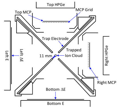
In the present work, the challenges associated with direct neutron detection are circumvented by instead studying the nuclear recoil from decay. Radioactive ions are suspended in vacuum as a 1-mm3 cloud at the center of the BPT. When a trapped ion undergoes decay, the recoil ion and emitted radiation emerge from the cloud with negligible scattering, allowing for their properties to be measured with radiation detectors arranged around the BPT as shown in Fig. 1. Two plastic-scintillator - telescopes, two microchannel-plate (MCP) detectors, and two high-purity germanium (HPGe) detectors are used to measure particles, recoil ions, and rays, respectively.
Beta-delayed-neutron spectroscopy is performed by recording the time of flight (TOF) of the recoil ions, which is determined from the time difference between the particle hitting a detector and the recoil ion hitting an MCP detector. Due to the additional momentum imparted by the neutron, ions from n emission have shorter TOFs than those from decay without neutron emission. The recoil-ion momentum can be reconstructed from the TOF and the distance the ion travels to the MCP surface. The neutron energy may then be obtained through conservation of energy and momentum. The resulting neutron-energy spectrum can be determined down to 100 keV; at lower energies, TOF cannot be used to identify n events because the corresponding recoil ions have energies comparable to those from decays without neutron emission. In this section, the ion production, transport, and confinement, as well as the detection of the decay particles are discussed.
II.1 Beam delivery at CARIBU
At CARIBU, fission fragments from a 100-mCi 252Cf source were thermalized in a large helium-filled gas catcher Savard et al. (2008), extracted primarily as ions, transported through an isobar separator Davids and Peterson (2008), and delivered to a radiofrequency-quadrupole buncher containing a small amount of helium gas to accumulate, cool, and bunch the beam. The isobar separator had a mass resolution of 14000, which allowed for some suppression of isobars one neutron away from the desired species and essentially complete removal of isobars more than one neutron away.
The optimal isobar-separator settings were selected by monitoring the distribution of isotopes present in the beam during tuning. The beam composition was characterized by using the two HPGe detectors surrounding the BPT and by performing mass scans with the Canadian Penning Trap (CPT) mass spectrometer Savard et al. (1997); Wang et al. (2004). The ion bunches were injected into the BPT at time intervals of int and accumulated over a length of time meas, after which the ions were ejected from the trap to measure backgrounds over a time period bkgd; this cycle was repeated throughout the entire run. The values of int, meas, and bkgd used for each isotope are given in Table 1 and were chosen based on the radioactive half-life of the isotope being studied and the distribution of isobaric contaminants present during the measurement. The total measurement times and average beam rates are also shown in Table 1.
| Isotope | Half-life | Measurement time | Average beam rate | int | meas | bkgd |
|---|---|---|---|---|---|---|
| (s) | (h) | (ions/s) | (s) | (s) | (s) | |
| 135Sb | 1.679(15) Singh et al. (2008) | 45.7 | 50 | 1.0 | 19.9 | 10.1 |
| 136Sb | 0.923(14) Sonzogni (2002) | 60.7 | 5 | 0.6 | 8.9 | 4.9 |
| 140I | 0.86(4) Nica (2007) | 35.3 | 5 | 0.6 | 8.3 | 4.3 |
II.2 Trapping with the BPT
Ion confinement was achieved by applying direct-current (DC) and time-varying, sinusoidal radiofrequency (RF) voltages to four sets of electrode plates extending to within 11 mm from the center of the trap as shown in Fig. 1. The DC voltages were used to produce a harmonic confining potential with a 5-V electrostatic valley in the axial direction, and the RF voltages, with a peak-to-peak amplitude of about 200 V and a frequency of 310 kHz, were used to confine ions in the radial direction. Higher harmonics at 620 and 930 kHz were observed with amplitudes less than 10% of the amplitude of the primary frequency. The trapped ions were thermalized in 5 Torr of helium gas.
Following decay, the charge state of the recoil ion is typically ; however, higher charge states can arise due to processes such as electron shakeoff, Auger-electron emission, and internal conversion. The stability conditions for the BPT, determined from the Mathieu equations Paul (1990), were chosen so that the decay daughters, which all have charge states higher than , were not confined in the trap.
II.3 Particle detection
Two plastic-scintillator - telescopes were used for spectroscopy. The detector was a 1-mm-thick, 10.6-cm-diameter disk that had a nearly 100% intrinsic detection efficiency for particles and only a 1% intrinsic detection efficiency for rays and neutrons. The detectors were placed 105 mm from the center of the BPT and each covered a solid angle of 5 of . The detectors were 10.2-cm-thick, 13.3-cm-diameter disks located immediately behind the detectors that were capable of stopping the particles. Each - telescope was contained in its own vacuum chamber (held below Torr) and separated from the BPT vacuum by a 10-m-thick aluminized-Kapton window. The Left and Bottom detectors had -energy thresholds of 76(24) keV and 62(30) keV, respectively, and a neutron detection threshold of 370(70) keV Czeszumska et al. .
Two 50.3 50.3 mm2 resistive-anode Chevron MCP detectors Wiza (1979) with 1-ns timing resolution and sub-mm position sensitivity were used for recoil-ion detection. The front face of each detector was biased to approximately kV to accelerate incoming ions and thereby provide a more uniform detection efficiency. Each detector was placed 4.5 mm behind a grounded 89%-transmission grid to help shield the detector from the RF fields of the BPT and to prevent the recoil-ion trajectories from being affected by the MCP bias voltage until they passed through the grid. The hit locations of the ions were reconstructed from the relative amounts of charge collected at the four corners of the anode Lampton and Carlson (1979). The central 46 46 mm2 region of each MCP detector had the best position resolution and was taken to be the fiducial area in the data analysis. Each detector was located 53.0(5) mm away from the trap center and subtended a solid-angle of 5 of .
The intrinsic efficiencies of the MCP detectors were determined to be 33.3(15)% and 29.3(14)% for the Right and Top detectors, respectively, from a detailed study of the decays of trapped 134Sb ions held in the BPT Siegl et al. (2018). The ion detection efficiencies also had to be corrected for additional loss of MCP pulses to electronic thresholds Czeszumska et al. ; Czeszumska (2016). For the Right MCP detector, this was a 3% correction. However, the Top MCP detector had a lower gain, resulting in a correction that ranged between 5–30% (depending on the impact energy of the ions) and showed some spatial dependence.
Two coaxial single-crystal p-type HPGe detectors were used to detect rays. The detectors, which had relative efficiencies of 80% and 140%, were located within 10 cm of the trapped-ion cloud behind the Right and Top MCP detectors, respectively. Standard -ray point sources (60Co, 133Ba, 137Cs, 152Eu) with activities determined to within 1.5-2.5% (at 1) were used to calibrate the photopeak detection efficiencies.
The data-acquisition system was triggered on a signal from any detector. A 22-s coincidence window was then opened, during which the amplitude and timing of each detected event was recorded along with the phase of the BPT RF voltage. The TOF for recoil ions was determined with a timing resolution of 3 ns FWHM. The nonparalyzable deadtime per event was 142 s.
III Analysis and Results
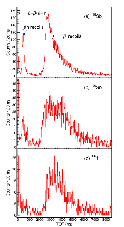
The TOF of the recoil ions was determined from -MCP detector coincidences and used to distinguish n decays from decays without neutron emission. The TOF spectra measured for 135,136Sb and 140I are shown in Fig. 2. The n events have TOFs primarily between 200 and 2000 ns, and -decay events without neutron emission have longer TOFs. A peak at 0 ns arose from -particle events in the detector that were in coincidence with a ray or scattered particle triggering the MCP detector.
The n energy spectra and branching ratios determined from these TOF spectra are discussed in this section. The Monte Carlo simulations of the decays and experimental setup needed to analyze the data are introduced first.
III.1 Monte Carlo simulations
The -decay kinematics were generated using simulation code originally developed in Ref. Scielzo et al. (2003) and later adapted for n decay Yee et al. (2013); Siegl et al. (2018); Munson et al. (2018). For each -decay transition, a distribution of and momenta was generated, assuming an allowed -spectrum shape. For excited states, the subsequent deexcitation to the ground state by the emission of rays, conversion electrons (CEs), and neutrons was also included. The resulting nuclear recoil was determined from the momentum imparted from each of these decay particles. For n emission, the transitions were assumed to be allowed Gamow-Teller and assigned a - angular correlation, , of . For transitions to states below the neutron-separation energy, an approximation was made that for a given isotope, all the were fixed to a single value, which was determined from the measured -ion coincidences using an approach described in detail in Ref. Munson et al. (2018). For 135Sb and 140I, this value of was and , respectively. For 136Sb, the presence of trapped 136Te ions complicated the analysis of the recoil ions and a value for could not be obtained.
The decays were spatially distributed with a 1-mm-FWHM Gaussian distribution in three dimensions, corresponding to the measured ion-cloud extent Siegl et al. (2018). The emitted particles, rays, CEs, and neutrons were propagated using GEometry ANd Tracking 4 (GEANT4) Agostinelli et al. (2003); Allison et al. (2006) version 4.10.0.p01 to model the scattering and energy loss of the particles within the apparatus. The energies deposited in the , , and HPGe detectors were recorded, and the electronic thresholds of the detectors were taken into account. Recoil ions of various charge states were propagated through the time-varying electric fields of the BPT using the SIMION 8.1 Manura and Dahl (2008) ion-optics code. The average charge states following the decay of 135,136Sb and 140I were determined to be 2.20, 2.51, and 2.16, respectively Munson et al. (2018), from the RF-phase dependence of the measured -ion coincidence rate using the approaches described in Ref. Siegl et al. (2018). For ions that struck an MCP detector, a threshold cut was applied Czeszumska et al. and the TOF, energy, velocity, and position at impact were recorded.
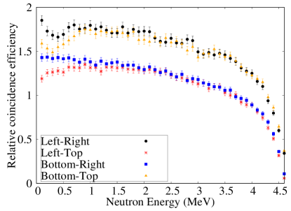
The efficiencies for detecting particles and -ion coincidences were determined using these simulations. Fig. 3 shows the -ion-coincidence detection efficiency as a function of neutron energy for 135Sb, with the product of the corresponding detector solid angles and MCP-detector intrinsic efficiency divided out. At the highest neutron energies, the coincidence-detection efficiency drops rapidly because of the limited energy available for the leptons, which results in fewer particles having energies above the detector thresholds. However, decays that populate highly-excited states are largely suppressed because of phase-space considerations. The two combinations (Left-Right and Bottom-Top) have higher efficiencies than the two combinations (Left-Top and Bottom-Right) primarily because of neutron-ion coincidences, which are present because the neutron and recoil ion are emitted with momenta nearly apart and therefore strike back-to-back detectors. The -ion-coincidence detection-efficiency curves for 136Sb and 140I have similar features.
III.2 Neutron energy spectra
The neutron energy was obtained by assuming the recoil ion and neutron had equal and opposite momenta. The momentum was approximated from the recoil-ion TOF and hit position on the MCP surface; the distance traveled by the recoil ion was approximated as a straight path from the trap center to the MCP grid, and effects due to the acceleration of the ion between the grid and the MCP surface were corrected for analytically. Background events were subtracted from the resulting spectrum, after which the neutron energy was adjusted to account for the contribution to the recoil-ion momentum from lepton emission. The spectrum was then scaled by the -ion coincidence efficiency. Each of these data analysis steps is explained below.
The background from accidental coincidences was determined from the TOF region between 15–20 s, which both data and simulation indicated had no true -ion coincidences. This subtraction resulted in a 3–9 correction, depending on the isotope.
After accounting for accidental coincidences, counts remained in the 50–200-ns time window where no -ion coincidences from trapped ions were expected. These counts were present both while the BPT was trapping ions and while the BPT was held empty following ejection of the trapped ions and were likely due to radioactivity that accumulated on the BPT and detector surfaces during data collection. The TOF distribution of these events was most pronounced between 50–200 ns and decreased with increasing TOF, extending into the n TOF region. The shape of this background, when converted into a neutron-energy distribution, closely resembled an exponential function. The subtraction of this background was performed by normalizing this exponential function to match the number of counts between 50-—200 ns collected when the BPT was trapping ions. This resulted in a 15–30 correction to the total number of observed n decays, depending on the isotope being analyzed.
During data collection, isobaric contaminants one neutron away from the isotope of interest were suppressed but not completely removed. For 135,136Sb and 140I, the more neutron-rich isobar (135,136Sn and 140Te, respectively) is a n emitter, but has a 252Cf-fission yield a couple orders of magnitude lower than the isotope of interest, making its contribution to the total number of n decays in the BPT negligible. For 135Sb and 140I, the more proton-rich isobar (135Te and 140Xe, respectively) does not decay by n emission and therefore cannot contribute n events. For 136Sb, the more proton-rich isobar, 136Te, has a n branching ratio roughly ten times smaller than that of 136Sb, but a fission yield 30 times larger. The suppression of 136Te by the isobar separator, together with the measurement cycle favoring the shorter-lived species, resulted in an average trapped-ion activity with about 15% more 136Sb than 136Te. The 136Te contribution to the total number of n coincidences was determined to be 5% based on the ratio of the 136Sb and 136Te activities, after accounting for the n branching ratios and the fraction of neutrons with energies above the 100-keV neutron threshold (estimated to be 0.6(2) for 136Te from the neutron-energy spectrum in Ref. Shalev and Rudstam (1974) and determined in Sec. III.3 to be 0.89(6) for 136Sb).
The neutron energy determined solely from the recoil-ion momentum was adjusted to account for the momentum imparted to the recoil ion from lepton emission. For the -ion coincidences measured by detectors apart, the neutron energy tended to be overestimated because the particle was emitted in approximately the same direction as the neutron and therefore contributed to the momentum of the nuclear recoil. Simulations showed that neglecting the leptons resulted in an overestimation of the inferred neutron energy of 25–30 at 100 keV, which steadily decreased to 10, 7, and 4% at neutron energies of 500 keV, 1000 keV, and above 2000 keV, respectively. For the -ion coincidences measured by detectors apart, the overestimation was only 1–2 for all neutron energies. For 136Sb and 140I, the neutron emission was assumed to directly populate the ground state, as there is currently no data indicating that excited states are populated. For 135Sb, however, decays to the ground state as well as the first, second, and third excited states (populated with probabilities of 62, 21, 11, and 6, respectively Hoff et al. (1989)) were taken into account, and the inclusion of the excited states led to a 1 shift in the inferred neutron energy.
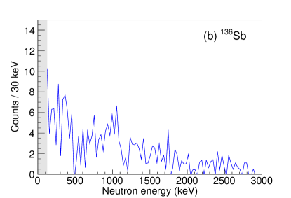
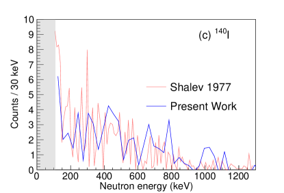
For each isotope, the neutron-energy spectrum obtained for each -MCP detector pair was corrected by the corresponding neutron-energy-dependent -ion coincidence efficiency. The results summed together for the four -MCP detector pairs are shown in Fig. 4 for 135,136Sb and 140I. For 136Sb, the contribution from 136Te isobaric contamination was removed by subtracting the 136Te neutron-energy spectrum measured in Ref. Shalev and Rudstam (1974), which was scaled by the activity and n branching ratio and broadened to account for the experimental energy resolution. The neutron-energy resolution in the present work was primarily determined by the spatial distribution of the ion cloud and the spread in recoil momentum resulting from the lepton emission. Simulations indicated that the FWHM energy resolution was 60 at a neutron energy of 100 keV and steadily decreased to 25, 15, and 9 at 500 keV, 1000 keV, and above 2000 keV, respectively. The neutron energy spectrum was determined down to 100 keV; below this energy, the recoil momentum imparted from the emission of the leptons and any accompanying rays was comparable to the momentum imparted from neutron emission.
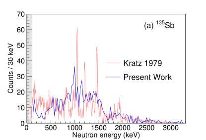
For 135Sb and 140I, the neutron-energy spectra are compared with direct neutron measurements by Kratz et al. Kratz et al. (1979) and Shalev and Rudstam Shalev and Rudstam (1977), respectively. For 136Sb, no previous measurement of the energy spectrum has been made. In the experiment by Kratz et al., n precursors were produced through neutron-induced fission of 235U at the Mainz TRIGA reactor, and two 3He ionization chambers, with energy resolutions of 12 keV for thermal neutrons and 20 keV for 1-MeV neutrons, were used to measure neutron energies. In the experiment by Shalev and Rudstam, n precursors were produced at the OSIRIS isotope-separator on-line facility. Neutron energies were measured with a neutron spectrometer that consisted of a cylindrical gridded ionization chamber filled with a 3He-argon gas mixture. The results obtained with the BPT for 135Sb and 140I have neutron-energy spectra and energy thresholds that are similar to the direct measurements. For 135Sb, the peaks in the spectrum obtained here are not as sharp because of the wider energy resolution; it also appears that there is a 4 energy shift in some of the features. The energy calibration in the present work depended primarily on the distance between the ion cloud and MCP detectors — this distance was determined with a fractional precision of 1, with consistent values obtained from both the measurement of the location of the trap electrodes and detectors and an analysis of the recoil-ion TOF spectra Siegl et al. (2018). This results in a 2 uncertainty in the neutron energy. An additional 1% uncertainty in the neutron energy was attributed to the energy shift applied to account for the lepton emission.
III.3 n branching ratios
The n branching ratios were obtained by comparing the number of detected -ion coincidences corresponding to decays that emitted a neutron with energy above 100 keV, , to the number of detected particles, , through the relation
| (1) |
where is the efficiency for detecting the -ion coincidences and is the -particle detection efficiency. The ratio was determined from simulations with an uncertainty of 5. The fraction of the n spectrum with neutron energies above 100 keV was obtained by assuming the neutron spectrum below threshold has the same intensity as the 100 keV above threshold, as there is currently no information on the neutron spectra at these low energies. This assumption yielded values of 0.95(3), 0.89(6), and 0.83(9) for 135,136Sb and 140I, respectively, and an uncertainty of half the difference from unity was assigned. Given that there do not appear to be pronounced features in the low-energy portions of these spectra, it is unlikely that there is large peak below 100 keV that would skew these estimates. To determine , the triggers originating from the trapped species of interest were isolated from those due to decays of isobaric contaminants and other backgrounds. This was accomplished by comparing the data to a model that takes into account the buildup and decay of the different species in the BPT over the course of the trapping cycle, while enforcing the decay-feeding relationships between the different populations Caldwell (2015). The results for were obtained with 5% precision.
For 135Sb and 140I, the n branching ratio was also obtained directly from the recoil-ion TOF spectrum by comparing to the number of -ion coincidences observed for decays without neutron emission, , using
| (2) |
where is the efficiency for detecting -ion coincidences without neutron emission and was determined in Ref. Munson et al. (2018). The results were obtained by selecting the events in the TOF region where decays without neutron emission are expected and subtracting the contribution from isobaric contaminants (when present) and accidental coincidences. For 135Sb and 140I, was determined with 3 and 6 precision, respectively. The ratio had an uncertainty of 5. For 136Sb, the trapped 136Te activity was substantial enough to make its subtraction from challenging, and therefore, a reliable value for could not be obtained.
| Isotope | () |
|---|---|
| 135Sb | 14.6 0.4 (stat) 1.0 (sys) |
| 136Sb | 17.6 1.0 (stat) 2.6 (sys) |
| 140I | 7.6 0.9 (stat) 2.6 (sys) |
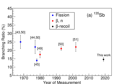
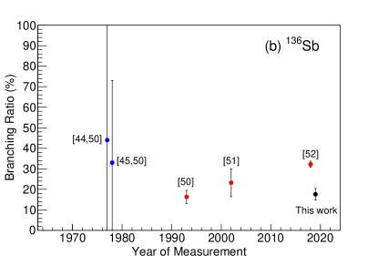
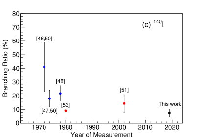
The n branching ratios were obtained from the weighted average of the results from the four -MCP detector pairs. For 135Sb and 140I, values of 14.6(16) and 8.1(34), respectively, were determined from Eq. 1, and values of 14.6(11) and 7.6(28), respectively, were determined from Eq. 2. For 136Sb, Eq. 1 yielded a of 17.6(28). In these approaches to determining , the systematic uncertainty due to the -particle detection efficiency largely cancels out. However, obtaining directly from the recoil-ion TOF spectrum yields a smaller total uncertainty because the systematic uncertainties due to the MCP solid angles and intrinsic efficiencies also cancel out. Therefore, for 135Sb and 140I, the n branching ratios obtained from the recoil-ion TOF spectrum are recommended; for 136Sb, only the value obtained from the comparison to detected particles is available. In Table 2, the recommended n branching-ratio results are provided. These values are compared with results obtained from previous direct measurements in Fig. 5. In the direct measurements, was determined either from the fission yield and neutrons-per-fission of the isotope Tomlinson and Hurdus (1968); Rudolph et al. (1977); Crançon et al. (1978); Schüssler and Herrmann (1972); Kratz and Herrmann (1974); Kratz (1978), or by counting particles and neutrons separately Lund et al. (1980); Rudstam et al. (1993); Pfeiffer et al. (2002); Caballero-Folch et al. (2018); Aleklett et al. (1980), usually with plastic scintillators and neutron detectors (e.g., BF3 tubes, 3He tubes), respectively. For each isotope, there is roughly a factor of two spread among the results, despite the fact that in many cases, the quoted uncertainties are significantly smaller than these differences. These discrepancies are evident even when comparing measurements that used similar experimental techniques, underscoring the challenging nature of performing n spectroscopy and indicating unforeseen systematic effects were likely responsible for these differences.
The results for 135,136Sb and 140I were determined in an analogous manner to the results for 137,138I and 144,145Cs in Ref. Czeszumska et al. . In Ref. Czeszumska et al. , the n branching ratios were obtained by comparing the number of -ion coincidences corresponding to n decay to the -decay activity, which was measured three different ways: (1) from the number of particles detected by the detectors, (2) from the number of -ion coincidences registered by the and MCP detectors, and (3) from the number of - coincidences registered by the and HPGe detectors. These three independent measures gave consistent results that were in excellent agreement with previous direct measurements. They also gave an opportunity to probe systematic effects and provided confidence that they were under control. In the present work, was obtained using methods (1) and (2), with limited statistics for -delayed -ray emission not allowing method (3). For 135Sb and 140I, where from methods (1) and (2) could be compared, consistent results were again obtained.
IV Summary and Conclusions
Beta-delayed-neutron spectroscopy was performed using the BPT instrumented with two plastic-scintillator - telescopes, two MCP detectors, and two HPGe detectors to measure particles, recoil ions, and rays, respectively. Both the n energy spectra and branching ratios were determined for the neutron-rich nuclei 135,136Sb and 140I. The n energy spectrum for 136Sb was measured for the first time, and the spectra for 135Sb and 140I were compared with results from direct neutron measurements by Kratz et al. Kratz et al. (1979) and Shalev and Rudstam Shalev and Rudstam (1977), respectively. The n energy spectra from the present work were similar in shape and had comparable energy thresholds to those obtained through direct neutron detection. The n branching ratios were obtained by comparing the number of -ion coincidences from n decays to the number of detected decays, which was determined from the number of particles registered by the detector and, when possible, the number of -ion coincidences. The latter approach to determining the number of detected decays was preferred when available, as it resulted in smaller systematic uncertainties in .
The neutron-energy spectra were obtained with -ion-coincidence efficiencies of 0.5%, which is several orders of magnitude larger than the neutron-detection efficiencies achievable with the 3He and gas-proportional detectors used for direct neutron spectroscopy. The ion-trap approach is therefore well suited for use at radioactive-beam facilities, where efficient techniques are desired to make the most of the delivered beam intensities. The n branching ratios for 136Sb and 140I were determined with beam intensities of only 5 ions/s, and with improvements to the detector array, results could be obtained with beams of less than 1 ion/s.
Upgrades to the BPT setup are currently in development. Plans include increasing the -recoil-coincidence detection efficiency using larger plastic scintillators and MCP detectors, and lowering the neutron energy threshold by further minimizing the impact of the electric fields on the trajectories of the recoil ions. The latter will be accomplished by bringing the electrodes closer to the center of the ion trap to allow for a lower-amplitude RF voltage to be applied, thus reducing the perturbation of the ion trajectories while the ions are in transit to the MCP detectors. Future experiments will also benefit from the increased intensities and purities of the beams delivered by the CARIBU facility Hirsh et al. (2016); Savard et al. (2016); since these measurements were performed, the beam intensities have increased by an order of magnitude. These improvements will allow n measurements to be performed for neutron-rich nuclei even further from stability, allowing access to many of the isotopes that significantly impact r-process nucleosynthesis.
V Acknowledgements
We acknowledge and appreciate the assistance of the ATLAS staff. This material is based upon work supported by the Department of Energy, National Nuclear Security Administration, under Award Numbers DE-NA0000979 (NSSC), DE-AC52-07NA27344 (LLNL), and DE-NA0002135 (SSGF); Office of Nuclear Physics Contract DE-AC02-06CH11357 (ANL); NEUP Project Number 13-5485 (University of California); Grant DE-FG02-94ER40834 (University of Maryland); Louisiana State Board of Regents Research Competitiveness Subprogram LEQSF(2016-19)-RD-A-09; NSERC, Canada, under Application No. 216974; NSF contract PHY-1419765; and the Department of Homeland Security.
References
- Burbidge et al. (1957) E. M. Burbidge, G. R. Burbidge, W. A. Fowler, and F. Hoyle, Rev. Mod. Phys. 29, 547 (1957).
- Cameron (1957) A. G. W. Cameron, Publ. Astron. Soc. Pac. 69, 201 (1957).
- Meyer et al. (1992) B. S. Meyer, G. J. Mathews, W. M. Howard, S. E. Woosley, and R. D. Hoffman, Astrophys. J. 399, 656 (1992).
- Farouqi et al. (2010) K. Farouqi, K.-L. Kratz, B. Pfeiffer, T. Rauscher, F.-K. Thielemann, and J. W. Truran, Astrophys. J. 712, 1359 (2010).
- Goriely et al. (2011) S. Goriely, A. Bauswein, and H.-T. Janka, The Astrophy. J. 738, L32 (2011).
- Pian et al. (2017) E. Pian, P. D’Avanzo, S. Benetti, M. Branchesi, E. Brocato, S. Campana, E. Cappellaro, S. Covino, V. D’Elia, J. P. U. Fynbo, et al., Nature 551, 67 (2017).
- Das (1994) S. Das, Prog. Nucl. Energy 28, 209 (1994).
- D’Angelo (2002) A. D’Angelo, Prog. Nucl. Energy 41, 1 (2002).
- Kratz (1984) K.-L. Kratz, Nucl. Phys. A 417, 447 (1984).
- Raman et al. (1983) S. Raman, B. Fogelberg, J. A. Harvey, R. L. Macklin, P. H. Stelson, A. Schröder, and K.-L. Kratz, Phys. Rev. C 28, 602 (1983).
- Hamilton et al. (1985) J. H. Hamilton, P. G. Hansen, and E. F. Zganjar, Rep. Prog. Phys. 48, 631 (1985).
- Winger et al. (2009) J. A. Winger, S. V. Ilyushkin, K. P. Rykaczewski, C. J. Gross, J. C. Batchelder, C. Goodin, R. Grzywacz, J. H. Hamilton, A. Korgul, W. Królas, et al., Phys. Rev. Lett. 102, 142502 (2009).
- Pappas and Sverdrup (1972) A. C. Pappas and T. Sverdrup, Nucl. Phys. A188, 48 (1972).
- Kratz et al. (1979) K.-L. Kratz, W. Rudolph, H. Ohm, H. Franz, M. Zendel, G. Herrmann, S. G. Prussin, F. M. Nuh, A. A. Shihab-Eldin, D. R. Slaughter, W. Halverson, and H. V. Klapdor, Nucl. Phys. A 317, 335 (1979).
- Kawano et al. (2008) T. Kawano, P. Möller, and W. B. Wilson, Phys. Rev. C 78, 054601 (2008).
- McCutchan et al. (2012) E. A. McCutchan, A. A. Sonzogni, T. D. Johnson, D. Abriola, M. Birch, and B. Singh, Phys. Rev. C 86, 041305 (2012).
- Yee et al. (2013) R. M. Yee, N. D. Scielzo, P. F. Bertone, F. Buchinger, S. Caldwell, J. A. Clark, C. M. Deibel, J. Fallis, J. P. Greene, S. Gulick, et al., Phys. Rev. Lett. 110, 092501 (2013).
- Scielzo et al. (2012) N. D. Scielzo, G. Li, M. G. Sternberg, G. Savard, P. F. Bertone, F. Buchinger, S. Caldwell, J. A. Clark, J. Crawford, C. M. Deibel, et al., Nucl. Instrum. Methods Phys. Res. A 681, 94 (2012).
- Scielzo et al. (2014) N. D. Scielzo, R. M. Yee, P. F. Bertone, F. Buchinger, S. A. Caldwell, J. A. Clark, A. Czeszumska, C. M. Deibel, J. P. Greene, S. Gulick, et al., Nucl. Data Sheets 120, 70 (2014).
- Savard et al. (2008) G. Savard, S. Baker, C. Davids, A. F. Levand, E. F. Moore, R. C. Pardo, R. Vondrasek, B. J. Zabransky, and G. Zinkann, Nucl. Instrum. Methods Phys. Res. B 266, 4086 (2008).
- (21) A. Czeszumska et al., (in preparation).
- Mumpower et al. (2016) M. R. Mumpower, R. Surman, G. C. McLaughlin, and A. Aprahamian, Prog. Part. Nucl. Phys. 86, 86 (2016).
- Davids and Peterson (2008) C. N. Davids and D. Peterson, Nucl. Instrum. Methods Phys. Res. B 266, 4449 (2008).
- Savard et al. (1997) G. Savard, R. C. Barber, D. Beeching, F. Buchinger, J. E. Crawford, S. Gulick, X. Feng, E. Hagberg, J. C. Hardy, V. T. Koslowsky, et al., Nucl. Phys. A 626, 353 (1997).
- Wang et al. (2004) J. C. Wang, G. Savard, K. S. Sharma, J. A. Clark, Z. Zhou, A. F. Levand, C. Boudreau, F. Buchinger, J. E. Crawford, J. P. Greene, et al., Nucl. Phys. A 746, 651 (2004).
- Singh et al. (2008) B. Singh, A. A. Rodionov, and Y. L. Khazov, Nucl. Data Sheets 109, 517 (2008).
- Sonzogni (2002) A. A. Sonzogni, Nucl. Data Sheets 95, 837 (2002).
- Nica (2007) N. Nica, Nucl. Data Sheets 108, 1287 (2007).
- Paul (1990) W. Paul, Rev. Mod. Phys. 62, 531 (1990).
- Wiza (1979) J. L. Wiza, Nucl. Instrum. Methods 162, 587 (1979).
- Lampton and Carlson (1979) M. Lampton and C. W. Carlson, Rev. Sci. Instrum. 50, 1093 (1979).
- Siegl et al. (2018) K. Siegl, N. D. Scielzo, A. Czeszumska, J. A. Clark, G. Savard, A. Aprahamian, S. A. Caldwell, B. S. Alan, M. T. Burkey, C. J. Chiara, et al., Phys. Rev. C 97, 035504 (2018).
- Czeszumska (2016) A. Czeszumska, Ph.D. thesis, University of California, Berkeley, Department of Nuclear Engineering (2016).
- Scielzo et al. (2003) N. D. Scielzo, S. J. Freedman, B. K. Fujikawa, and P. A. Vetter, Phys. Rev. A 68, 022716 (2003).
- Munson et al. (2018) J. M. Munson, K. Siegl, N. D. Scielzo, B. S. Alan, A. Czeszumska, G. Savard, A. Aprahamian, S. A. Caldwell, C. J. Chiara, J. A. Clark, et al., Nucl. Instrum. Methods Phys. Res. A 898, 60 (2018).
- Agostinelli et al. (2003) S. Agostinelli, J. Allison, K. Amako, J. Apostolakis, H. Araujo, P. Arce, M. Asai, D. Axen, S. Banerjee, G. Barrand, et al., Nucl. Instrum. Methods Phys. Res. A 506, 250 (2003).
- Allison et al. (2006) J. Allison, K. Amako, J. Apostolakis, H. Araujo, P. Arce Dubois, M. Asai, G. Barrand, R. Capra, S. Chauvie, R. Chytracek, et al., IEEE Trans. Nucl. Sci. 53, 270 (2006).
- Manura and Dahl (2008) D. Manura and D. Dahl, SIMION 8.0/8.1 User Manual (Scientific Instrument Services, Inc. Ringoes, NJ 08551) (2008).
- Shalev and Rudstam (1974) S. Shalev and G. Rudstam, Nucl. Phys. A 230, 153 (1974).
- Hoff et al. (1989) P. Hoff, B. Ekström, and B. Fogelberg, Z. Phys. A 332, 407 (1989).
- Shalev and Rudstam (1977) S. Shalev and G. Rudstam, Nucl. Phys. A 275, 76 (1977).
- Caldwell (2015) S. A. Caldwell, Ph.D. thesis, University of Chicago, Department of Physics (2015).
- Tomlinson and Hurdus (1968) L. Tomlinson and M. H. Hurdus, J. Inorg. Nucl. Chem. 30, 1649 (1968).
- Rudolph et al. (1977) W. Rudolph, K.-L. Kratz, and G. Herrmann, J. Inorg. Nucl. Chem. 39, 753 (1977).
- Crançon et al. (1978) J. Crançon, C. Ristori, H. Ohm, W. Rudolph, K.-L. Kratz, and M. Asghar, Z. Phys. A 287, 45 (1978).
- Schüssler and Herrmann (1972) H.-D. Schüssler and G. Herrmann, Radiochim. Acta 18, 123 (1972).
- Kratz and Herrmann (1974) K.-L. Kratz and G. Herrmann, Nucl. Phys. A 229, 179 (1974).
- Kratz (1978) K.-L. Kratz, Radiochim. Acta 25, 1 (1978).
- Lund et al. (1980) E. Lund, P. Hoff, K. Aleklett, O. Glomset, and G. Rudstam, Z. Phys. A 294, 233 (1980).
- Rudstam et al. (1993) G. Rudstam, K. Aleklett, and L. Sihver, At. Data Nucl. Data Tables 53, 1 (1993).
- Pfeiffer et al. (2002) B. Pfeiffer, K.-L. Kratz, and P. Möller, Prog. Nucl. Energy 41, 39 (2002).
- Caballero-Folch et al. (2018) R. Caballero-Folch, I. Dillmann, J. Agramunt, J. L. Taín, A. Algora, J. Äystö, F. Calviño, L. Canete, G. Cortès, C. Domingo-Pardo, et al., Phys. Rev. C 98, 034310 (2018).
- Aleklett et al. (1980) K. Aleklett, P. Hoff, E. Lund, and G. Rudstam, Z. Phys. A 295, 331 (1980).
- Hirsh et al. (2016) T. Y. Hirsh, N. Paul, M. Burkey, A. Aprahamian, F. Buchinger, S. Caldwell, J. A. Clark, A. F. Levand, L. L. Ying, S. T. Marley, et al., Nucl. Instrum. Methods Phys. Res. B 376, 229 (2016).
- Savard et al. (2016) G. Savard, A. F. Levand, and B. J. Zabransky, Nucl. Instrum. Methods Phys. Res. B 376, 246 (2016).