Light-induced breathing in photochromic yttrium oxy-hydrides
Abstract
When exposed to air, metallic yttrium dihydride YH2 films turn into insulating and transparent yttrium oxy-hydride (YHO). The incorporation of oxygen causes the lattice expansion of YH and the emergence of photochromic properties. However, the oxidation of YH2 is not completely irreversible: under illumination some oxygen atoms move towards the YHO surface, leaving behind an oxygen-deficient bulk (responsible for the photochromic darkening and observed lattice contraction). Under illumination, and according to experimental evidence, some oxygen atoms can effectively leave the film, being replaced by other oxygen atoms once the illumination has stopped, i.e., YHO ‘breathes’ when subjected to illumination/darkness cycling. Based on this ‘breathing’, YHO films become more hydrophobic under illumination conditions than when kept in darkness. Ab-initio calculations point to the light-induced weakening of the Y-O bond as the possible mechanism for explaining these experimental observations.
.
I Introduction
Yttrium hydride and other rare-earth hydrides are extremely reducing agents, a feature that complicates considerably their study. For their adequate handling in air, rare-earth hydride thin films are usually protected against oxidation by, for example, Pd capping layers Huiberts et al. (1996). However, the incorporation of oxygen in rare-earth hydrides after intentional exposure to air Nafezarefi et al. (2017); Montero et al. (2018); La et al. (2018), or even through accidental contamination Miniotas et al. (2000), leads to the formation of oxy-hydrides, which exhibit very interesting properties. One of the pioneer works on this family of materials was carried out by Miniotas et al. Miniotas et al. (2000), who reported gigantic electrical resistivity in oxygen-containing gadolinium hydride. Later, Mongstad et al. Mongstad et al. (2011a) reported photochromic properties in oxygen-containing yttrium hydride, a feature observed very recently by Nafezarefi et al. Nafezarefi et al. (2017) in other rare-earth oxy-hydrides such as dysprosium, gadolinium or erbium oxy-hydrides. The photochromism in Y-related compounds can be traced back to Ohmura et. al Ohmura et al. (2007), who observed light-induced reversible darkening in yttrium hydride thin films subjected to high pressures ( GPa). Despite the importance of the discovery, the emergence of this new inorganic photochromic material went unnoticed at that time, presumably because the pressure range required is not suitable for practical applications. Today, however, it is known that yttrium oxy-hydride –hereinafter referred in the text simply as YHO, a notation that, in principle, is not related to the stoichiometry of the compound, which will be discussed later– as well as other rare-earth oxy-hydrides, are photochromic at room temperature and at ambient pressure; hence, YHO, as an inorganic photochromic material has multitude of potential applications Towns (2016). However, the origin of the photochromic mechanism in YHO is still open to debate Montero et al. (2017), and in this sense, the wettability of the YHO surface under illumination and darkness conditions has been found to provide valuable insights: the present work reports on the light-induced hydrophobicity enhancement in YHO, i.e., the reduction of the surface energy under illumination. All oxides and nitrides of low-electronegativity metals can exhibit hydrophobicity Zenkin et al. (2014); Azimi et al. (2013). It can be, therefore, expected that YHO exhibits hydrophobic properties as well. However, while the surface of yttrium oxy-hydride increases its hydrophobicity when illuminated, other metal oxides turn into hydrophilic under UV illumination. In the latter case, the formation of electron-hole pairs under illumination leads to the creation of defect sites, where hydroxyl groups can be adsorbed, leading to hydrophilic properties Ho et al. (2007). Generally, when metal oxides are stored in darkness during periods of time ranging from 7 to 50 days Feng et al. (2004); Yadav et al. (2016), oxygen replaces back the adsorbed hydroxyl groups, giving raise to hydrophobicity. In the present work, the unexpected behavior observed in YHO, i.e., the enhancement of the hydrophobic properties under illumination, has been found to be caused by the same reason, that is, the oxygen-enrichment of the surface under illumination. This behavior provides new insights on the photochromic mechanism in yttrium oxy-hydride: under illumination, oxygen atoms are released from the lattice (in consequence the YHO lattice contracts); some of these O atoms can reach the surface, causing the enhancement of the hydrophobic properties. The displaced oxygen atoms leave behind an oxygen-deficient structure responsible for the optical darkening of the film. In darkness, the YHO lattice expands back as a consequence of the filling of the oxygen vacancies by oxygen atoms, allowing the film to bleach back to its original state. Since YHO expands/contracts reversibly under dark/illumination cycling (produced by the displacement inwards/outwards of oxygen atoms) we refer to this process as breathing. The breathing hypothesis presented in this work explains the photochromic behavior as well as the wettability change in YHO and it is supported by both theoretical and experimental results.
II Methods
Oxygen containing yttrium hydride thin films were prepared onto glass substrates following a two-step deposition process consisting on the fabrication by magnetron sputtering of YH2 metallic films (in a Leybold Optics A550V7 sputter unit) followed by a post-deposition oxidation process in air. Further details on the synthesis process of photochromic YHO can be found elsewhere Montero et al. (2018); Montero and Karazhanov (2018). A cold white LED array from Thorlabs (colour temperature 4600-9000 K) was used as illumination source for the photo-darkening experiments.The crystallographic structure of the obtained films was characterized by using x-ray diffraction (XRD) in a Bruker Siemens D500 spectrophotometer (CuK radiation, parallel beam geometry). The composition and surface oxidation states were studied by x-ray photoelectron spectroscopy (XPS) in an Ulvac PHI Quantera II instrument. Surface roughness characterizations were performed using Atomic Force Microscopy with area of 5 m2 from Photonic-tech Picostation. The optical transmittance (T) of the YHO films in the clear and photodarkened state was measured using an Ocean Optics spectrophotometer QE65000 and a Perkin-Elmer Lambda-900 with integrating sphere. Contact angle (CA) measurements were performed using KSV Attension Optical Tensiometer under air. 5 l drop volume was used for each CA measurement and three different sessile droplets were measured on several substrates for each value and averaged with a standard deviation of 2. CA values in the equilibrium () for water, ethylene glycol (EG), and methylene iodide (MeI)–both EG and MeI from Sigma-Aldrich–were used to calculate surface free energies of Yttrium oxy-hydride films at clear and photo-darkened state using the van Oss-Good-Chaudhury method van Oss et al. (1988); van Oss (1993).
The calculations were performed with the Vienna Ab initio Simulation Package (VASP) code G and J (1996a, b, 1994), based on density functional theory (DFT) using a plane-wave pseudopotential method together with the potential projector augmented-wave (PAW) Blöchl (1994); Kresse and Joubert (1999); G and J (1993). The generalized gradient approximation (GGA) in the scheme of Perdew-Burke-Ernzerhof (PBE) is used to describe the exchange-correlation functional G and J (1994). To describe the electron-ion interaction standard PAW-PBE pseudopotentials Perdew et al. (1996) are used with 1s1 for H, 2s22p4 for O and 4s24p64d15s2 for Y atoms as the valence-electron configuration. The plane wave functions of valence electrons are expanded in a plane wave basis set, and the use of PAW pseudopotentials allows a plane wave energy cutoff (). Only plane waves with kinetic energies smaller than are used in the expansion. Reciprocal-space integration over the Brillouin zone is approximated through a careful sampling at finite number of k-points using a Monkhorst-Pack mesh G and J (1993). We choose the energy cutoff to be 700 eV, and the Brillouin-zone sampling mesh parameters for the k-points set are 888. In the optimisation process the energy change is set to eV. The charge densities are converged to eV in the self-consistent calculation. The range-separated hybrid Heyd-Scuseria-Ernzerhof (HSE06) functional is used for density of states calculations Heyd et al. (2003); Krukau et al. (2006); Henderson et al. (2011). The hybrid functional requires a standard value of the (short-range) Hartree-Fock exchange (21%) mixed with a portion of PBE exchange (79%), also known as the HSE06 hybrid functional Krukau et al. (2006); Henderson et al. (2011). Selection of the parameter has been performed as an inverse value of infinity dielectric constant that is valid, if the energy band gap of these systems is larger than 3 eV.
III Results
III.1 Hydrophobicity control through light illumination
| (Water) | (EG) | (MeI) | ||||||
|---|---|---|---|---|---|---|---|---|
| (∘) | (∘) | (∘) | (mJ/m2) | (mJ/m2) | (mJ/m2) | (mJ/m2) | (mJ/m2) | |
| Clear state | 95 | 81 | 43 | 46.55 | 38.07 | 8.98 | 2.98 | 6.76 |
| Photo-darkened State | 115 | 81 | 60 | 28.88 | 28.58 | 0.31 | 0.18 | 0.13 |
| Clear State | Photo-darkened State | |
|---|---|---|
| % | % | |
| Carbon | 48 | 36 |
| Oxygen | 36 | 48 |
| Yttrium | 16 | 16 |
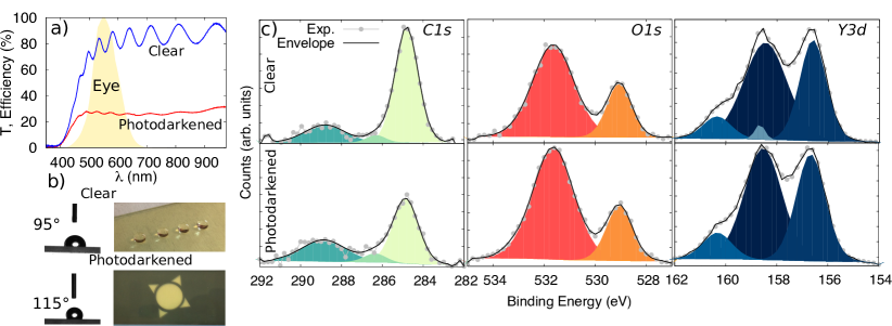
YHO thin films exhibit photochromic properties, that is, YHO films undergo a reversible decrease of their optical transmittance when illuminated with light of adequate energy and intensity Montero and Karazhanov (2018). Figure 1 (a) shows the transmittance in the clear and photodarkened state for 1400 nm-thick YHO film. This film decreased its luminous transmittance, from 78.5% to 26.7% after illumination (for a definition of and how to calculate it, visit ref. Li et al. (2010)). The luminous efficiency of the human eye (photopic vision) is presented in Figure 1 (a) for comparison Li et al. (2010). How to obtain such optical contrast by illumination will be discussed in detail in next section.
Non-illuminated (clear) YHO thin films show hydrophobicity with equilibrium contact angle () values of 95∘ for water (see Table 1); however, values increased to 115∘ after illumination (again in the case of water), see Table 1 and Figure 1 (b). Table 1 also shows for ethylene glycol (EG) and methylene iodine (MeI) for the clear and photo-darkened states. In the case of MeI, also increases after illumination, from 43∘ to 60∘, while remain constant for EG. AFM studies performed in such films revealed a relatively smooth surface, being the RMS value of around 8 nm.
The observed initial hydrophobicity of the YHO films (clear state) can be explained by the electronic structure of rare earth elements: according to a detailed experimental analysis of the entire rare earth oxide series carried out by Azimi et al. Azimi et al. (2013), the unfilled 4f orbitals shielded by a full octet of electrons (from the 5s2p6 shell) which is characteristic of rare earth oxides, results in a lower tendency of such compounds to form hydrogen bonds with the adjacent water molecules Azimi et al. (2013); Imanaka (2004). However, hydrophobicity is not exclusive of the lanthanide f-shell group, but it can be achievable in any metal oxide provided that the electronegativity of such metal is low enough Zenkin et al. (2014). The low electronegativity of Y and the fact that the surface is composed mainly by yttrium oxide Moldarev et al. (2018), explains the high shown in Table 1.
Under illumination, a decrease of the hydrophobicity, caused by the creation of electron-hole pairs, would be expected, as observed in other metal oxides Ho et al. (2007); Feng et al. (2004, 2005); Wang et al. (2006); Zhu et al. (2006); de Sun et al. (2001). However, this is not the tendency observed in YHO, in which, as described before, the hydrophobicity is enhanced under illumination. In this case, the observed light-induced decrease of wettability has to be explained through changes in surface composition, namely the oxygen-to-metal ratio. In metal oxides, coordinatively unsaturated oxygen atoms work as a Lewis base while the metal cations work as a Lewis acid. Combined Lewis acid and base orientation of the surface causes high affinity towards water molecules Kung (1989) and, therefore, the oxygen-to-metal ratio in the surface is crucial for understanding the wettability properties Khan et al. (2015).
In order to study the compositional changes of the YHO surface, XPS measurements were performed before and after illumination and the results presented for C1s, O1s and Y3d in Figure 1 (c). See Table 2 for the quantification of the different elements by XPS. The carbon (adventitious) C1s signal can be deconvoluted into three different contributions; the signal corresponding to C-C has been established at 284.8 eV as a charge correction reference. Other contributions are C-O-C at 286.3 eV and O-C=O at 288.8 eV Beamson and Briggs (1993). After illumination, the carbon content on the surface decreases, see Table 2. This decrease is accused mainly by the C-C contribution, Figure 1 (c). Since the content of C in the surface decreases, the increase of adsorbed hydrocarbons is ruled out, in our case, as the possible cause for the light-induced enhancement of the hydrophobicity Preston et al. (2014); Lundy et al. (2017); Külah et al. (2017).
The O1s signal is composed by two contributions at 529.0 eV and 531.2 eV; the former can be attributed to O atoms bound to Y atoms, whereas the latter can be assigned to physisorbed O Craciun et al. (1999). After illumination, the O content of the surface increases, see Table 2. This increase takes place both for O bound to Y as well as for physisorbed O. However, the largest increase is observed in the later, Figure 1 (c).
The obtained results for Y3d correspond very well to the Y2O3 stoichiometry, i.e., at the top surface consist of Y2O3, in agreement with our previous studies Moldarev et al. (2018). The Y3d has well resolved spin-orbit components, namely Y3d3/2 and Y3d5/2. These components can be deconvoluted into yttrium carbonates, with small contributions at 158.6 eV and 160.3 eV, as well as the main Y2O3 contribution at 156.6 eV and 158.4 eV, Figure 1 (c) Craciun et al. (1999). Not surprisingly there is no change in Y content during illumination, but the carbonate component does decrease in favour of the Y2O3 component.
Surface energy calculations, performed using the Oss-Chaudhury-Good method van Oss et al. (1988); van Oss (1993), confirm the lower wettability through reduction (under illumination) of the total surface energy (), see Table 1. The enrichment in oxygen of the surface, confirmed by XPS, reduces the Lewis sites as the surface approaches the Y2O3 stoichometry. The nonpolar Liftshitz-van der Waals surface energy component, , also decreases from 38.07 mJ/m2 to 28.57 mJ/m2 while the polar acid-base component, [where , being and the Lewis acid and base parameters of surface tension, respectively], decreases from 8.98 mJ/m2 to 0.31 mJ/m2 after illumination.
Hydrophobic yttrium-based oxides have been reported in the past Zenkin et al. (2014); Barshilia et al. (2012); in this case as the Y2O3-x coatings approached the Y2O3 stoichiometry, they showed increased contact angles Barshilia et al. (2012) which is consistent with metal-to-oxygen ratio of surface studies Khan et al. (2015). Consequently, the enrichment in oxygen of the surface under illumination causes the light-induced hydrophobicity enhancement observed in YHO thin films. In the next section, the exchange of oxygen atoms between the film and the atmosphere, induced by illumination, is demonstrated.
III.2 Light-induced “breathing”
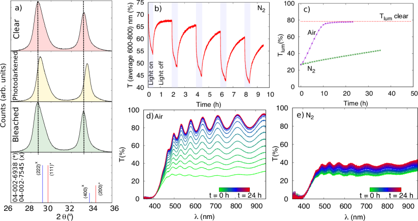
As described above, photochromic yttrium oxy-hydride can be obtained by the oxidation in air of reactively sputtered metallic YH2 thin films. The incorporation of oxygen in the YH2 lattice causes the increase of the lattice constant from 5.20 to 5.34 Å Mongstad et al. (2011b); Nafezarefi et al. (2017); Montero et al. (2018) and hence the displacement of the diffraction peaks towards lower angles. However under illumination, the lattice of the YHO films contracts back, but without reaching the original oxygen-free YH2 lattice constant Maehlen et al. (2013): Figure 2 (a) shows grazing incidence XRD patterns corresponding to an yttrium oxy-hydride sample in its initial (clear), illuminated (photodarkened) and recovered (bleached) states. The standard diffraction peaks for YH and Y2O3 according to the Joint Committee of Powder Diffraction Standards (JCPDS) card nums. 04-002-6938 and 04-002-7545 are also shown for comparison.
The analysis of the XRD patterns revealed how the films undergo an accordion-like transformation, i.e., the YHO lattice contracts and expands when subjected to illumination/darkness cycles, respectively. In addition to these observations, our previous studies performed from a purely optical perspective pointed to the formation, under illumination, oxygen-deficient YHO domains within the YHO lattice Montero et al. (2017). The filling factor (ff) of such domains was predicted to be very small Montero et al. (2017) although it increased with illumination time Montero et al. (2017). The fact that the metallic domains are diluted in the dielectric YHO structure (Maxwell-Garnett effective medium approximation holds) explains why the darkened samples exhibit higher optical absorption than high optical reflectance Mongstad et al. (2011a). In view of the above we postulate that, in YHO samples under illumination, some of the oxygen atoms are pushed towards the surface, leaving behind oxygen-deficient (metallic) domains which are responsible for the overall darkening of the film; in other words, YHO can be partially reverted to oxygen-defficient YHO by illumination. Therefore, the photochromic change is caused by very subtle changes in oxygen content–as predicted by to our effective medium calculations Montero et al. (2017)–presumably difficult to detect experimentally. However, if this hypothesis is correct, after long enough illumination/darkness cycling in an oxygen-free atmosphere, a small amount of oxygen atoms could effectively leave the YHO film every cycle, affecting the photochromic recovery; this is indeed the case as demonstrated by the following experiment: YHO thin films were subjected to 2 h period cycles (0.5 h illumination followed by 1.5 h darkness) inside a glove box filled with N2. The O2 and H2O content within the glove box was below 0.1 and 1.4 ppm, respectively. The average transmittance of the film was measured between 600 and 800 nm during cycling and plotted in Figure 2 (b). In the absence of air, the films lost part of their initial transparency in each cycle, not being able to recover fully. After 4 weeks of continuous cycling within the glove box, the luminous transmittance of the samples decreased from 78.5 % in the non-illuminated state, to 26.7%. This heavily photodarkened films were allowed to bleach in total darkness, both in air and in N2 atmosphere (glove box); the evolution of Tlum is presented in Figure 2 (c). It is evident from this figure that the bleaching speed in darkness of the photodarkened samples was much slower inside the globe box than in air. In addition, a series of transmittance measurements have been performed during the recovery stage along a lapse of 24 hours both, in air Figure 2 (d) and inside the glove-box (N2 atmosphere), Figure 2 (d). Note that, in both cases, the films were kept in total darkness (except for illumination with the spectrophotometer probe: 1 s each hour for performing the measurements). The films kept in air recover totally their initial transparency after few hours (T, presented in Figure 2 (c) as an horizontal dashed line) while the films in N2 recovered very little in the same period of time. Since there are not significant differences between the temperature inside and outside of the glove-box (both at 20∘ C), the data presented in Figure 2 (c, d and e) strongly indicates that a source oxygen from the ambient is crucial for the adequate recovering of the photo-darkened films. This is consistent with the de-oxygenation hypothesis: a source oxygen is necessary in order to replace the oxygen atoms released during illumination; this points to a light-induced exchange of oxygen atoms between the film and the environment.
Considering the low electronegativity of Y, the idea of oxygen being pushed out of the YHO lattice by illumination may look counterintuitive at first. However, it is important to consider that for achieving a noticeable darkening only very small amounts of oxygen are required to leave the film. Indeed, a ff of just 2 % of oxygen-deficient YHO is known to cause a drop of the visible transmittance larger than 30% Montero and Karazhanov (2018). A theoretical model for understanding the light-induced oxygen release in YHO films is presented below.
III.3 Theoretical considerations
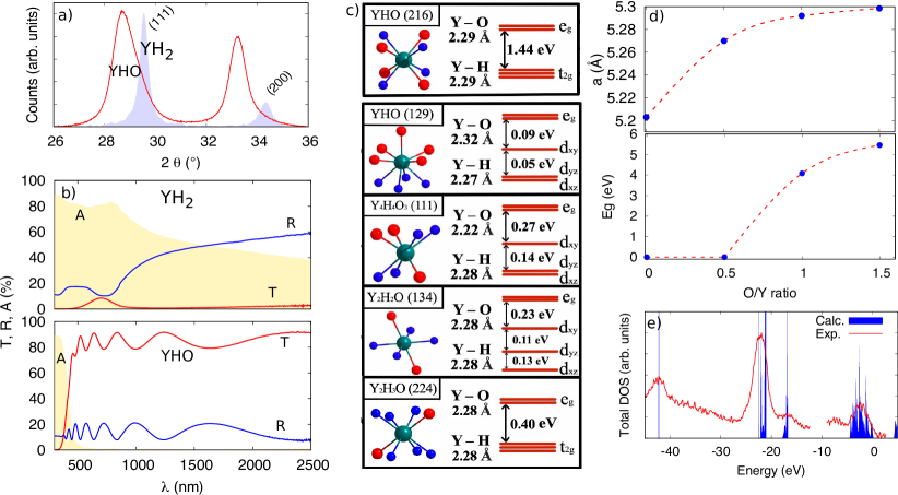
Experimental evidence, presented above, points to light-induced oxygen exchange between the film and the atmosphere. In the present section, this question is addressed by DFT modelling (ab initio calculations using the software VASP). It is known that photochromic YHO coatings are obtained experimentally by the partial oxidation of YH2 films in air. As discussed before, the incorporation of oxygen into YH2 results in the expansion of the YH2 lattice, i.e. the lattice parameter (a) increases, and hence the XRD peaks corresponding to YHO appear displaced towards lower angles when compared to oxygen-free YH2, Figure 3 (a). The oxygen intake also causes the band gap opening: Figure 3 (b) shows the experimental transmittance (T), reflectance (R) and absorbance (A) corresponding to YH2 and compared to photochromic YHO. YH2 presents the optical behaviour of a metal but, after the incorporation of oxygen, turns into YHO, a wide-bandgap semiconductor. Taking the crystalline structure of YH2 as the starting point [fm-3m and space group symmetry number (SPGN) 225] diverse YHO lattices of stoichiometry YHxOy have been obtained, Figure 3 (c). In particular, multiscale modelling Pishtshev et al. (2018) predicted the possibility of lattices of stoichiometry: (i) Y4H4O3 111 P2m and (ii) Y2H2O 134 P42/nmm Pishtshev et al. (2018), as well as (iii), YHO and SPGN 129 P4/nmm, 215 P3m, 224 Pnm and 216 P3m, amongst others. Systematic theoretical and experimental studies Montero et al. (2018) pointed to P3m with SPGN 216 as the most energetically favourable yttrium oxy-hydride lattice, i.e., stoichiometry and in YHxOy. Please note that YH2 (225), Y4H6O2 (224), Y2H2O (134) as well as Y4H4O3 (111), present a metallic character, whereas YHO (216) is predicted to be a wide-band gap semiconductor, as expected experimentally. Besides, the and stoichiometry is consistent with the results obtained by ion beam analysis Moldarev et al. (2018). According to these results, YHO crystallises into a cubic structure with a lattice constant a = 5.29 Å, which corroborates the lattice expansion that takes place in YH2 (a = 5.20 Å) when exposed to air. In particular, the expansion of a as well as the opening of the bandgap after air exposure is predicted by DTF, see Figure 3 (d), where calculated lattice constant and bandgap are plotted as a function of the Y/O ratio. The predicted value of a for YHO is, however, slightly smaller than the experimental value observed (a = 5.34 Å) Mongstad et al. (2011b); Montero et al. (2018); discrepancies may arise due to the thin-film nature of the experimentally studied specimens as well as to other factors (lattice strain, defects, etc.). In this energetically favourable YHO (216) lattice, Y, O and H atoms occupy the Wyckoff positions 4c (1/4; 1/4; 1/4), 4a (0, 0, 0) and 4b (1/2, 1/2, 1/2) respectively. Since H and O atoms share tetrahedral sites in the lattice, YHO belong to the emerging family of materials called oxy-hydrides Montero et al. (2018). The partial oxidation of YH2, and hence the formation of YHO, triggers the expansion of the unit cell volume Pishtshev et al. (2018). As a consequence of the lattice expansion, the bond distances in YHO will be subjected to oxygen-induced elongation –see Figure 3 (c), where Y-O and Y-H bond lengths, as well as the splitting of the Y 3 states at the conduction band minimum for different YHxOy stoichiometries is presented–.
The experimental XPS data and the calculated total density of states of YHO (216) are in good agreement, as shown in Figure 3 (c). The opening of a wide band bap as the oxygen atoms are incorporated in the YH2 structure is also predicted by the ab initio calculations, Figure 3 (d). However, the model overestimates the band gap, being 4.9 eV the calculated value for YHO (216), approximately 1 eV larger than the experimental band gap determined in the photochromic films by optical methods Montero et al. (2018); Montero and Karazhanov (2018). It should be noted that the YHO films, obtained by the oxidation of YH2 previously prepared by reactive sputtering, are polycrystalline and multiphase in nature–note the widening of the XRD peaks of YHO when compared to YH2 in Figure 3 (a)–. Therefore, the energy band diagram of the material most likerly corresponds to an heterostructure of type-II with staggered band gap.
The projected DOS for YHO (216) revealed that both O and H atoms strongly contribute to the topmost valence band states. However, they are not hybridized because both H and O atoms are connected to the Y atoms independently from each other. On the other hand, the bottommost conduction band, which in the case of an ideal lattice is three times degenerate, and it is formed mostly by Y d-states, in particular t2g states, Figure 3 (c). This result suggests that the light-induced O release from the film can be caused by the pseudo Jahn-Teller distortion effect: Y atoms are located at the centre of the tetrahedral H and O sublattices and, under illumination, the transfer of electrons from the valence band to the t2g bands will turn the YHO (216) lattice unstable Bersuker (2013). As the p orbitals of O atoms are hybridized with the Y d orbitals, the degeneracy of the t2g states can be avoided by the removal of oxygen atoms. As a result, an O-deficient unit cell, with smaller lattice constant will be created. As reported in Pishtshev et al. Pishtshev et al. (2018) there are many O-deficient structural arrangements that can be obtained from YHO, being Y4H6O2 (224) a likely candidate of predicted metallic character and lattice constant = 5.27 Å (Figure 3). In summary, as a result of illumination, metallic domains of smaller lattice constant will be created in the YHO (216) lattice, which result in the in the photohromic effect and the lattice contraction observed experimentally. The material seems to be able to host the out-diffused O atoms which, in some cases can reach the surface being detected by XPS or even leave the film as demonstrated before. After stopping the illumination, the released O atoms can return to their former positions and the initial optical transparency will be restored.
IV Conclusions
When exposed to air the YH2 lattice expands from 5.20 to 5.34 Å due to the incorporation of oxygen. Beyond the lattice expansion, YH2 turns into YHO which is transparent and photochromic. However, the incorporation of oxygen can be, at least partially, reverted by illumination. According to theoretical calculations, the pseudo Jan-Teller effect may cause the weakening of the Y-O bond by the action of light of adequate wavelength (UV and blue); as a result, some oxygen atoms can be released leaving behind an oxygen deficient structure responsible for the photochromic darkening of the YHO films. Released oxygen atoms move towards the film surface, causing an enhancement of the hydrophobic properties, being some O atoms able to leave the film. For this reason, the adequate bleaching of the photo-darkened films has to take place in an oxygen rich atmosphere. In summary, YHO “breathes” as a consequence of illumination: the YHO lattice contraction/expansion under illumination/darkness is accompanied by oxygen release/intake which causes the observed change in the optical properties.
Acknowledgements
This work has been supported by the Norwegian Research Council through the FRINATEK project 287545, internal project of the Institute for Energy Technology and Turkish Council of Higher Education Board 100/2000 PhD scholarship. The computations have been performed by using the Norwegian Notur supercomputing facilities through the project nn4608k.
V BIBLIOGRAPHY
References
- Huiberts et al. (1996) J. N. Huiberts, R. Griessen, J. H. Rector, R. J. Wijngaarden, J. P. Dekker, D. G. D. Groot, and N. J. Koeman, Nature 380, 231 (1996).
- Nafezarefi et al. (2017) F. Nafezarefi, H. Schreuders, B. Dam, and S. Cornelius, Applied Physics Letters 111, 3 (2017).
- Montero et al. (2018) J. Montero, F. A. Martinsen, M. Lelis, S. Z. Karazhanov, B. C. Hauback, and E. S. Marstein, Solar Energy Materials and Solar Cells 177, 106 (2018).
- La et al. (2018) M. La, N. Li, R. Sha, S. Bao, and P. Jin, Scripta Materialia 142, 36 (2018).
- Miniotas et al. (2000) A. Miniotas, B. Hjörvarsson, L. Douysset, and P. Nostell, Applied Physics Letters 76, 2056 (2000).
- Mongstad et al. (2011a) T. Mongstad, C. Platzer-Björkman, J. P. Maehlen, L. P. A. Mooij, Y. Pivak, B. Dam, E. S. Marstein, B. C. Hauback, and S. Z. Karazhanov, Solar Energy Materials and Solar Cells 95, 3596 (2011a).
- Ohmura et al. (2007) A. Ohmura, A. MacHida, T. Watanuki, K. Aoki, S. Nakano, and K. Takemura, Applied Physics Letters 91 (2007).
- Towns (2016) A. Towns, Applied Photochemistry. In: Applied Photochemistry: When Light Meets Molecules (Springer International Publishing, Switzerland, 2016) pp. 227–79.
- Montero et al. (2017) J. Montero, F. A. Martinsen, M. García-Tecedor, S. Z. Karazhanov, D. Maestre, B. Hauback, and E. S. Marstein, Physical Review B 95, 1 (2017).
- Zenkin et al. (2014) S. Zenkin, S. Kos, and J. Musil, Journal of the American Ceramic Society 97, 2713 (2014).
- Azimi et al. (2013) G. Azimi, R. Dhiman, H. M. Kwon, A. T. Paxson, and K. K. Varanasi, Nature Materials 12, 315 (2013).
- Ho et al. (2007) S. L. Ho, D. Kwak, Y. L. Dong, G. L. Seung, and K. Cho, Journal of the American Chemical Society 129, 4128 (2007).
- Feng et al. (2004) X. Feng, L. Feng, M. Jin, J. Zhai, L. Jiang, and D. Zhu, Journal of the American Chemical Society 126, 62 (2004).
- Yadav et al. (2016) K. Yadav, B. R. Mehta, S. Bhattacharya, and J. P. Singh, Scientific Reports 6 (2016).
- Montero and Karazhanov (2018) J. Montero and S. Z. Karazhanov, Physica Status Solidi (A) Applications and Materials Science 215, 1 (2018).
- van Oss et al. (1988) C. J. van Oss, M. K. Chaudhury, and R. J. Good, Chemical Reviews 88, 927 (1988).
- van Oss (1993) C. J. van Oss, Colloids and Surfaces A: Physicochemical and Engineering Aspects (1993).
- G and J (1996a) K. G and F. J, Phys Rev B - Condens Matter Mater Phys 54, 11169 (1996a).
- G and J (1996b) K. G and F. J, Comput Mater Sci 6, 15 (1996b).
- G and J (1994) K. G and H. J, Phys Rev B 49, 14251 (1994).
- Blöchl (1994) P. E. Blöchl, Physical Review B 50, 17953 (1994).
- Kresse and Joubert (1999) G. Kresse and D. Joubert, Physical Review B 59, 1758 (1999).
- G and J (1993) K. G and H. J (1993) pp. 558–561.
- Perdew et al. (1996) J. P. Perdew, K. Burke, and M. Ernzerhof, Physical Review Letters 77, 3865 (1996).
- Heyd et al. (2003) J. Heyd, G. E. Scuseria, and M. Ernzerhof, J Chem Phys 118, 8207 (2003).
- Krukau et al. (2006) A. V. Krukau, O. A. Vydrov, A. F. Izmaylov, and G. E. Scuseria, The Journal of Chemical Physics 125, 224106 (2006).
- Henderson et al. (2011) T. M. Henderson, J. Paier, and G. E. Scuseria, physica status solidi (b) 248, 767 (2011).
- Li et al. (2010) S. Y. Li, G. A. Niklasson, and C. G. Granqvist, Journal of Applied Physics 108 (2010).
- Imanaka (2004) N. Imanaka, Binary Rare Earth Oxides , 111 (2004).
- Moldarev et al. (2018) D. Moldarev, D. Primetzhofer, C. C. You, S. Z. Karazhanov, J. Montero, F. Martinsen, T. Mongstad, E. S. Marstein, and M. Wolff, Solar Energy Materials and Solar Cells 177, 66 (2018).
- Feng et al. (2005) X. Feng, J. Zhai, and L. Jiang, Angewandte Chemie - International Edition 44, 5115 (2005).
- Wang et al. (2006) S. Wang, X. Feng, J. Yao, and L. Jiang, Angewandte Chemie - International Edition 45, 1264 (2006).
- Zhu et al. (2006) W. Zhu, X. Feng, L. Feng, and L. Jiang, Chemical Communications , 2753 (2006).
- de Sun et al. (2001) R. de Sun, A. Nakajima, A. Fujishima, T. Watanabe, and K. Hashimoto, The Journal of Physical Chemistry B 105, 1984 (2001).
- Kung (1989) H. H. Kung, in Transition Metal Oxides, Studies in Surface Science and Catalysis, Vol. 45 (Elsevier, The Netherlands, 1989) pp. 53 – 71.
- Khan et al. (2015) S. Khan, G. Azimi, B. Yildiz, and K. K. Varanasi, Applied Physics Letters 106, 061601 (2015).
- Beamson and Briggs (1993) G. Beamson and D. Briggs, Journal of Chemical Education 70, A25 (1993).
- Preston et al. (2014) D. J. Preston, N. Miljkovic, J. Sack, R. Enright, J. Queeney, and E. N. Wang, Applied Physics Letters 105, 011601 (2014).
- Lundy et al. (2017) R. Lundy, C. Byrne, J. Bogan, K. Nolan, M. N. Collins, E. Dalton, and R. Enright, ACS Applied Materials and Interfaces 9, 13751 (2017).
- Külah et al. (2017) E. Külah, L. Marot, R. Steiner, A. Romanyuk, T. A. Jung, A. Wäckerlin, and E. Meyer, Scientific Reports 7, 1 (2017).
- Craciun et al. (1999) V. Craciun, J. Howard, E. Lambers, R. Singh, D. Craciun, and J. Perriere, Applied Physics A 69, S535 (1999).
- Barshilia et al. (2012) H. C. Barshilia, A. Chaudhary, P. Kumar, and N. T. Manikandanath, Nanomaterials (Basel, Switzerland) 2, 65 (2012).
- Mongstad et al. (2011b) T. Mongstad, C. Platzer-Björkman, S. Karazhanov, A. Holt, J. Maehlen, and B. Hauback, Journal of Alloys and Compounds 509, S812 (2011b).
- Maehlen et al. (2013) J. P. Maehlen, T. T. Mongstad, C. C. You, and S. Karazhanov, Journal of Alloys and Compounds 580, S119 (2013).
- Pishtshev et al. (2018) A. Pishtshev, E. Strugovshchikov, and S. Karazhanov, Chemrxiv (2018), 10.26434/chemrxiv.6950138.v1.
- Bersuker (2013) I. B. Bersuker, Chem Rev 3, 1351–1390 (2013).