Structural analysis of nuclear spin clusters
via two-dimensional nanoscale nuclear magnetic resonance spectroscopy
Abstract
Two-dimensional Nuclear Magnetic Resonance (NMR) is essential in molecular structure determination. The Nitrogen-Vacancy (NV) center in diamond has been proposed and developed as an outstanding quantum sensor to realize NMR in nanoscale. In this work, we develop a scheme for two-dimensional nanoscale NMR spectroscopy based on quantum controls on an NV center. We carry out a proof of principle experiment on a target of two coupled nuclear spins in diamond. A COSY-like sequences is used to acquire the data on time domain, which is then converted to frequency domain with the fast Fourier transform (FFT). With the two-dimensional NMR spectrum, the structure and location of the set of nuclear spin are resolved. This work marks a fundamental step towards resolving the structure of a single molecule.
Molecular structure analysis is a cornerstone of biology, chemistry and medicine. Among three vastly used techniques for structure analysis, X-ray algara-siller_square_2015 , electron microscopy adrian_cryo-electron_1984 , and nuclear magnetic resonance (NMR) aue_twodimensional_1976 ; oschkinat_three-dimensional_1988 ; wuthrich_way_2001 , NMR is the promising technique to reveal the structure information with nondestructive in vivo detection at room temperature. However, the conventional NMR relays on a large ensemble of molecules to obtain a sufficient good signal-to-noise ratio, which will average out some individual properties. NMR for single molecule structure analysis is of paramount importance, and a crucial step of this is single molecular two-dimensional NMR spectroscopy (2D NMR).
Thank to high sensitive atomical-scale NV centers balasubramanian_nanoscale_2008 ; maze_nanoscale_2008 , nanoscale magnetic resonance spectroscopy has been developed rapidly over the past years. One-dimensional nanoscale NMR staudacher_nuclear_2013 ; mamin_nanoscale_2013 ; lovchinsky_magnetic_2017 , single spin sensitivity NMR muller_nuclear_2014 , single molecule magnetic resonance shi_single-protein_2015 ; lovchinsky_nuclear_2016 , the detection of macroscopic-scale chemical shift and J-coupling aslam_nanoscale_2017 ; glenn_high-resolution_2018 , the 2D spectrum of NV-nuclear spin system boss_one-_2016 ; abobeih_atomic-scale_2019 and microscopic 2D NMRsmits_two-dimensional_2019 have been realized with NV centers. While these works on nanoscale NMR make it possible to provide unprecedented insight into molecule structure, 2D nanoscale NMR ajoy_atomic-scale_2015 ; ma_proposal_2016 is critical for the unambiguous determination of molecule structure (Fig. 1(a)).
In the present work, we propose a scheme for structure reconstruction of single molecule by 2D nanoscale NMR based on the NV center in diamond. We carry out an experiment to characterize a pair of coupled nuclear spins in diamond and demonstrate experimentally 2D nanoscale NMR spectroscopy. The 2D information obtained offers high-confident analysis of three-dimensional molecular geometry structure. In the end, we evaluate the performance of our scheme on a typical single protein.
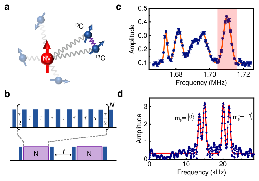
We use a single NV center in a CVD-grown diamond with a natural abundance (1.1 %) of 13C nuclear spins (Fig. 1(a)). The NV electron spin is used as a quantum sensor to probe 13C nuclear spins cluster, with the pulse sequence plotted in Fig. 1(b), through dynamical decoupling spectroscopy du_preserving_2009 ; zhao_sensing_2012 . The NV electron spin is prepared in the superposition state . During the controlled evolution, the coherence of NV electron spin decreases, leading to a dip on resonance by , where is the Larmor frequency of nuclear spin and is the parallel component of hyperfine interaction between NV electron spin and nuclear spin. The dips of the spectrum in Fig. 1(c) reveal nuclear spins with different hyperfine interactions. The high resolution correlation spectroscopy laraoui_high-resolution_2013 ; kong_towards_2015 is then applied to the NV and nuclear spins system to resolve the fine structure of spectrum by focusing on the red region in Fig. 1(c) with center frequency 1.71 MHz. Dynamical decoupling protocol with time interval resonant with is applied, and the correlation between the first and the second dynamical decoupling protocols is recorded. During the free evolution in between, the nuclear spins’ evolution are dependent on the electron spin states . Thus, the spectrum contains different peaks centered at and . Actually, the spectrum in Fig. 1(d) shows two peaks for both and subspaces.
However, such a spectrum in Fig. 1(d) alone could allow different interpretations. It may correspond to a two coupling nuclear spin cluster or three isolated nuclear spins (see SM section IIA). The one-dimensional NMR spectrum alone cannot discriminate these different interpretations. A two-dimensional correlation map is then necessary to provide such important information.
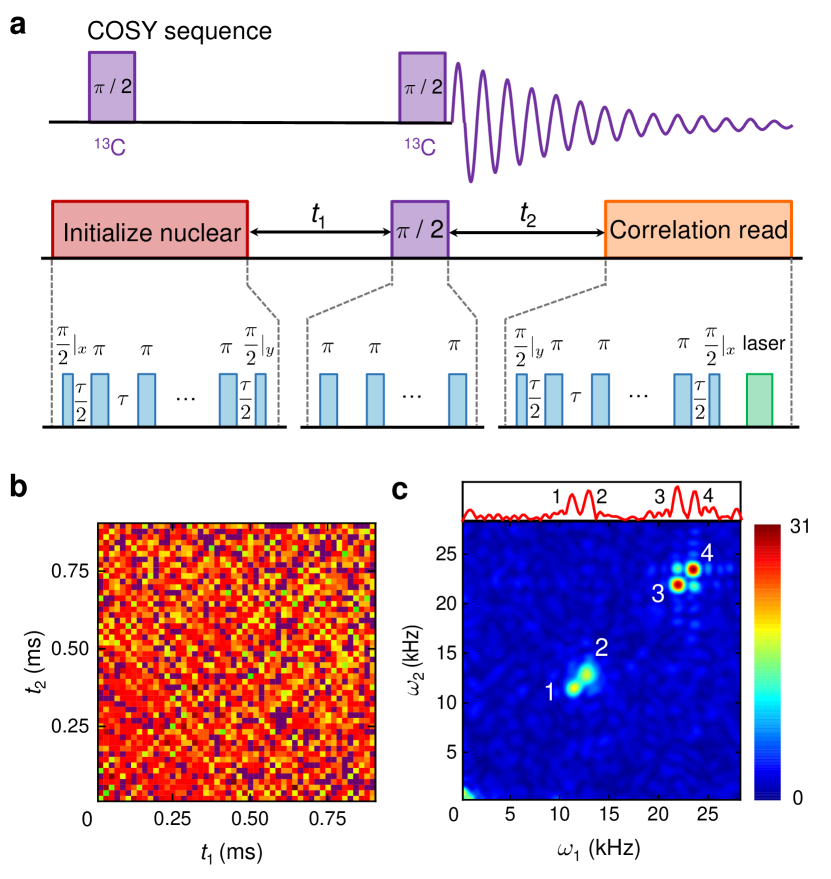
In analog to the COSY spectroscopy in conventional NMR, we develop a two-dimensional protocol to investigate such correlation map of target nuclear spins (Fig. 2(a)). The Hamiltonian for the system of an NV sensor and two nuclear spins is , where is the NV electron spin, is the th nuclear spin, is the hyperfine coupling between NV and nuclear, is dipolar coupling interaction between two nuclear spins. The NV sensor is driven with the dynamical decoupling sequence which is in resonant with the red region center frequency in Fig. 1(b). Then the target nuclear spin is initialized to the transverse direction. During an interval of 0 to , the two nuclear spins and interact and a phase is accumulated in the first nuclear spin, where is the parallel component of the hyperfine interaction of the NV sensor with , and is the component of the coupling between nuclear spin and . Then a series of =40 dynamical decoupling pulse is applied, which corresponds to half pulse on the nuclear spin. A free evolution of duration comes afterwards, another phase accumulates. In the end, another dynamical decoupling sequence is applied to read out the transverse component of the nuclear spin.
Sweeping the duration time and from 4 s to 0.9 ms, the correlation map is realized, as shown in Fig. 2(b). The 2D FFT spectrum in Fig. 2(c) clearly shows the correlations of peaks of the one-dimensional spectrum, i.e. which peaks belong to the same spin system. In detail, the cross peaks between the \nth3 and \nth4 peaks indicate that they belong to a coupled spin system. The \nth3 and \nth4 peaks correspond to the condition , while the \nth1 and \nth2 peaks correspond to .
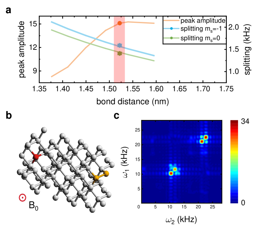
From the spectrum, we can obtain the bond length of off-axis 13C-13C nuclear spins. The optimally estimated bond length is 1.52 0.02 Å (Fig. 3(a)), (SM section II D). We also resolve the position of the nuclear spin cluster in the diamond lattice as shown in Fig. 3(b). The position is uniquely determined to be one of a set of symmetrically equivalent positions. Using these parameters, the simulated 2D spectrum is shown in Fig. 3(b), which fits well with the experiment spectrum (minor difference in the \nth1 and \nth2 peaks due to field fluctuation).
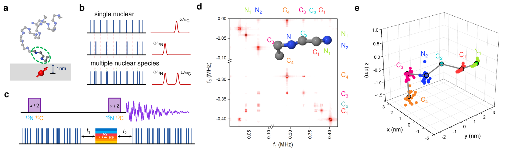
The demonstrated two-dimensional NMR technique, with potential improvements in the sensor fabrication, may enable the capability to resolve the structure of a single molecule. We consider as an example the single avian pancreatic polypeptide molecular placed at a similar distance as the nuclear spin cluster to the NV center. Without loss of generality, two amino acid are labeled with and as shown in Fig. 4(a). To resolve the molecule structure, and are observed at the same time to get heteronuclear coupling. A non-periodical multi-nuclear species correlation sequence is introduced here (Fig. 4(b), SM secrion III B). It consists of pulses inserted on zeros of between two pulses to observe nuclear spins with all different frequency . Unlike in an ensemble NMR experiment, the strong gradient field (about 0.1 mT/nm) from NV center muller_nuclear_2014 splits the homonuclear spins spectrum of a single molecule. With an artificially fabricated antena jakobi_measuring_2017 , a much larger gradient up to 10 mT/nm is possible. Utilized by multi-nuclear species correlation sequence, the 2D heteronuclear spin resonance sequence is shown in Fig. 4(d) in analog to the homonuclear one (Fig. 3(a)). Then the simulated two-dimensional NMR spectrum is demonstrated in Fig. 4(d), the corresponding nuclear spins frequencies are labeled along the axis. The off-diagonal non-zero terms correspond to the coupling between different nuclear spins. By rotation the polar angle between the external magnetic field and the NV center, the dipolar interaction tensors are measured to characterize the distances between nuclear spins (SM section III D). By solving the discretizable molecular distance geometry problem, the molecule conformation is resolved (Fig. 4(e)) with a resolution 0.3 nm.
Overall, the 2D nanoscale NMR spectrum of a single cluster of two coupled nuclear spins is demonstrated by using a quantum sensor of the NV center in diamond. Equipped with a high sensitive NV sensor, the length of chemical bonds is resolved under 2D nanoscale NMR in diamond. Although the structure of a single cluster of two nuclear spins has previously been resolved shi_sensing_2014 , a 2D correlation spectroscopy is still necessary to resolve the structure of more complicated molecule or nanostructure. The 2D nanoscale NMR spectroscopy reported here, together with previous work on nanoscale NMR, can yield valuable structural information as opposed to bulk NMR, where the couplings typically hamper structure analysis. The spectral resolution can be improved by, for example, the Qdyne technique schmitt_submillihertz_2017 ; boss_quantum_2017 and weak measurement readout cujia_watching_2018 ; pfender_high-resolution_2018 . The sensitivity can be improved by more efficient readout method shields_efficient_2015 . With further improvements, our 2D nanoscale NMR method has the potential to characterize the structure of a single molecule or conformational dynamics at single molecule level that is not accessible by conventional methods.
In the process of preparing the manuscript, a similar work just appears on arXiv abobeih_atomic-scale_2019 .
I Acknowledgments
We thank Fedor Jelezko for helpful discussions. The authors at University of Science and Technology of China are supported by the National Key RD Program of China (Grant No. 2018YFA0306600, No. 2016YFA0502400, No. 2018YFF01012501, and No. 2017YFA0305000), the National Natural Science Foundation of China (Grants No. 81788101, No. 91636217, No. 11761131011, No. 11722544, and No. 11775209), the CAS (Grants No. GJJSTD20170001, No. QYZDY-SSW-SLH004, and No. YIPA2015370), Anhui Initiative in Quantum Information Technologies (Grant No. AHY050000), the CEBioM, the Thousand-Young-Talent Program of China, and the Fundamental Research Funds for the Central Universities, the Innovative Program of Development Foundation of Hefei Center for Physical Science and Technology (Grants No. 2017FXCX005).
Supplementary materials
Supplementary Text
Figs. S1 to S11
References
- (1) G. Algara-Siller, O. Lehtinen, F. C. Wang, R. R. Nair, U. Kaiser, H. A. Wu, A. K. Geim, and I. V. Grigorieva, Nature 519, 443 (2015).
- (2) M. Adrian, J. Dubochet, J. Lepault, and A. W. Mc- Dowall, Nature 308, 32 (1984).
- (3) W. P. Aue, E. Bartholdi, and R. R. Ernst, The Journal of Chemical Physics 64, 2229 (1976).
- (4) H. Oschkinat, C. Griesinger, P. J. Kraulis, O. W. Sørensen, R. R. Ernst, A. M. Gronenborn, and G. M. Clore, Nature 332, 374 (1988).
- (5) K. Wüthrich, Nature Structural Biology 8, 3 (2001).
- (6) G. Balasubramanian, I. Y. Chan, R. Kolesov, M. Al- Hmoud, J. Tisler, C. Shin, C. Kim, A. Wojcik, P. R. Hemmer, A. Krueger, T. Hanke, A. Leitenstorfer, R. Bratschitsch, F. Jelezko, and J. Wrachtrup, Nature 455, 648 (2008).
- (7) J. R. Maze, P. L. Stanwix, J. S. Hodges, S. Hong, J. M. Taylor, P. Cappellaro, L. Jiang, M. V. G. Dutt, E. Togan, A. S. Zibrov, A. Yacoby, R. L. Walsworth, and M. D. Lukin, Nature 455, 644 (2008).
- (8) T. Staudacher, F. Shi, S. Pezzagna, J. Meijer, J. Du, C. A. Meriles, F. Reinhard, and J. Wrachtrup, Science 339, 561 (2013).
- (9) H. J. Mamin, M. Kim, M. H. Sherwood, C. T. Rettner, K. Ohno, D. D. Awschalom, and D. Rugar, Science 339, 557 (2013).
- (10) I. Lovchinsky, J. D. Sanchez-Yamagishi, E. K. Urbach, S. Choi, S. Fang, T. I. Andersen, K. Watanabe, T. Taniguchi, A. Bylinskii, E. Kaxiras, P. Kim, H. Park, and M. D. Lukin, Science 355, 503 (2017).
- (11) C. Müller, X. Kong, J.-M. Cai, K. Melentijević, A. Stacey, M. Markham, D. Twitchen, J. Isoya, S. Pezzagna, J. Meijer, J. F. Du, M. B. Plenio, B. Naydenov, L. P. McGuinness, and F. Jelezko, Nature Communications 5, 4703 (2014).
- (12) F. Shi, Q. Zhang, P. Wang, H. Sun, J. Wang, X. Rong, M. Chen, C. Ju, F. Reinhard, H. Chen, J. Wrachtrup, J. Wang, and J. Du, Science 347, 1135 (2015).
- (13) I. Lovchinsky, A. O. Sushkov, E. Urbach, N. P. d. Leon, S. Choi, K. D. Greve, R. Evans, R. Gertner, E. Bersin, C. Müller, L. McGuinness, F. Jelezko, R. L. Walsworth, H. Park, and M. D. Lukin, Science 351, 836 (2016).
- (14) N. Aslam, M. Pfender, P. Neumann, R. Reuter, A. Zappe, F. F. d. Oliveira, A. Denisenko, H. Sumiya, S. Onoda, J. Isoya, and J. Wrachtrup, Science 357, 67 (2017).
- (15) D. R. Glenn, D. B. Bucher, J. Lee, M. D. Lukin, H. Park, and R. L. Walsworth, Nature 555, 351 (2018).
- (16) J. M. Boss, K. Chang, J. Armijo, K. Cujia, T. Rosskopf, J. R. Maze, and C. L. Degen, Physical Review Letters 116 197601 (2016).
- (17) M. H. Abobeih, J. Randall, C. E. Bradley, H. P. Bartling, M. A. Bakker, M. J. Degen, M. Markham, D. J. Twitchen, and T. H. Taminiau, arXiv:1905.02095 (2019).
- (18) J. Smits, J. Damron, P. Kehayias, A. F. McDowell, N. Mosavian, I. Fescenko, N. Ristoff, A. Laraoui, A. Jarmola, and V. M. Acosta, arXiv:1901.02952 (2019).
- (19) A. Ajoy, U. Bissbort, M. D. Lukin, R. L. Walsworth, and P. Cappellaro, Physical Review X 5, 011001 (2015).
- (20) W.-L. Ma and R.-B. Liu, Phys. Rev. Applied 6, 054012 (2016).
- (21) J. Du, X. Rong, N. Zhao, Y. Wang, J. Yang, and R. B. Liu, Nature 461, 1265 (2009).
- (22) N. Zhao, J. Honert, B. Schmid, M. Klas, J. Isoya, M. Markham, D. Twitchen, F. Jelezko, R.-B. Liu, H. Fedder, and J. Wrachtrup, Nature Nanotechnology 7, 657 (2012).
- (23) A. Laraoui, F. Dolde, C. Burk, F. Reinhard, J. Wrachtrup, and C. A. Meriles, Nature Communications 4, 1651 (2013).
- (24) X. Kong, A. Stark, J. Du, L. P. McGuinness, and F. Jelezko, Physical Review Applied 4, 024004 (2015).
- (25) See Supplementary Material for detail.
- (26) I. Jakobi, P. Neumann, Y. Wang, D. B. R. Dasari, F. El Hallak, M. A. Bashir, M. Markham, A. Edmonds, D. Twitchen, and J. Wrachtrup, Nature Nanotechnology 12, 67.
- (27) F. Shi, X. Kong, P. Wang, F. Kong, N. Zhao, R.-B. Liu, and J. Du, Nature Physics 10, 21 (2014).
- (28) S. Schmitt, T. Gefen, F. M. Stürner, T. Unden, G. Wolff, C. Müller, J. Scheuer, B. Naydenov, M. Markham, S. Pezzagna, J. Meijer, I. Schwarz, M. Plenio, A. Retzker, L. P. McGuinness, and F. Jelezko, Science 356, 832 (2017).
- (29) J. M. Boss, K. S. Cujia, J. Zopes, and C. L. Degen, Science 356, 837 (2017).
- (30) K. S. Cujia, J. M. Boss, K. Herb, J. Zopes, and C. L. Degen, arXiv:1806.08243 [quant-ph].
- (31) M. Pfender, P. Wang, H. Sumiya, S. Onoda, W. Yang, D. B. R. Dasari, P. Neumann, X.-Y. Pan, J. Isoya, R.- B. Liu, and J. Wrachtrup, Nature Communications 10, 594.
- (32) B. Shields, Q. Unterreithmeier, N. de Leon, H. Park, and M. Lukin, 114, 136402.