A RIXS investigation of the crystal-field splitting of Sm3+ in SmB6
Abstract
The crystal-field (CF) splitting of the Hund’s rule ground state of Sm3+ in the strongly correlated topological insulator SmB6 has been determined with high resolution resonant inelastic x-ray scattering (RIXS) at the Sm M5 edge. The valence selectivity of RIXS allows isolating the crystal-field-split excited multiplets of the Sm3+ (4) configuration from those of Sm2+ (4) in intermediate valent SmB6. The very large energy range of RIXS allows the crystal-field analysis of a high lying multiplet at about 2.4 eV that has the same total angular momentum as the ground state so that ambiguities due to the elastic tail can be avoided. We find that the doublet and quartet of the Hund’s rule ground state are split by = 2010 meV which sets an upper limit for the 4 band width. This indicates an extremely large mass renormalization from the band structure value, pointing out the need to consider the coefficients of fractional parentage for the hopping of the 4 electrons.
SmB6 is an intermediate valent Kondo insulator in which the hybridization of localized 4 electrons and the conduction band (-hybridization) leads to the formation of a gap Menth et al. (1969); Cohen et al. (1970); Allen et al. (1979); Gorshunov et al. (1999); Riseborough (2000) of the order of 20 meV Xu et al. (2013); Zhu et al. (2013); Neupane et al. (2013); Jiang et al. (2013); Denlinger et al. (2013, 2014). Accordingly, the resistivity increases with decreasing temperature but instead of diverging it reaches a plateau below about 10 K. Surface states could be an explanation for the finite low temperature conductivity Hatnean et al. (2013); Zhang et al. (2013); Kim et al. (2013); Wolgast et al. (2013); Kim et al. (2014a); Wolgast et al. (2015); Thomas et al. (2016); Nakajima et al. (2016) and indeed it was theoretically predicted that SmB6 has all the ingredients, like strong spin-orbit coupling and electrons of opposite parity ( and ), for being a strongly correlated topological insulator Dzero et al. (2010); Takimoto (2011); Lu et al. (2013); Alexandrov et al. (2013). This prediction initiated many studies like angle-resolved photoelectron spectroscopy (ARPES) Xu et al. (2013); Zhu et al. (2013); Neupane et al. (2013); Jiang et al. (2013); Frantzeskakis et al. (2013); Denlinger et al. (2013, 2014); Xu et al. (2014a, b); Hlawenka et al. (2018); Ohtsubo et al. (2019), scanning tunneling spectroscopy Ruan et al. (2014); Rössler et al. (2014, 2016); Jiao et al. (2016), or de Haas-van Alphen (dHvA) Li et al. (2014); Tan et al. (2015); Thomas et al. (2018). Yet, despite all these efforts, the exciting question whether these surface states are topologically non-trivial still remains to be answered.
The -hybridization is also responsible for the intermediate valent character of Sm in SmB6. At low temperatures valences of 2.5 to 2.7 have been reported Allen et al. (1980); Tarascon et al. (1980); Mizumaki et al. (2009); Butch et al. (2016); Utsumi et al. (2017) so that the electronic configuration of Sm is described by the Hund’s rule ground states of the Sm (2+) and Sm (3+) configurations.
The surface topology is a bulk property so that it is indispensable to have knowledge of the parities, symmetries and near ground state energy scales of the participating bulk states. The two low lying Sm2+ 4 multiplets with the total angular momenta = 0 and = 1 are not CF split and their wave functions are spherical Lea et al. (1962). In contrast, the cubic CF splitting of the quartet and doublet of the lowest energy multiplet of Sm3+ 4 has eluded its determination till today.
Band structure calculations have been very successful in the field of semiconducting topological insulators, but they are not adequate for the rare earths because of correlations, nor are they accurate enough because the energy scales are much smaller. For example, several density functional theory calculations imply that the hole of the 4 configuration resides in the doublet Lu et al. (2013); Yanase and Harima (1992); Antonov et al. (2002); Kang et al. (2015); Singh and Lee (2018) but a recent hard x-ray - inelastic x-ray scattering (NIXS) investigation Sundermann et al. (2018); foo (a) by some of the authors of the present study reveals that the ground-state symmetry of the Sm3+ configuration is the quartet. Band structure calculations also suggest energy scales of the order of hundred meV for (see e.g. Kang et al. (2015); Singh and Lee (2018)) although the extrapolation of the CF parameters within the REB6 series suggests a splitting of the order of 15 meV; an extrapolation that is, of course, only valid in diluted systems Loewenhaupt and Prager (1986); Frick and Loewenhaupt (1986). Along the same line, band structure calculations produce 4 band widths of several hundred meV, while so far no 4 dispersions in ARPES have been observed within the experimental resolution Xu et al. (2013); Zhu et al. (2013); Neupane et al. (2013); Jiang et al. (2013); Frantzeskakis et al. (2013); Denlinger et al. (2013, 2014); Xu et al. (2014a, b); Hlawenka et al. (2018); Ohtsubo et al. (2019). The inclusion of correlation effects using Gutzwiller or dynamical mean field approaches (DMFT) Lu et al. (2013); Kim et al. (2014b) does produce narrower bands and smaller CF splittings but it is not clear whether the mass renormalizations used or found are realistic.
Inelastic neutron scattering (INS) is the obvious technique for tackling this problem, but although providing very useful information on SmB6, INS has not been successful in finding . The strong neutron absorption of Sm and B even in double isotopic samples, the superposition of both Sm configurations, and the presence of -hybridization cause serious complications. Nevertheless, the following pieces of information have been obtained by INS: The spin orbit transitions and at 35 meV and 130 meV have been observed Alekseev et al. (1993, 1995) (see near-ground state multiplets in red and blue boxes of Fig. 1 (b)). At low temperatures a long living spin resonance at about 14 meV shows up in the poly- and single crystalline data Alekseev et al. (1993, 1995) at the and high symmetry points with a form factor that is not 4-like Fuhrman et al. (2015). More recent INS data show that the spin resonance with an intrinsic width of about 0.1 meV (FWHM) decays above 30 K and that at low temperatures no magnetic intensity is observed below the energy of this resonance which is typical for a spin gap Fuhrman et al. (2018). At 100 K i.e. well above the temperature at which the many body spin resonance disappears, quasielastic magnetic intensity (/2 10 meV HWHM) following the Sm3+ magnetic form factor has been extracted by Alekseev et al. after a very careful intensity examination Alekseev et al. (2016). This is suggestive of the recovery of the single ion, though broadened, magnetic spectrum, but the size the CF splitting of the Sm3+ Hund’s rule ground state remains undetermined. We note that also the high resolution ARPES studies so far have not been able to detect the CF splitting, unlike in for example YbIr2Si2 Vyalikh et al. (2010), which maybe due to the complications of the SmB6 surface Zhu et al. (2013); Rössler et al. (2014, 2016).
Here resonant inelastic x-ray scattering (RIXS) is a promising option Amorese et al. (2018a, b). RIXS is not only element, it is also configuration selective. This is well known from studying valences at the rare earth -edge in the so called partial florescence yield mode Kotani and Shin (2001); Dallera et al. (2002). Here we use the configuration selectivity at the -edge (3 4) to distinguish the excitation spectra of the two Sm configurations.
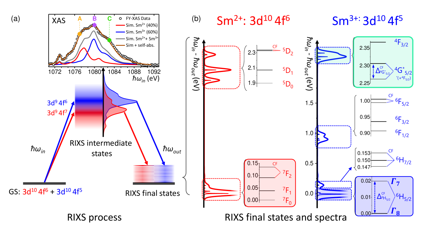
Figure 1 shows the -edge RIXS process for SmB6. The initial state configuration is an admixture of Sm2+: 34 (red) and Sm3+: 34 (blue). The resonant absorption of an 1090 eV x-ray photon at the M5 edge (3 4) creates a core hole. In this intermediate state the absorption lines of the two configurations are split in energy due to the different impact of the core hole potential on either configuration. Finally, in RIXS spectroscopy the intensity of the photons emitted by the resonant radiative decay is monitored as a function of the outgoing photon energy () so that energy transfer spectra can be measured. In principle the decay process in the RIXS process of SmB6 yields the superposition of two multiplet spectra (see simulations of two independent configurations in Fig. 1 (b)) but the choice of the incident photon energy along the XAS edge allows enhancing the signal of one of the two configurations. It is possible to resolve the CF splittings in a RIXS experiment because the large life time broadening of the intermediate state does not enter, i.e. the life time broadening in RIXS that matters is that of the final state Haak et al. (1978); Jensen et al. (1989); Hämäläinen et al. (1991); Carra et al. (1995).
Figure 1 (b) shows calculation of RIXS spectra for pure Sm2+ (red) and pure Sm3+ (blue). The photon-in photon-out RIXS process yields the selection rule = 0, 1, 2 so that multiplets with = 0, 1, 2 (for Sm2+) and = 1/2, 3/2, 5/2, 7/2, and 9/2 (for Sm3+) are accessible, the latter ones being so weak that they are not shown. In the cubic point symmetry of SmB6 only multiplets with 2 are CF split as shown on an enlarged energy scale in the colored boxes of Fig. 1 (b).
Apart from the valence selectivity, another advantage of RIXS is that the transferred energy is, in contrast to INS, practically unlimited, i.e. with RIXS we can study higher lying multiplets instead of the strongly hybridized Hund’s rule ground state of Sm3+ (blue box in Fig. 1 (b)). We will show that we can take advantage of the CF effect on the multiplet at about 2.4 eV (see the green box in Fig. 1 (b)). The asterisk indicates that due to the particularly strong intermultiplet mixing acting on this level, is no longer a good quantum number so that the multiplet labeling is not strictly valid. The total angular momentum = 5/2, however, remains a good quantum number for CF splittings smaller than the SO splittings. and the Hund’s rule ground state have the same so that the same CF parameter foo (b) (together with ) determines the CF splitting. The size of the splitting is given by JLS whereby JLS is something like a Stevens factor that is calculated within the full multiplet routine, while is determined experimentally. Hence, we can gain information on the splitting of the lowest energy multiplet by fitting the RIXS signal of the multiplet. For the JLS factor is larger than for (approximately double) so that the CF splitting is larger and less hampered by the limited energy resolution at the Sm -edge. In addition, the signal is free of the strong tail of the elastic peak (at 0 eV) and of the signal from other low energy excitations.
The SmB6 Kim et al. (2014a) -edge RIXS experiment at 20 K was performed at the ERIXS spectrometer of the ID32 beamline Brookes et al. (2018) at the European Synchrotron Radiation Facility (ERSF), Grenoble, France with a resolution of 45 meV at the Sm -edge ( 1090 meV). Data were taken with two different scattering angles, namely 2 = 90∘ and 150∘. Further details of the set-up are given in the Appendix. Simulation were performed with the full multiplet code Quanty Haverkort et al. (2012); Haverkort (2016). Atomic parameters were taken from the Cowan code Cowan (1981) and the reduction factor of the Slater integrals = = 0.86 were used (see Appendix). These values are in agreement with those in Ref. Sundermann et al. (2018).
The inset of Fig 1 (a) shows the bulk-sensitive experimental fluorescence-yield XAS (FY-XAS) data of the Sm -edge of SmB6 at 20 K, with the photon polarization parallel to the 100 direction (black line). These data have been simulated by calculating an XAS spectrum (gray line) containing Sm3+ (40% ) and Sm2+ (60%) spectral weights according to the SmB6 valence at low Allen et al. (1980); Tarascon et al. (1980); Mizumaki et al. (2009); Butch et al. (2016); Utsumi et al. (2017). Then self-absorption effects were included in the simulation (see brown line) Pellegrin et al. (1993) and compared with the FY-XAS data. Note, for reasons of graphical clarity the XAS data have been rescaled by a factor of three. The orange, purple and green dots marked A, B and C indicate the incident energies that were used for the RIXS experiment.
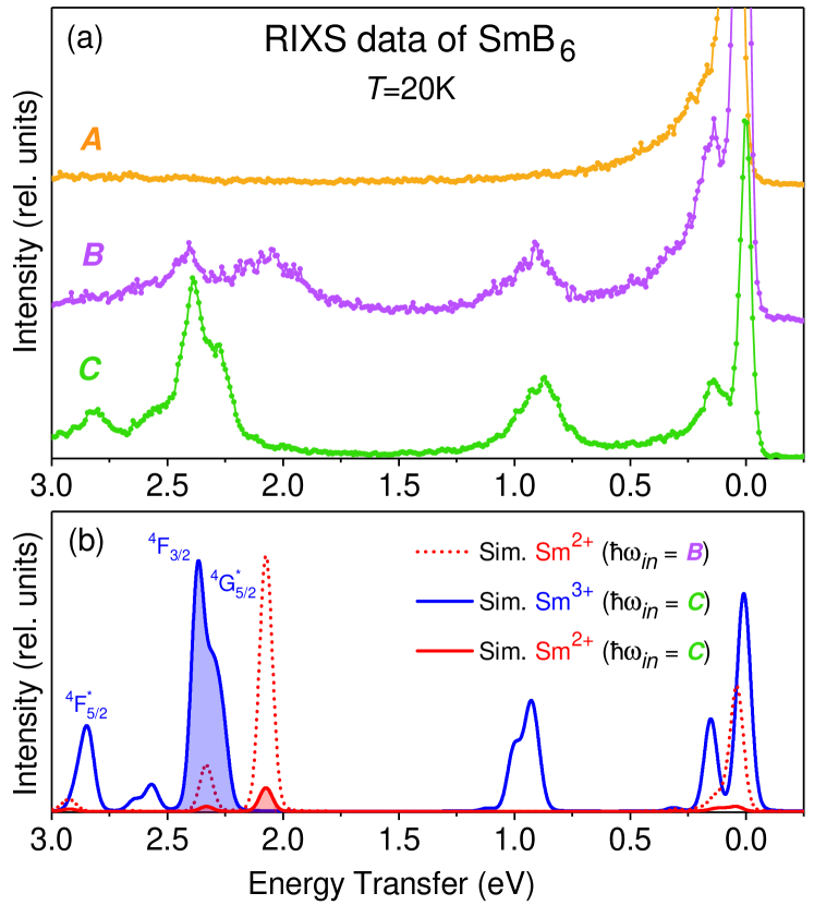
Figure 2 (a) shows the RIXS data at = 20 K up to 3 eV taken with the three different incident energies = A,B, and C and a scattering angle of = 90∘. = A corresponds to the pre-edge region where the absorption process is dominated by the ground state of Sm2+ . The asymmetric intensity close to the elastic line is indicative for the low energy transitions at 35 meV and some at about 150 meV, whereas higher energy transfers have no cross-section because they require larger incident energies due to selection rules. At = C the absorption arises mainly from the ground state of Sm3+. Energy B is in-between, i.e. the RIXS spectrum shows features characteristic of both valences but is not simply the superposition of spectrum A and C because of the incident energy dependence of the accessible excitations. Figure 2 (b), shows full multiplet RIXS calculations for the same spectrometer configuration for Sm2+ with the incident energies B (dotted red line) and C (red solid line) and and for Sm3+ with incident energy C (solid blue line) . The comparison of both panels demonstrates the energy selectivity of the RIXS signal and it confirms that the spectrum measured with = C resembles almost purely Sm3+ multiplets. We will therefore focus on the region of the and multiplets measured with this incident energy for further analysis of the crystal-field problem of Sm3+ (see colored regions in Fig. 2 (b).
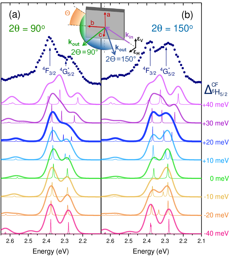
The top of Fig. 3 (a) and (b) show the RIXS data of the and multiplets (dark blue dots) at around 2.4 eV energy transfer (C) measured with horizontal () polarization and two different scattering angles, 2 = 90∘ and 150∘, thus taking advantage of the cross-section dependence on the scattering geometry. We recall that the multiplet is not affected by the CF because of 2 but that a finite CF splits the multiplet into two levels.
Figure 3 also shows simulations broadened with a 45 meV Gaussian resolution function for different CF splittings. We find that, for the same CF parameter, is about 2.2 times larger than . We show the simulations for = meV in steps of 10 meV, whereby the positive numbers refer to a and the negative ones to a ground state. Here the narrow thin lines correspond to the same CF simulation but with an unrealistic small resolution in order to visualize the details of the CF splittings. The simulation with = 0 (green lines) shows two main peaks, the multiplet and the about 80 meV higher in energy. We learn from these simulations that for CF splittings of less than 40 meV the intermixing of the two multiplets is negligible. We now compare in detail data and simulations: For 0 meV CF (green lines) and for 10 and 10 meV splitting (light blue and yellow lines) and 2 = 150∘ the intensity would be stronger than the 4F3/2 peak, see Fig. 3 (b). This is not the case in the experiment. Hence, the CF splitting of the ground state must be larger that 10 meV. For a negative CF splitting only one CF excitation would have intensity in the 2 =90∘ configuration, thus leading to a deep valley between the two multiplets which has not been observed. see Fig. 3 (a). We therefore conclude that the splitting must be positive, i.e. we confirm the results of previous directional dependent NIXS data Sundermann et al. (2018). For 40 meV CF splitting the spectral shape has changed considerably for both scattering geometries so we also exclude this possibility as well. It turns out that peak shapes and intensity ratios of both scattering configurations are best reproduced with = +20 meV.
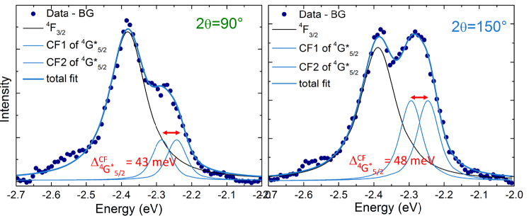
Figure 4 shows the same RIXS data as in Fig. 3 but after subtracting a linear background. The lines represent an empirical fit with three Voigt profiles whereby the Gaussian contribution is kept fixed to the experimental resolution. The Lorentzian widths, the line positions and intensities were varied with the simplification that lifetime broadening and intensity of the two CF excitations are identical. The best fits yield = 43 and 48 meV for the 2 = 90∘ and 150∘ scattering configuration, respectively, corresponding to a splitting of 20 and 22 meV of the ground state multiplet . Other trials with larger crystal-field splittings no longer reproduce the data, see Appendix. We learn form this exercise that () should be 66 meV (30 meV). Summarizing, we thus find = 2010 meV.
The present RIXS result agrees surprisingly well with the CF splitting that is expected from the extrapolation within the REB6 series Loewenhaupt and Prager (1986). The result also explains the lineshape of the lowest state signal as measured in photoemission Denlinger et al. (2014); we can now propose to describe it in terms of two Lorenzian lines, one twice as strong as the other according to a quartet ground state and a excited doublet, that are about 20 meV apart. Furthermore, the present data confirm the - inelastic x-ray scattering result of SmB6 that also finds a quartet ground state Sundermann et al. (2018); foo (a).
The finding that the CF splitting is 10 meV 30 meV in combination with the NIXS result that the ground state is not a highly mixed and state Sundermann et al. (2018) indicates that the 4 band width is small and less than 30 meV. Considering the fact that the band width from band structure calculations is several hundred meV, we infer that the mass renormalization is extremely large. This also gives credit to the idea that coefficients of fractional parentage should be considered for removing or adding an electron from/to the lowest Sm or multiplet states Sawatzky and Green (2016): a reduction factor of 0.033 can be found for the – hopping. A Gutzwiller study uses a somewhat less strong reduction factor Lu et al. (2013), while a DMFT calculation Kim et al. (2014b) found indeed the extremely narrow bands. It should be noted however, that the sign of the CF splitting and thus also its magnitude used or found in these many body calculations,Lu et al. (2013); Kim et al. (2014b) is different from the experiment. It would be highly desirable if these calculations could be tuned in a way that they reproduce the experimental values.
The electronic configuration selectivity, the large accessible energy transfers, and the cross-section dependence on the scattering configuration in RIXS have been instrumental to observe finger prints of the CF splitting of the Sm3+ Hund’s rule ground state in SmB6. We find = 2010 meV describes the data well, thereby setting limits to the 4 band width.
I Appendix
I.1 Sample and Experiment
The RIXS experiment was performed on aluminum flux grown single crystals Kim et al. (2014a) that were aligned by Laue prior to the experiment. The data were cleaved under vacuum, then transferred to the main chamber and measured at 20 K. Data were acquired for about 5 hours for each spectrum (only 3 hours for the spectrum B). The instrument 45 meV-FWHM Gaussian response function was estimated by measuring a carbon tape. The measurements were performed with horizontal polarization () of the incident photons, two different scattering angles, = 90∘ and = 150∘, a sample angle of and with the b and c directions of the sample in the scattering plane (see inset of Fig. 3).

I.2 Simulation
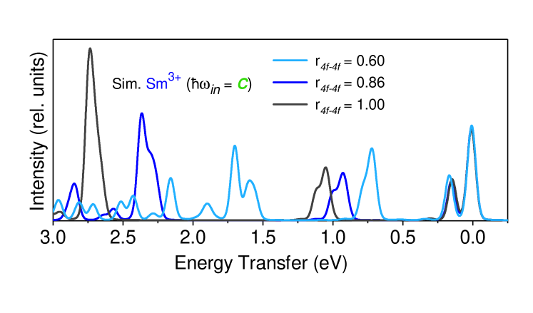
Simulations were performed with the full multiplet code Quanty Haverkort et al. (2012); Haverkort (2016). Atomic parameters were taken from the Cowan code Cowan (1981). Figure 5 shows that the energy positions of the multiplets depend are very sensitive to changes of the reduction factor of the Slater integrals so that was determined by adjusting the energy positions of the multiplet excitations. The reduction factor , on the other hand, affects only slightly the relative intensities of the RIXS peaks. We find that = = 0.86 provides a very good fit of the relative RIXS intensities and line positions, and to the XAS data. These values are in agreement with those in Ref. Sundermann et al. (2018).
Figure 6 shows empirical descriptions of the and multiplets with three Voigt profiles, one for the and two for the crystal-field split multiplet. The Gaussian contribution was kept fixed to the instrumental resolution of 45 meV, while the Lorentzian line widths were varied. Here the constraint was imposed that the two crystal-field excitations have the same line width and also the same intensity. The position of the three lines was varied with the limitation that the separation of the crystal-field excitations was set to specific values (see panels of Fig. 6). For = 55 meV ( = 25 meV) both configurations are still well described with the three Voigt profiles, for 66 meV ( 30 meV) it is no longer possible to describe the data with the scattering angle of 2 = 150∘. This shows that the crystal-field splitting of the ground state must be smaller than 30 meV.
II Acknowledgment
A.A. and A.S. gratefully acknowledge the financial support of the Deutsche Forschungsgemeinschaft under project SE 1441-4-1.
References
- Menth et al. (1969) A. Menth, E. Buehler, and T. H. Geballe, “Magnetic and semiconducting properties of SmB6,” Phys. Rev. Lett. 22, 295–297 (1969).
- Cohen et al. (1970) R. L. Cohen, M. Eibschütz, and K. W. West, “Electronic and magnetic structure of SmB6,” Phys. Rev. Lett. 24, 383–386 (1970).
- Allen et al. (1979) J. W. Allen, B. Batlogg, and P. Wachter, “Large low-temperature Hall effect and resistivity in mixed-valent SmB6,” Phys. Rev. B 20, 4807–4813 (1979).
- Gorshunov et al. (1999) B. Gorshunov, N. Sluchanko, A. Volkov, M. Dressel, G. Knebel, A. Loidl, and S. Kunii, “Low-energy electrodynamics of SmB6,” Phys. Rev. B 59, 1808–1814 (1999).
- Riseborough (2000) P. S. Riseborough, “Heavy fermion semiconductors,” Adv. Phys. 49, 257–320 (2000).
- Xu et al. (2013) N. Xu, X. Shi, P. K. Biswas, C. E. Matt, R. S. Dhaka, Y. Huang, N. C. Plumb, M. Radović, J. H. Dil, E. Pomjakushina, K. Conder, A. Amato, Z. Salman, D. McK. Paul, J. Mesot, H. Ding, and M. Shi, “Surface and bulk electronic structure of the strongly correlated system SmB6 and implications for a topological Kondo insulator,” Phys. Rev. B 88, 121102 (2013).
- Zhu et al. (2013) Z.-H. Zhu, A. Nicolaou, G. Levy, N. P. Butch, P. Syers, X. F. Wang, J. Paglione, G. A. Sawatzky, I. S. Elfimov, and A. Damascelli, “Polarity-driven surface metallicity in SmB6,” Phys. Rev. Lett. 111, 216402 (2013).
- Neupane et al. (2013) M. Neupane, N. Alidoust, S-Y. Xu, T. Kondo, Y. Ishida, D. J. Kim, Chang Liu, I. Belopolski, Y. J. Jo, T-R. Chang, H-T. Jeng, T. Durakiewicz, L. Balicas, H. Lin, A. Bansil, S. Shin, Z. Fisk, and M. Hasan, “Surface electronic structure of the topological kondo-insulator candidate correlated electron system SmB6,” Nat. Commun. 4, 2991 (2013).
- Jiang et al. (2013) J. Jiang, S. Li, T. Zhang, Z. Sun, F. Chen, Z.R. Ye, M. Xu, Q.Q. Ge, S.Y. Tan, X.H. Niu, M. Xia, B.P. Xie, Y.F. Li, X.H. Chen, H.H. Wen, and D.L. Feng, “Observation of possible topological in-gap surface states in the Kondo insulator SmB6 by photoemission,” Nature Comm. 4, 3010 (2013).
- Denlinger et al. (2013) J. D. Denlinger, J. W. Allen, J.-S. Kang, K. Sund, J.-W. Kim, J. H. Shim, B. I. Min, D.-J. Kim, and Z. Fisk, “Temperature dependence of linked gap and surface state evolution in the mixed valent topological insulator SmB6,” arXiv:1312.6637 (2013).
- Denlinger et al. (2014) J. D. Denlinger, J. W. Allen, J.-S. Kang, K. Sun, B.-II Min, D. J-. Kim, and Z. Fisk, “SmB6 Photoemission: Past and Present,” JPS Conf. Proc. 3, 017038 (2014).
- Hatnean et al. (2013) M. C. Hatnean, M. R. Lees, D. M. K. Paul, and G. Balakrishnan, “Large, high quality single-crystals of the new topological Kondo insulator, SmB6,” Sci. Rep. , 3403 (2013).
- Zhang et al. (2013) X. Zhang, N. P. Butch, P. Syers, S. Ziemak, R. L. Greene, and J. Paglione, “Hybridization, inter-ion correlation, and surface states in the Kondo insulator SmB6,” Phys. Rev. X 3, 011011 (2013).
- Kim et al. (2013) D. J. Kim, S. Thomas, T. Grant, J. Botimer, Z. Fisk, and J. Xia, “Surface hall effect and nonlocal transport in SmB6: Evidence for surface conduction,” Sci. Rep. 3, 3150 (2013).
- Wolgast et al. (2013) S. Wolgast, C. Kurdak, K. Sun, J. W. Allen, D. J. Kim, and Z. Fisk, “Low-temperature surface conduction in the Kondo insulator SmB6,” Phys. Rev. B 88, 180405 (2013).
- Kim et al. (2014a) D. J. Kim, J. Xia, and Z. Fisk, “Topological surface state in the Kondo insulator samarium hexaboride,” Nat. Mater. 13, 466–470 (2014a).
- Wolgast et al. (2015) S. Wolgast, Y. S. Eo, T. Öztürk, G. Li, Z. Xiang, C. Tinsman, T. Asaba, B. Lawson, F. Yu, J. W. Allen, K. Sun, L. Li, C. Kurdak, D.-J. Kim, and Z. Fisk, “Magnetotransport measurements of the surface states of samarium hexaboride using corbino structures,” Phys. Rev. B 92, 115110 (2015).
- Thomas et al. (2016) S. Thomas, D. J. Kim, S. B. Chung, T. Grant, Z. Fisk, and Jing Xia, “Weak antilocalization and linear magnetoresistance in the surface state of SmB6,” Phys. Rev. B 94, 205114 (2016).
- Nakajima et al. (2016) Y. Nakajima, P. Syers, X. Wang, R. Wang, and J. Paglione, “One-dimensional edge state transport in a topological Kondo insulator,” Nature Phys. 12, 213–217 (2016).
- Dzero et al. (2010) M. Dzero, K. Sun, V. Galitski, and P. Coleman, “Topological Kondo insulators,” Phys. Rev. Lett. 104, 106408 (2010).
- Takimoto (2011) T. Takimoto, “SmB6: A promising candidate for a topological insulator,” J. Phys. Soc. Jpn. 80, 123710 (2011).
- Lu et al. (2013) F. Lu, J. Z. Zhao, H. Weng, Z. Fang, and X. Dai, “Correlated topological insulators with mixed valence,” Phys. Rev. Lett. 110, 096401 (2013).
- Alexandrov et al. (2013) V. Alexandrov, M. Dzero, and P. Coleman, “Cubic topological Kondo insulators,” Phys. Rev. Lett. 111, 226403 (2013).
- Frantzeskakis et al. (2013) E. Frantzeskakis, N. de Jong, B. Zwartsenberg, Y. K. Huang, Y. Pan, X. Zhang, J. X. Zhang, F. X. Zhang, L. H. Bao, O. Tegus, A. Varykhalov, A. de Visser, and M. S. Golden, “Kondo hybridization and the origin of metallic states at the (001) surface of SmB6,” Phys. Rev. X 3(4), 041024 (2013).
- Xu et al. (2014a) N. Xu, C. E. Matt, E. Pomjakushina, X. Shi, R. S. Dhaka, N. C. Plumb, M. Radović, P. K. Biswas, D. Evtushinsky, V. Zabolotnyy, J. H. Dil, K. Conder, J. Mesot, H. Ding, and M. Shi, “Exotic Kondo crossover in a wide temperature region in the topological Kondo insulator SmB6 revealed by high-resolution ARPES,” Phys. Rev. B 90, 085148 (2014a).
- Xu et al. (2014b) N. Xu, P. K. Biswas, J. H. Dil, R. S. Dhaka, G. Landolt, S. Muff, C. E. Matt, X. Shi, N. C. Plumb, M. Radovic, E. Pomjakushina, K. Conder, A. Amato, S. V. Borisenko, R. Yu, H.-M. Weng, Z. Fang, X. Dai, J. Mesot, H. Ding, and M. Shi, “Direct observation of the spin texture in SmB6 as evidence of the topological Kondo insulator,” Nat. Commun. 5, 4566 (2014b).
- Hlawenka et al. (2018) P. Hlawenka, K. Siemensmeyer, E. Weschke, A. Varykhalov, J. Sánchez-Barriga, N. Y. Shitsevalova, A. V. Dukhnenko, V. B. Filipov, S. Gabáni, S. Flachbart, O. Rader, and E. D. L. Rienks, “Samarium hexaboride: A trivial surface conductor,” Nat. Comm. 9, 517 (2018).
- Ohtsubo et al. (2019) Y. Ohtsubo, Y. Yamashita, K. Hagiwara, S. Ideta, K. Tanaka, R. Yukawa, K. Horiba, H. Kumigashira, K. Miyamoto, T. Okuda, W. Hirano, F. Iga, and S. Kimura, “Non-trivial surface states of samarium hexaboride at the (111) surface,” Nat. Comm. 10, 2298 (2019).
- Ruan et al. (2014) W. Ruan, C. Ye, M. Guo, F. Chen, X. Chen, G..-M. Zhang, and Y. Wang, “Emergence of a coherent in-gap state in the SmB6 Kondo insulator revealed by scanning tunneling spectroscopy,” Phys. Rev. Lett. 112, 136401 (2014).
- Rössler et al. (2014) S. Rössler, T. H. Jang, D. J. Kim, Tjeng. L. H., Z. Fisk, and S Steglich, F. Wirth, “Hybridization gap and fano resonance in SmB6,” Proc. Nat. Acad. Science. U.S.A. 111, 4798 (2014).
- Rössler et al. (2016) S. Rössler, Lin Jiao, D. J. Kim, S. Seiro, K. Rasim, F. Steglich, L. H. Tjeng, Z. Fisk, and S. Wirth, “Surface and electronic structure of SmB6 through scanning tunneling microscopy,” Phil. Mag. 96, 3262–3273 (2016).
- Jiao et al. (2016) L. Jiao, S. Rössler, D. J. Kim, L. H. Tjeng, Z. Fisk, F. Steglich, and S. Wirth, “Hybridization gap and fano resonance in SmB6,” Nat. Commun. 7, 13762 (2016).
- Li et al. (2014) Z. Li, J. Li, P. Blaha, and N. Kioussis, “Predicted topological phase transition in the SmS Kondo insulator under pressure,” Phys. Rev. B 89, 121117 (2014).
- Tan et al. (2015) B. S. Tan, Y.-T. Hsu, B. Zeng, M. Ciomaga Hatnean, N. Harrison, Z. Zhu, M. Hartstein, M. Kiourlappou, A. Srivastava, M. D. Johannes, T. P. Murphy, J.-H. Park, L. Balicas, G. G. Lonzarich, G. Balakrishnan, and Suchitra E. Sebastian, “Unconventional Fermi surface in an insulating state,” Science 349, 287–290 (2015).
- Thomas et al. (2018) S. M. Thomas, Xiaxin Ding, F. Ronning, V. Zapf, J. D. Thompson, Z. Fisk, and P.F.S. Rosa, “Quantum oscillations in flux-grown SmB6 with embedded aluminium,” (2018), arXiv:1806.00117.
- Allen et al. (1980) J. W. Allen, L. I. Johansson, I. Lindau, and S. B. Hagstrom, “Surface mixed valence in Sm and SmB6,” Phys. Rev. B 21, 1335–1343 (1980).
- Tarascon et al. (1980) J. M. Tarascon, Y. Ishikawa, B. Chevalier, J. Etourneau, P. Hagenmuller, and Kasaya M., “Temperature dependence of the samarium oxidation state in SmB6 and Sm1-xLaxB6,” J. Physique 41, 1141 (1980).
- Mizumaki et al. (2009) M. Mizumaki, S. Tsutsui, and Iga F., “Temperature dependence of Sm valence in SmB6 studied by x-ray absorption spectroscopy,” J. Phys.: Conf. Ser. 176, 012034 (2009).
- Butch et al. (2016) N. P. Butch, J. Paglione, P. Chow, Y. Xiao, C. A. Marianetti, C. H. Booth, and J. R. Jeffries, “Pressure-resistant intermediate valence in the Kondo insulator SmB6,” Phys. Rev. Lett. 116, 156401 (2016).
- Utsumi et al. (2017) Y. Utsumi, D. Kasinathan, K.-T. Ko, S. Agrestini, M. W. Haverkort, S. Wirth, Y.-H. Wu, K.-D. Tsuei, D.-J. Kim, Z. Fisk, A. Tanaka, P. Thalmeier, and L. H. Tjeng, “Bulk and surface electronic properties of SmB6: A hard x-ray photoelectron spectroscopy study,” Phys. Rev. B 96, 155130 (2017).
- Pellegrin et al. (1993) E. Pellegrin, N. Nücker, J. Fink, S. L. Molodtsov, A. Gutiérrez, E. Navas, O. Strebel, Z. Hu, M. Domke, G. Kaindl, S. Uchida, Y. Nakamura, J. Markl, M. Klauda, G. Saemann-Ischenko, A. Krol, J. L. Peng, Z. Y. Li, and R. L. Greene, “Orbital character of states at the Fermi level in La2-xSrxCuO4 and R2-xCexCuO4 (R=Nd,Sm),” Phys. Rev. B 47, 3354–3367 (1993).
- Lea et al. (1962) K. R. Lea, M. J. M. Leask, and W. P. Wolf, “The raising of angular momentum degeneracy of f-electron terms by cubic crystal fields,” J. Phys. Chem. Solids 23, 1381 – 1405 (1962).
- Yanase and Harima (1992) A. Yanase and H. Harima, “Band calculations on YbB12, SmB6 and CeNiSn,” Prog. Theo. Phys. Supp. 108, 19–25 (1992).
- Antonov et al. (2002) V. N. Antonov, B. N. Harmon, and A. N. Yaresko, “Electronic structure of mixed-valence semiconductors in the LSDA+ approximation. II. SmB6 and YbB12,” Phys. Rev. B 66, 165209 (2002).
- Kang et al. (2015) C. J. Kang, J. Kim, K. Kim, J. Kang, J. D. Denlinger, and B. Il Min, “Band symmetries of mixed-valence topological insulator: SmB6,” J. Phys. Soc. Jpn. 84, 024722 (2015).
- Singh and Lee (2018) C. N. Singh and W. C. Lee, “Importance of orbital fluctuations for the magnetic dynamics in the heavy-fermion compound SmB6,” Phys. Rev. B 97, 241107 (2018).
- Sundermann et al. (2018) M. Sundermann, H. Yavas, K. Chen, D. J. Kim, Z. Fisk, D. Kasinathan, M. W. Haverkort, P. Thalmeier, A. Severing, and L. H. Tjeng, “ crystal field ground state of the strongly correlated topological insulator SmB6,” Phys. Rev. Lett. 120, 016402 (2018).
- foo (a) (a), Non-resonant inelastic x-ray scattering (NIXS) specifically targets the CF ground state symmetry, not the CF splitting.
- Loewenhaupt and Prager (1986) M. Loewenhaupt and M. Prager, “Crystal fields in PrB6 and NdB6,” Z. Phys. B Cond. Mat. 62, 195–199 (1986).
- Frick and Loewenhaupt (1986) B. Frick and M. Loewenhaupt, “Crystal field spectroscopy by inelastic neutron scattering,” Z. Phys. B Cond. Mat. 63, 213–230 (1986).
- Kim et al. (2014b) J. Kim, K. Kim, C.-J. Kang, S. Kim, H. C. Choi, J.-S. Kang, J. D. Denlinger, and B. I. Min, “Termination-dependent surface in-gap states in a potential mixed-valent topological insulator: SmB6,” Phys. Rev. B 90, 075131 (2014b).
- Alekseev et al. (1993) P. A. Alekseev, V. N. Lazukov, R. Osborn, B. D. Rainford, I. P. Sadikov, E. S. Konovalova, and Yu. B. Paderno, “Neutron scattering study of the intermediate-valent ground state in SmB6,” Euro. Phys. Lett. 23, 347 (1993).
- Alekseev et al. (1995) P. A. Alekseev, J. M. Mignot, J. Rossat-Mignod, V. N. Lazukov, I. P. Sadikov, E. S. Konovalova, and Yu B. Paderno, “Magnetic excitation spectrum of mixed-valence SmB6 studied by neutron scattering on a single crystal,” J. Physics: Cond. Matt. 7, 289 (1995).
- Fuhrman et al. (2015) W. T. Fuhrman, J. Leiner, P. Nikolić, G. E. Granroth, M. B. Stone, M. D. Lumsden, L. DeBeer-Schmitt, P. A. Alekseev, J.-M. Mignot, S. M. Koohpayeh, P. Cottingham, W. A. Phelan, L. Schoop, T. M. McQueen, and C. Broholm, “Interaction driven subgap spin exciton in the Kondo insulator SmB6,” Phys. Rev. Lett. 114, 036401 (2015).
- Fuhrman et al. (2018) W. T. Fuhrman, J. R. Chamorro, P. A. Alekseev, J.-M. Mignot, T. Keller, J. A. Rodriguez-Rivera, Y. Qiu, P. Nikolić, T. M. McQueen, and C. L. Broholm, “Screened moments and extrinsic in-gap states in samarium hexaboride,” Nature Comm. 9, 1539 (2018).
- Alekseev et al. (2016) P. A. Alekseev, J. M. Mignot, P. S. Savchenkov, and V. N. Lazukov, “First evidence for a Sm3+-type contribution to the magnetic form factor in the quasielastic spectral response of intermediate valence SmB6,” J. Exp. and Theo. Phys. Lett. 103, 636–642 (2016).
- Vyalikh et al. (2010) D. V. Vyalikh, S. Danzenbächer, Yu. Kucherenko, K. Kummer, C. Krellner, C. Geibel, M. G. Holder, T. K. Kim, C. Laubschat, M. Shi, L. Patthey, R. Follath, and S. L. Molodtsov, “ dependence of the crystal-field splittings of states in rare-earth systems,” Phys. Rev. Lett. 105, 237601 (2010).
- Amorese et al. (2018a) A. Amorese, N. Caroca-Canales, S. Seiro, C. Krellner, G. Ghiringhelli, N. B. Brookes, D. V. Vyalikh, C. Geibel, and K. Kummer, “Crystal electric field in CeRh2Si2 studied with high-resolution resonant inelastic soft x-ray scattering,” Phys. Rev. B 97, 245130 (2018a).
- Amorese et al. (2018b) A. Amorese, K. Kummer, N. B. Brookes, O. Stockert, D. T. Adroja, A. M. Strydom, A. Sidorenko, H. Winkler, D. A. Zocco, A. Prokofiev, S. Paschen, M. W. Haverkort, L. H. Tjeng, and A. Severing, “Determining the local low-energy excitations in the Kondo semimetal CeRu4Sn6 using resonant inelastic x-ray scattering,” Phys. Rev. B 98, 081116 (2018b).
- Kotani and Shin (2001) A. Kotani and S. Shin, “Resonant inelastic x-ray scattering spectra for electrons in solids,” Rev. Mod. Phys. 73, 203–246 (2001).
- Dallera et al. (2002) C. Dallera, M. Grioni, A. Shukla, G. Vankó, J. L. Sarrao, J. P. Rueff, and D. L. Cox, “New spectroscopy solves an old puzzle: The Kondo scale in heavy fermions,” Phys. Rev. Lett. 88, 196403 (2002).
- Haak et al. (1978) H. W. Haak, G. A. Sawatzky, and T. D. Thomas, “Auger-photoelectron coincidence measurements in copper,” Phys. Rev. Lett. 41, 1825–1827 (1978).
- Jensen et al. (1989) E. Jensen, R. A. Bartynski, S. L. Hulbert, E. D. Johnson, and R. Garrett, “Line narrowing in photoemission by coincidence spectroscopy,” Phys. Rev. Lett. 62, 71–73 (1989).
- Hämäläinen et al. (1991) K. Hämäläinen, D. P. Siddons, J. B. Hastings, and L. E. Berman, “Elimination of the inner-shell lifetime broadening in x-ray-absorption spectroscopy,” Phys. Rev. Lett. 67, 2850–2853 (1991).
- Carra et al. (1995) P. Carra, M. Fabrizio, and B. T. Thole, “High resolution x-ray resonant raman scattering,” Phys. Rev. Lett. 74, 3700–3703 (1995).
- foo (b) (b), Here , with the expectation value of the radial part of the matrix element.
- Brookes et al. (2018) N.B. Brookes, F. Yakhou-Harris, K. Kummer, A. Fondacaro, J.C. Cezar, D. Betto, E. Velez-Fort, A. Amorese, G. Ghiringhelli, L. Braicovich, R. Barrett, G. Berruyer, F. Cianciosi, L. Eybert, P. Marion, P. van der Linden, and L. Zhang, “The beamline ID32 at the ESRF for soft x-ray high energy resolution resonant inelastic x-ray scattering and polarisation dependent x-ray absorption spectroscopy,” Nucl. Instrum. Methods Phys. Res. A 903, 175 – 192 (2018).
- Haverkort et al. (2012) M. W. Haverkort, M. Zwierzycki, and O. K. Andersen, “Multiplet ligand-field theory using Wannier orbitals,” Phys. Rev. B 85, 165113 (2012).
- Haverkort (2016) M. W. Haverkort, “Quanty for core level spectroscopy - excitons, resonances and band excitations in time and frequency domain,” J. Phys.: Conf. Ser. 712, 012001 (2016).
- Cowan (1981) R.D. Cowan, The theory of atomic structure and spectra. (University of California, Berkley, 1981).
- Sawatzky and Green (2016) G. Sawatzky and R. Green, “Quantum materials: Experiments and theory,” edt. E. Pavarini, E. Koch, J. van den Brink and G. Sawatzky (Forschungszentrum Julich), 1.1 (2016).