Shape anisotropy of magnetic nanoparticles in (Co86Nb12Ta2)x(SiO2)1-x composite films revealed by grazing-incidence small-angle X-ray scattering
Abstract
Structure of amorphous nangranular composite (Co86Nb12Ta2)x(SiO2)1-x films was studied by means of grazing-incidence small-angle X-ray scattering as a function of atomic concentration of the metal fraction in vicinity of the percolation threshold. It has been established that the average size of magnetic nanoparticles increases with and this increment exhibit the anisotropic character. It was found that in a whole concentration range the particles are elongated along the growth direction perpendicular to the film plane. For the lowest concentration clusters with isotropic shape and average radius of 1 nm were observed, while for higher concentrations up to the average cluster length along the film normal is nm and the cross-section is nm.
I Introduction
Magnetic nanogranular composites (or nanocomposites) are promising materials that combine characteristic structural and magnetic properties of nanoparticles and thin films providing an opportunity to optimize an anisotropy and dimensionality of magnetic properties by changing the composition knobel2008superparamagnetism ; yao2014magnetic . Nanocomposites consists of metallic nanoparticles incorporated in an insulating matrix. The interest in such materials is maintained due to a wide range of controlled conductive, magnetic, optical and other physical properties, which can be used in microwave devices, spintronics, medicine and biology gerber1997magnetoresistance ; Stuart ; kodama1999magnetic ; fujimori2006spintronics ; vzutic2004spintronics ; atwater2010plasmonics ; suber2011approaches ; lambert2014all . High-frequency devices is one of the key applications of magnetic nanocomposites due to their exceptional soft-magnetic properties. One of the most important parameters that determines the soft-magnetic characteristics of composite is the magnetic anisotropy of nanoparticles. In the case of nanocrystalline grains the anisotropy constant is determined mostly by the crystal anisotropy of the material, while a random orientation of crystallographic axes of each particle causes a high dispersion of magnetic anisotropy axes. In case of amorphous nanoparticles as a metal phase it is possible to establish a control of both magnitude and dispersion of magnetic anisotropy fields by tuning the particles concentration in a composite material. Previously the presence of the magnetic anisotropy perpendicular to the film plane was observed in (Co86Nb12Ta2)x(SiO2)1-x film, and elongation of the (Co86Nb12Ta2) magnetic nanoclusters in the film growth direction was proposed. In this work, we further investigate this feature for (Co86Nb12Ta2)x(SiO2)1-x nanocomposite films with different concentrations of metal by means of grazing-incidence small-angle X-ray scattering (GISAXS).
II Experiment and discussion
The granular nanocomposite films (Co86Nb12Ta2)x(SiO2)1-x with a thickness of 5 m were prepared in Voronezh State Technical University (Russia) by ion beam co-sputtering of the composite target, which consists from plates of SiO2 fixed on a surface of amorphous ferromagnetic alloy Co86Nb12Ta2. The ratio was controlled by choosing the ratio between surface areas of SiO2 and metal targets. Magnetic percolation threshold in (Co86Nb12Ta2)x(SiO2)1-x composites corresponds to . All peculiarities of the deposition procedure and choice of components are described in works pisarev2003magnetooptical ; stognei2010anisotropy ; gridnev2012nonlinear .
Grazing-incident geometry improves the surface sensitivity compared to the transmissional small-angle X-ray scattering thus adapting this technique for investigation of surfaces and buried interfaces, thin films and multilayers. Furthermore, GISAXS is a non-destructive technique and unlike the real-space cross-sectional visualization techniques, like transmission electron microscopy, does not require any specific sample preparation. The GISAXS experiment was carried out at ID10 beamline of European Synchrotron Radiation Facility (ESRF, France). A detailed description of the GISAXS technique can be found in the review renaud2009probing . The geometry of experiment is determined by grazing-incidence angle of the X-ray beam on the sample surface and two scattering angles, and . The angles , and determine the values of components of the momentum transfer vector: , , :
Thus, by measuring the component of the momentum transfer perpendicular to the sample plane, one can get the information on the distribution of electron density in direction, while by measuring the component in the sample plane , it is possible to study the lateral structure of the sample.
In present experiment the collimated X-ray beam with the size m2 and the wavelength Å falling on the surface of the sample at the grazing angle was used for the GISAXS experiment, what allowed to probe the sample volume depth in approximately nm below the surface. The scattering intensity was measured by the two-dimensional position-sensitive detector PILATUS-300K. The detector was protected from the hight-intensity direct and specularly reflected beams by lead absorber.
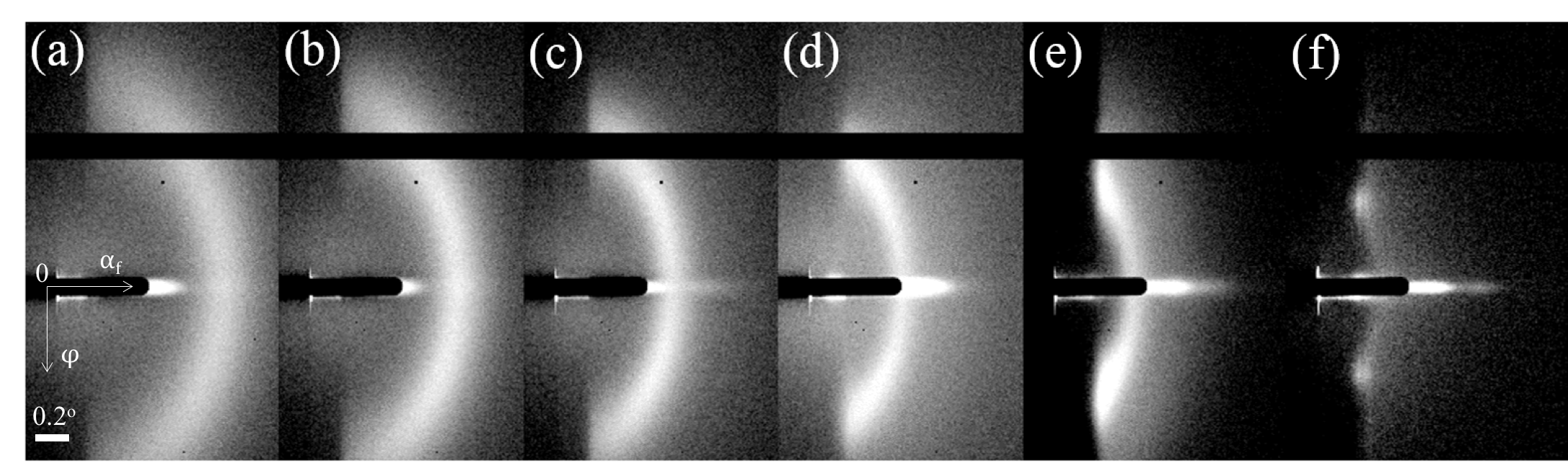
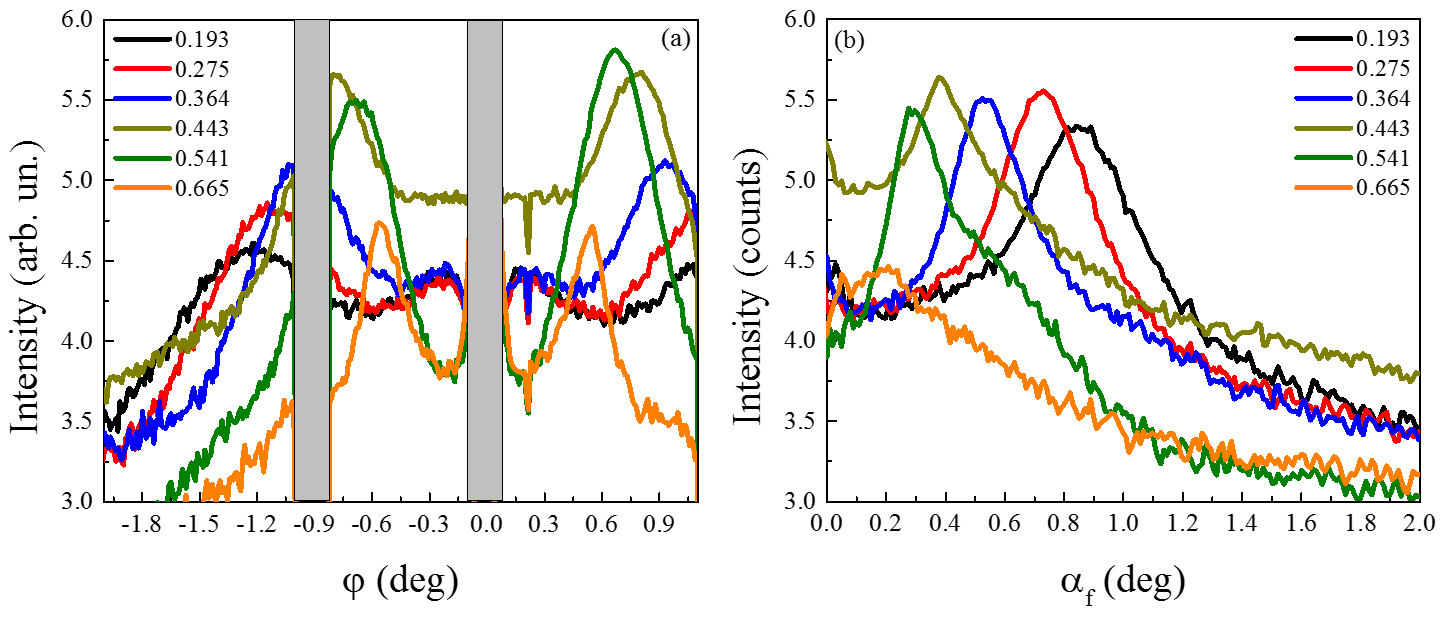
The two-dimensional GISAXS intensity maps from the sample (Co86Nb12Ta2)x(SiO2)1-x with varying from 0.193 to 0.665 are shown in Fig. 1. For all the measured samples the GISAXS patterns are typical to the disordered three-dimensional net of nanoparticles. The radius of the characteristic arc in the horizontal and vertical directions corresponds to the maxima of the correlation function in the plane of the film and perpendicular to the plane of the sample, respectively. As the concentration increases (Figures 1a–f), the maximum of the scattering intensity along shifts towards to the transmitted beam (), which corresponds to an increase in the size of the scattering objects in the real-space. The diffraction ring shown in Fig. 1a for the sample with lowest concentration of metal corresponds to the scattering from a system of grains (magnetic nanoparticles) with the in-plane radius , where is the radius of the ring along direction expressed in momentum transfer units and radius perpendicular to the sample plane , where is the radius of the ring along direction, correspondingly. The scattering is almost isotropic in plane indicating isotropic shape of nanoparticles with . To calculate the mean sizes of the granules for all concentrations the cross-sections of the two-dimensional intensity maps were taken along direction at (Fig. 2a) and along direction at (Fig. 2b).
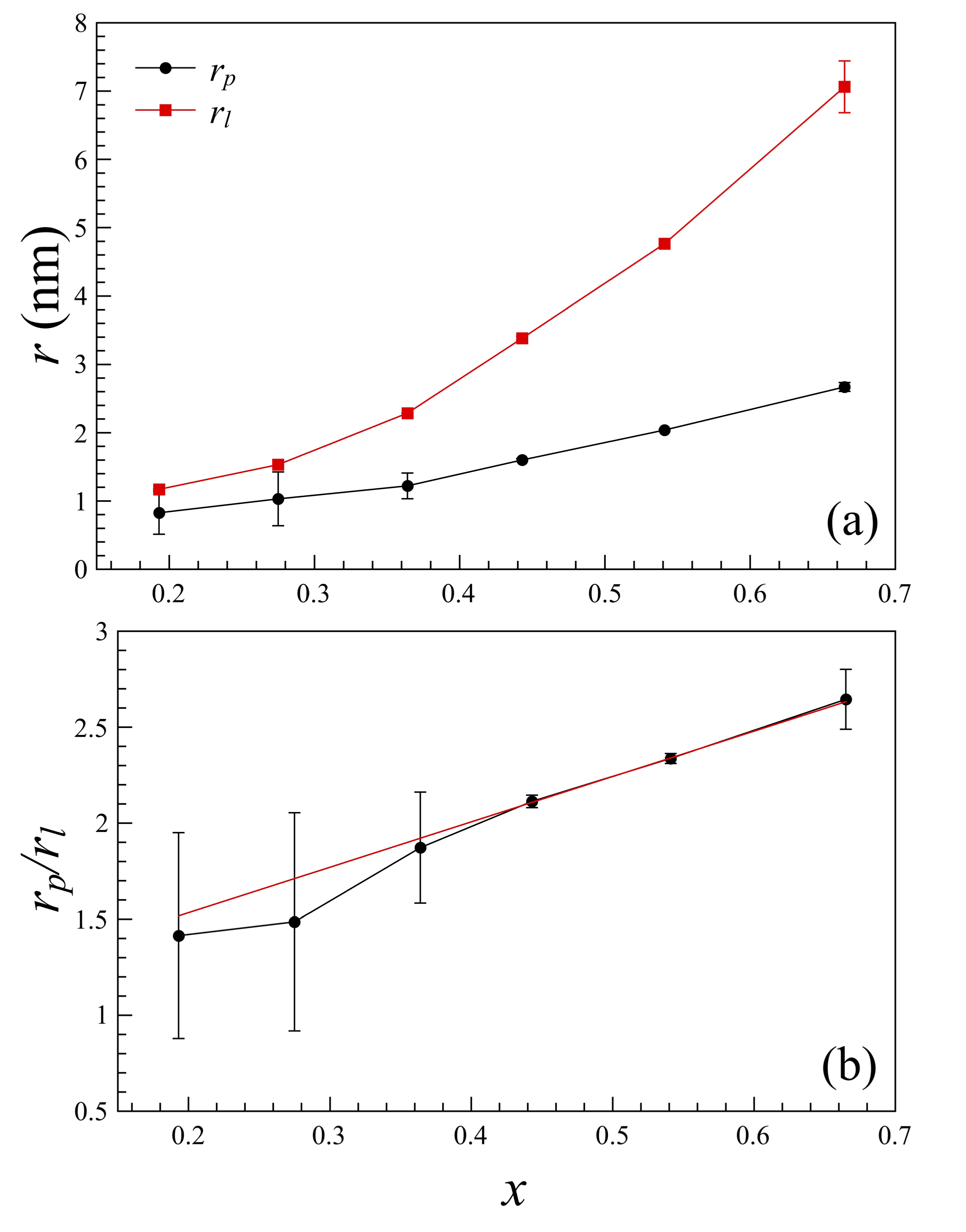
As it seen from Fig. 1 the ring-shaped SAXS intensity tends to shrink in the direction as the concentration increases. This is a clear indication of the Co86Nb12Ta2 particles elongation along the -axis (growth direction), while the lateral size increases moderately. The size of the nanoparticles in the in-plane of the sample and in the growth direction as a function of metal fraction are summarized in Fig. 3a. Indeed, as it was judged in work stognei2010anisotropy based on magnetic anisotropy data, the structural shape anisotropy of nanoclusters is manifested for the samples with metal concentration above the percolation threshold. Dependence of the shape anisotropy parameter can be reasonably well described by the linear function (Fig. 3b).
III Conclusion
In conclusion, in this non-destructive experiment we have verified that nanocomposites (Co86Nb12Ta2)x(SiO2)1-x contain anisotropic metal clusters. For a whole concentration range from to in vicinity of the percolation threshold the nanoparticles are elongated along the growth direction perpendicular to the film plane. For the lowest concentration clusters with almost isotropic shape and average radius of 1 nm were observed, while for higher concentrations the average cluster length along the film normal is nm and the cross-section is nm. Therefore the grain shape anisotropy must be included into consideration of the magnetic properties of this system, such as ferromagnetic resonance studies lutsev2002spin ; vyzulin2006special or absolute values of the magnetic anisotropy constant evaluation stognei2010anisotropy .
Acknowledgements.
Authors thank European Synchrotron Radiation Facility for the provided experimental opportunities. We would like to acknowledge A. Vorobiev and O. Konovalov for the technical assistance and O.V. Stognei for provided samples.References
- (1) M. Knobel, W. Nunes, L. Socolovsky, E. De Biasi, J. Vargas, J. Denardin, Journal of nanoscience and nanotechnology 8(6), 2836 (2008)
- (2) X. Yao, W. Zhong, C. Au, Y. Du, Magnetic Nanoparticles and Granular Thin Films (Springer, 2014)
- (3) A. Gerber, A. Milner, B. Groisman, M. Karpovsky, A. Sulpice, Thin solid films 304(1), 319 (1997)
- (4) H.R. Stuart, D.G. Hall, Applied Physics Letters 73(26), 3815 (1998). DOI http://dx.doi.org/10.1063/1.122903. URL http://scitation.aip.org/content/aip/journal/apl/73/26/10.1063/1.122903
- (5) R. Kodama, Journal of Magnetism and Magnetic Materials 200(1), 359 (1999)
- (6) H. Fujimori, S. Ohnuma, N. Kobayashi, T. Masumoto, Journal of magnetism and magnetic materials 304(1), 32 (2006)
- (7) I. Žutić, J. Fabian, S.D. Sarma, Reviews of modern physics 76(2), 323 (2004)
- (8) H.A. Atwater, A. Polman, Nature materials 9(3), 205 (2010)
- (9) L. Suber, D. Peddis, Approaches to synthesis and characterization of spherical and anisometric metal oxide magnetic nanomaterials (Wiley Online Library, 2011)
- (10) C.H. Lambert, S. Mangin, B.C.S. Varaprasad, Y. Takahashi, M. Hehn, M. Cinchetti, G. Malinowski, K. Hono, Y. Fainman, M. Aeschlimann, et al., Science 345(6202), 1337 (2014)
- (11) R. Pisarev, A. Rzhevskii, Y.E. Kalinin, A. Sitnikov, O. Stognei, F. Bentivegna, T. Rasing, et al., Physics of the Solid State 45(2), 283 (2003)
- (12) O. Stognei, A. Sitnikov, Physics of the solid state 52(12), 2518 (2010)
- (13) S. Gridnev, Y.E. Kalinin, A. Sitnikov, O. Stognei, Nonlinear Phenomena in Nano-and Microheterogeneous Systems (Binom Moscow, 2012)
- (14) G. Renaud, R. Lazzari, F. Leroy, Surface Science Reports 64(8), 255 (2009)
- (15) L. Lutsev, Physics of the Solid State 44(1), 102 (2002)
- (16) S. Vyzulin, Y.E. Kalinin, G. Kopytov, E. Lebedeva, A. Sitnikov, N. Syr’ev, Russian Physics Journal 49(3), 285 (2006)