Higher Order Quantum Ghost Imaging with Ultra-Cold Atoms
Abstract
Ghost imaging is a quantum optics technique that uses correlations between two beams to reconstruct an image in one beam from photons that do not interact with the object being imaged. While pairwise (second order) correlations are usually used to create the image, higher order correlations can be utilized to improve the performance of ghost imaging. In this paper, we demonstrate higher order atomic ghost imaging, using entangled ultracold metastable helium atoms from an s-wave collision halo. We construct higher order ghost images up to 5th order and show that using higher order correlations can improve the visibility of the images without impacting the resolution. This is the first demonstration of higher order ghost imaging with massive particles and the first higher order ghost imaging protocol of any type using a quantum source.
Ghost imaging is an unconventional imaging method from quantum optics Erkmen and Shapiro (2010); Shapiro and Boyd (2012), which uses two correlated beams of photons. One beam interacts with the object, after which the arrival time (only) of each photon from the beam is detected on a ‘bucket’ detector, while the second photon from each pair is detected with full 3D spatial and temporal resolution on a multi-pixel detector, but never interacts with the object. Using the correlations between the two beams, the image of the object can be reconstructed. While the technique was first theoretically proposed Klyshko (1988); Belinskii and Klyshko (1994) and experimentally demonstrated Pittman et al. (1995); Strekalov et al. (1995) using light, it has also recently been extended to X-rays Pelliccia et al. (2016), cold atoms Khakimov R. I. et al. (2016) and electrons Li et al. (2018), along with a recent proposal involving neutrons Chen, K and Han, S (2018). The correlated nature of ghost imaging means that in certain circumstances, such as for weakly absorbing objects Brida et al. (2010) or at low light levels Morris et al. (2015), it can out-perform conventional imaging. Ghost imaging has applications in a number of areas including optical encryption Kong et al. (2013); Yuan et al. (2016), improved telecommunications Ryczkowski et al. (2016) and remote sensing Erkmen (2012); Hardy and Shapiro (2013). It also has the potential to reduce the dosage rates in imaging Pelliccia et al. (2016) and tomography Kingston et al. (2018) using radiation such as X-rays, where potential damage to the sample from the radiation is a concern.
There are two distinct types of ghost imaging that have been demonstrated. Thermal or classical ghost imaging uses thermal or pseudothermal light, relying on semi-classical Hanbury Brown-Twiss (HBT) correlations to produce the images. In contrast, quantum ghost imaging uses a quantum source of correlated pairs, which for photons is usually created via spontaneous parametric down conversion (SPDC). While the SPDC pairs are entangled, entanglement is not necessary for ghost imaging Bennink et al. (2002); Erkmen and Shapiro (2008), although the performance of the imaging by some measures may be improved with entanglement Bennink et al. (2004).
While the majority of ghost imaging implementations utilise pairwise (second order) correlations, the basic schemes can be extended to employ higher order correlations between more particles by incorporating additional detectors into the setup. This has been shown to be able to improve the quality of key imaging parameters such as the visibility and contrast-to-noise ratio (CNR) Chen et al. (2010); Li et al. (2012), as well as the resolution Zhou et al. (2012), when implemented for ghost imaging with thermal light. However, due to the lower probability of higher order correlated events being detected, in practical cases such improvements do not necessarily translate to performance gains over second order ghost imaging when additional imaging improvements such as background subtraction are taken into account Chan et al. (2010). The use of higher order correlations to enhance the performance of ghost imaging is similar to the increase in visibility of multi-photon interference from thermal light via higher order correlations Agafonov et al. (2008). However, despite the body of work on higher order thermal ghost imaging, to the best of our knowledge there has been no demonstration of ghost imaging for quantum light. This is partly because the relatively small two photon bunching amplitude for thermal ghost imaging limits the achievable visibility to , meaning that there is a significant improvement attainable using higher order ghost imaging. For quantum ghost imaging using SPDC sources, the low occurrence of high order correlations would make higher order ghost imaging challenging.
Here we demonstrate higher order ghost imaging using correlated pairs of ultra cold metastable helium (He*) atoms Vassen et al. (2012) from an s-wave scattering halo of two colliding Bose-Einstein condensates Perrin et al. (2007); Jaskula et al. (2010). As shown in our previous experiment Khakimov R. I. et al. (2016), by imposing a mask on one of the atoms in each pair and detecting those atoms with full 3D resolution, we construct ghost images using the time correlations between those atoms and their corresponding correlated partners that do not pass through the mask. In addition to the two-atom correlations due to pairwise scattering, the complex many-body correlations in the halo leads to a complex hierarchy of correlations Hodgman et al. (2017), and we use a range of these higher order correlations to construct higher order ghost images up to 5th order. The quality of the resulting images was characterised via the visibility and resolution, with higher order imaging shown to be able to produce images with improved visibility at no detriment to resolution.
The experimental procedure for our higher order ghost imaging is similar to our previous method for 2nd order ghost imaging Khakimov R. I. et al. (2016), with the starting point being a Bose-Einstein Condensate (BEC) of helium atoms in the sublevel of the long lived metastable state Hodgman et al. (2009), confined in a magnetic trap. A Raman pulse then transfers nearly all atoms into the untrapped state, while also imparting a momentum of in the downwards direction (with gravity), where and nm is the wavelength of the Raman laser, and allowed to fall under gravity. The untrapped BEC is then split into 12 momentum components, also in the direction, by a second diffraction pulse operating in the Kapitza-Dirac regime Khakimov R. I. et al. (2016). Each pair of BECs in adjacent momentum components then collide, producing a spherical halo with a radius (in the momentum space frame of reference centred on the two BEC components) of comprising pairs of back-to-back correlated Perrin et al. (2007); Hodgman et al. (2017) and entangled Shin et al. (2018) atoms, analogous to the pairs of photons produced from a SPDC source in quantum optics. Each of the 11 scattering halos has a radial gaussian width of with an average mode occupancy varying from to across the halos, depending on the relative fraction of BEC atoms transferred to each of the momentum modes. The multiple halos technique is used to reduce our data acquisition time, with the ghost imaging implemented for each halo separately. Within each halo there is a complex hierarchy of higher order correlations, with correlations due to both the scattering collision and HBT style multi-particle interference (see Hodgman et al. (2017) for details). In general, the degree of correlation increases for higher orders of correlations.

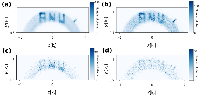
The halos fall under gravity mm onto a multi-channel plate and delay-line detector, while also expanding during the fall time. This detector allows the position of the atoms at the detector, corresponding to the atomic momenta in the collision halo, to be measured in full 3D, with an resolution of m and a resolution in the direction of m (s in arrival time) Henson et al. (2018). The detector is divided in half centred on the halo, with one half assigned as the ‘bucket’ port and the other the ‘multi-pixel’ port (see Fig. 1). A software mask mas is imposed on atoms arriving in the bucket port, so that only atoms that arrive in a defined region (the open area of the mask) are recorded. All the spatial information of these bucket port atoms is then discarded, with only their arrival times retained. In the basic scheme of second order ghost imaging, for each of these bucket port atoms (which have momentum ), any atom in the multi-pixel port which arrives within an arrival time interval set by the correlation length of the scattered pairs ( Hodgman et al. (2017)) centred around - is then added to the ghost image. The image is created from all correlated ghost image atoms across all 11 halos from 45,000 different experimental runs. Fig. 2(a) shows a sample image of a mask of the letters ‘ANU’.
For th order ghost imaging the procedure is similar, with total atoms required to be detected distributed in some particular combination around and , with all . If this condition is met then all atoms around are added to the image. Each has different possible combinations with varying degrees of correlation Hodgman et al. (2017). For example, in third order ghost imaging one atom is always in each of the mask and the image, with the choice for the extra atom to be added to either port. Higher order extensions follow the same pattern. The two third order cases are shown in Fig. 2(b) and (c), while the fourth order case of three atoms in the image and one in the mask port shown in Fig. 2(d).
To quantitatively analyse and compare the performance of the different orders and combinations of ghost imaging, two widely used measures were calculated for each case: the visibility and the resolution. A rectangle mask of dimensions (0.220.18) was used for both of these, to simplify the analysis, although the results are similar for other mask shapes.
The visibility of the ghost image is defined as
| (1) |
is the total number of atoms in the region of the ghost image corresponding to the location of open area of the mask on the opposite side of the halo, while is the total number of atoms in the region of the semi-circular arc corresponding to the halo in the rest of ghost image. The non-halo regions, where almost no atoms will be found, are excluded. Both and are normalized individually by the size of their respective areas.
The experimentally measured visibility for 8 different ghost imaging cases is shown in Fig. 3, representing all combinations up to 5th order (the two cases of fifth order with 1 and 2 atoms in the image port are not shown, as there were insufficient counts to produce a meaningful result). As the plot shows, the best visibility for each order is achieved for the cases with one atom in the image port and the rest in the mask (blue points). For these cases increases with , while for the opposite case (one atom in mask port and the rest in the image port) shown in yellow, decreases with . For the intermediate cases (shown in green) the results are somewhere in the middle. The insets in Fig. 1 show schematically the process (for the three atom case) of how higher order ghost imaging can improve the visibility. In this case, the higher degree of correlation for three atoms means that a second atom (blue) detected in the bucket port is more likely to be found within a correlation length of the first (green). Intuitively, this increased detection probability results from HBT style bunching of the two particles in the bucket port. Due to the mask, these will both by definition lie within the image, as potential arrival locations outside the mask (indicated by the red hatched area in Fig. 1) are excluded as a possible location for additional atoms. An atom detected in the image port, however, is more likely to be found within a radius around - of on the opposite side of the halo (cyan area). The two atoms detected confine this to a narrower window with (on average) a larger overlap on the image than the area allowed around for a single bucket port atom only (green dashed line).
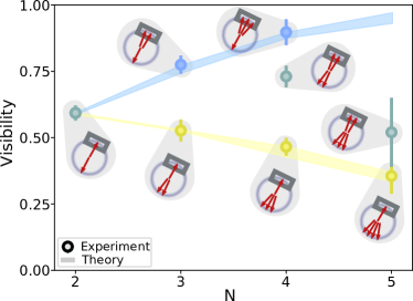
In both of the extreme cases (blue and yellow in Fig. 3), the degree of correlation is the same, although times as many atoms are detected in the image port for the case with one atom in the image port. However, for higher order schemes some intermediate (green) configurations are more correlated than others (e.g. for fourth order the degree of correlation scales as in the case of two atoms on each side of the halo but as for the cases of three atoms on each side Hodgman et al. (2017), which is smaller for the values of in our halos). This would suggest that the change in is not primarily due to an increased degree of correlation, but rather the relative number of atoms in the image port compared to the bucket port. The cause of this is that in the best case more atoms in the mask increases the probability that the only atom in the image will be a correlated hit and not a background atom, as the atoms in the mask are limited to a smaller area. In contrast, there is a much larger area where atoms in the ghost image can possibly fall, and thus a lot more background atoms are included. The trend of the extreme cases is similar to that observed with optics for multi-photon interference Agafonov et al. (2008).
This interpretation of extra atoms in the image causing increased blurring due to finite , and this effect being more dominant than the relative bunching amplitude, is supported by numerical simulations of our ghost imaging SOM . The visibilities generated for our experimental parameters are shown as yellow and blue bands in Fig. 3, which agree well with the experimental results. Importantly, the simulations show that while an increased bunching amplitude does improve visibility, above some value this effect mostly saturates, whereas increasing always decreases the visibility. For our experiments we are always in the regime SOM , and thus the optimal visibility is always with the maximum number of atoms in the bucket port.
The other image quality measure implemented was the resolution, which is dependent on the finite correlation length of the correlated atoms Khakimov R. I. et al. (2016). To measure the resolution the ghost image of a size rectangle for each case is integrated along the axis. A Gaussian convolved with a top-hat function of the size of the square mask was then fitted to the data and the width of the fitted Gaussian , which corresponds to the resolution of the image, extracted.
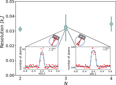
The measured resolutions for the different cases are shown in Fig. 4, along with some sample plots showing the raw data. As can be seen, the measured resolution for all cases is close to . There is also little difference in the measured resolution for the different ghost imaging methods, although the resolution for higher order cases with more atoms in the image may be slightly worse.
In conclusion, we have demonstrated higher order ghost imaging with atoms up to 5th order, the first such demonstration with massive particles and the first higher order ghost imaging experiment using a quantum source, as to the best of our knowledge all previous demonstrations were for semi-classical ghost imaging implemented with thermal or pseudo-thermal light. The visibility was seen to improve with higher orders for the cases with only 1 atom in the ghost image, while the reverse occurred for cases with only one atom in the mask. In all cases the resolution did not change significantly. The improved visibility makes higher order ghost imaging better for applications with demanding visibility or signal to flux ratio requirements, such as in X-ray ghost imaging Pelliccia et al. (2016) and tomography Kingston et al. (2018), where higher-order schemes could be used to produce images with improved visibility at lower dosage rates, or similar damage sensitive schemes with atoms such as atomic lithography. By extending the higher order atomic ghost imaging scheme presented here, complex fundamental tests of quantum mechanics with massive particles could also be possible, such as multi-atom entanglement Kofler et al. (2012) or Bell’s inequality measurement schemesJack et al. (2009) using 3 or more particles.
Acknowledgements.
The authors would like to thank Bryce Henson and David Shin for technical assistance. This work was supported through Australian Research Council (ARC) Discovery Project grants DP120101390, DP140101763 and DP160102337. SSH is supported by ARC Discovery Early Career Researcher Award DE150100315.References
- Erkmen and Shapiro (2010) B. I. Erkmen and J. H. Shapiro, Adv. Opt. Photon. 2, 405 (2010).
- Shapiro and Boyd (2012) J. H. Shapiro and R. W. Boyd, Quant. Inf. Proc. 11, 949 (2012).
- Klyshko (1988) D. Klyshko, JETP 67, 1131 (1988).
- Belinskii and Klyshko (1994) A. V. Belinskii and D. N. Klyshko, JETP 78, 259 (1994).
- Pittman et al. (1995) T. B. Pittman, Y. H. Shih, D. V. Strekalov, and A. V. Sergienko, Phys. Rev. A 52, R3429 (1995).
- Strekalov et al. (1995) D. V. Strekalov, A. V. Sergienko, D. N. Klyshko, and Y. H. Shih, Phys. Rev. Lett. 74, 3600 (1995).
- Pelliccia et al. (2016) D. Pelliccia, A. Rack, M. Scheel, V. Cantelli, and D. M. Paganin, Phys. Rev. Lett. 117, 113902 (2016).
- Khakimov R. I. et al. (2016) Khakimov R. I., Henson B. M., Shin D. K., Hodgman S. S., Dall R. G., Baldwin K. G. H., and Truscott A. G., Nature 540, 100 (2016).
- Li et al. (2018) S. Li, F. Cropp, K. Kabra, T. J. Lane, G. Wetzstein, P. Musumeci, and D. Ratner, Phys. Rev. Lett. 121, 114801 (2018).
- Chen, K and Han, S (2018) Chen, K and Han, S, ArXiv e-prints (2018), arXiv:arXiv:1801.10046 .
- Brida et al. (2010) G. Brida, M. Genovese, and I. Ruo Berchera, Nature Photon. 4, 227 (2010), arXiv:1004.1274 .
- Morris et al. (2015) P. A. Morris, R. S. Aspden, J. E. C. Bell, R. W. Boyd, and M. J. Padgett, Nature Commun. 6, 5913 (2015), arXiv:1408.6381 .
- Kong et al. (2013) L.-J. Kong, Y. Li, S.-X. Qian, S.-M. Li, C. Tu, and H.-T. Wang, Phys. Rev. A 88, 013852 (2013).
- Yuan et al. (2016) S. Yuan, J. Yao, X. Liu, X. Zhou, and Z. Li, Opt. Commun. 365, 180 (2016).
- Ryczkowski et al. (2016) P. Ryczkowski, M. Barbier, A. T. Friberg, J. M. Dudley, and G. Genty, Nature Photon. (2016), 10.1038/nphoton.2015.274.
- Erkmen (2012) B. I. Erkmen, J. Opt. Soc. Am. A 29, 782 (2012).
- Hardy and Shapiro (2013) N. D. Hardy and J. H. Shapiro, Phys. Rev. A 87, 023820 (2013).
- Kingston et al. (2018) A. M. Kingston, D. Pelliccia, A. Rack, M. P. Olbinado, Y. Cheng, G. R. Myers, and D. M. Paganin, Optica 5, 1516 (2018).
- Bennink et al. (2002) R. S. Bennink, S. J. Bentley, and R. W. Boyd, Phys. Rev. Lett. 89, 113601 (2002).
- Erkmen and Shapiro (2008) B. I. Erkmen and J. H. Shapiro, Phys. Rev. A 77, 043809 (2008).
- Bennink et al. (2004) R. S. Bennink, S. J. Bentley, R. W. Boyd, and J. C. Howell, Phys. Rev. Lett. 92, 033601 (2004).
- Chen et al. (2010) X.-H. Chen, I. N. Agafonov, K.-H. Luo, Q. Liu, R. Xian, M. V. Chekhova, and L.-A. Wu, Opt. Lett. 35, 1166 (2010).
- Li et al. (2012) H. Li, J. Shi, Z. Chen, and G. Zeng, J. Opt. Soc. Am. A 29, 2256 (2012).
- Zhou et al. (2012) Y. Zhou, J. Liu, J. Simon, and Y. Shih, J. Opt. Soc. Am. B 29, 377 (2012).
- Chan et al. (2010) K. W. C. Chan, M. N. O’Sullivan, and R. W. Boyd, Opt. Express 18, 5562 (2010).
- Agafonov et al. (2008) I. N. Agafonov, M. V. Chekhova, T. S. Iskhakov, and A. N. Penin, Phys. Rev. A 77, 053801 (2008).
- Vassen et al. (2012) W. Vassen, C. Cohen-Tannoudji, M. Leduc, D. Boiron, C. I. Westbrook, A. Truscott, K. Baldwin, G. Birkl, P. Cancio, and M. Trippenbach, Rev. Mod. Phys. 84, 175 (2012).
- Perrin et al. (2007) A. Perrin, H. Chang, V. Krachmalnicoff, M. Schellekens, D. Boiron, A. Aspect, and C. I. Westbrook, Phys. Rev. Lett. 99, 150405 (2007).
- Jaskula et al. (2010) J.-C. Jaskula, M. Bonneau, G. B. Partridge, V. Krachmalnicoff, P. Deuar, K. V. Kheruntsyan, A. Aspect, D. Boiron, and C. I. Westbrook, Phys. Rev. Lett. 105, 190402 (2010).
- Hodgman et al. (2017) S. S. Hodgman, R. I. Khakimov, R. J. Lewis-Swan, A. G. Truscott, and K. V. Kheruntsyan, Phys Rev Lett 118, 240402 (2017).
- Hodgman et al. (2009) S. S. Hodgman, R. G. Dall, L. J. Byron, K. G. H. Baldwin, S. J. Buckman, and A. G. Truscott, Phys. Rev. Lett. 103, 053002 (2009).
- Shin et al. (2018) D. K. Shin, B. M. Henson, S. S. Hodgman, T. Wasak, J. Chwedenczuk, and A. G. Truscott, ArXiv e-prints (2018), arXiv:1811.056815 .
- Henson et al. (2018) B. M. Henson, X. Yue, S. S. Hodgman, D. K. Shin, L. A. Smirnov, E. A. Ostrovskaya, X. W. Guan, and A. G. Truscott, Phys. Rev. A 97, 063601 (2018).
- (34) The mask is imposed in software to allow greater flexibility in analysing with different masks, since to install a mask physically into the system requires bringing the ultra-high vacuum detection chamber up to atmospheric pressure and then re-sealing and baking, meaning each mask installation requires nearly a week of experimental downtime. This software mask procedure has been shown to yield equivalent results to a physical mask when a mask was implemented with the same dimensions as in our previous ghost imaging experimental setup Khakimov R. I. et al. (2016).
- (35) See Supplementary Material for details.
- Kofler et al. (2012) J. Kofler, M. Singh, M. Ebner, M. Keller, M. Kotyrba, and A. Zeilinger, Phys. Rev. A 86, 032115 (2012).
- Jack et al. (2009) B. Jack, J. Leach, J. Romero, S. Franke-Arnold, M. Ritsch-Marte, S. M. Barnett, and M. J. Padgett, Phys. Rev. Lett. 103, 1 (2009).
I Supplementary Information
Numerical Simulation
To further investigate the experimentally observed trends in the ghost imaging visibility and gain additional insights into the factors contributing to this, we performed a simple Monte Carlo simulation that aims to emulate the key features of ghost imaging. The simulations used the same rectangle mask (0.220.18) used to produce the experimental visibility plot shown in Fig. 3. For two atom ghost imaging, a pair of random 2 dimensional coordinates were generated within the range of the mask, representing the atom recorded in the bucket port. In the neighborhood centred on the inverse of each of these coordinates (i.e. the opposite side of the halo), a pair of random coordinates was generated following a 2D Gaussian distribution with width , representing the correlated atoms detected in the image port. To account for uncorrelated atoms, a proportion of image port atoms were chosen to be randomly distributed over the entire image port, with the exact fraction corresponding to the degree of correlation () in the experimental data. Once sufficient random co-ordinates were generated, the visibility was calculated in the same manner as for the experimental data. 20 datasets were randomly generated for each parameter set, to produce an uncertainty spread of the range of visibilities for each case. The parameters varied were the correlation length () and ratio of correlated atoms to atoms in the background (i.e. the degree of correlation).
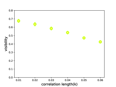
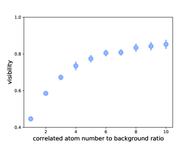
Fig. 5 shows the results of the first parameter investigated, using the rectangle mask with 2 correlated atoms. As the correlation length increases, naturally more atoms in correlated pairs would fall outside of the corresponding image area, which would increase the background atom numbers resulting in a decrease of visibility of the image. Note that the experimental halo has .
Setting and varying the number of atoms in the background, Fig. 6 shows the dependence of visibility on the degree of correlation, represented theoretically by the ratio of correlated image atoms to uniformly distributed background atoms. The degree of correlation corresponds to the experimental observable of the second order correlation function , which is directly related to the ratio between correlated atom number and background atom number. This conversion is for every correlated pairs if one atom is added to the background then , since in the simulations the background and image areas are same size. While Fig. 6 exhibits a trend of increasing visibility as the degree of correlation increases, it asymptotically approaches a limit for high values (for this parameter set around ). This limit is due to factors such as the values of the correlation length, the ratio of uncorrelated to correlated atoms, the size of the mask etc. Since these factors will somewhat depend on the experiment, to compare with the experimental data in Fig. 3 the degree of correlation is adjusted as a free parameter in the two atom data to ensure the generated visibility matches the experimental value, then kept consistent for the higher order data.
With set to the experimental value and the degree of correlation adjusted to match the experimental 2-atom result, the various different permutations of higher order ghost imaging were simulated. For these higher order cases, additional atoms were added to the image and/or bucket ports for each correlated event as required, with the previously described gaussian distributions with width centered around the original bucket port atom or the opposite side of the halo. The results for the two limiting cases of one atom in the bucket port (yellow line) and one atom in the image (blue line) are shown in Fig. 3, with the shaded region indicating the spread of numerical values generated. As can be seen the results match the experimental data very well for all orders, suggesting that our simple simulation captures the key aspects of the process.