Flagellar number governs bacterial spreading and transport efficiency
Abstract
Peritrichous bacteria synchronize and bundle their flagella to actively swim while disruption of the bundle leads to tumbling. It is still not known whether the number of flagella represents an evolutionary adaptation towards optimizing bacterial navigation. Here, we study the swimming dynamics of differentially flagellated Bacillus subtilis strains in a quasi-two-dimensional system. We find that decreasing the number of flagella N reduces the average turning angle between two successive run phases and enhances the duration and directional persistence of the run phase. As a result, having less flagella is beneficial for long-distance transport and fast spreading, while having a lot of flagella is advantageous for the processes which require localization and slow dynamics, such as biofilm formation. We develop a two-state random walk model that incorporates spontaneous switchings between the states and yields exact analytical expressions for transport properties, in remarkable agreement with experiments. The results of numerical simulations based on our two-state model suggest that the efficiency of searching and exploring the environment is optimized at intermediate values of N. The optimal choice of N, for which the search time is minimized, decreases with increasing the size of the environment in which the bacteria swim.
INTRODUCTION
Many bacterial species swim by the rotation of flagella Berg04 . Several flagellation patterns can be distinguished according to the flagellar arrangement on the cell body, ranging from polar (one flagellum at one pole) to peritrichous (helical arrangement on the whole cell body) Schuhmacher15 . Each flagellum is anchored within the cell membrane to a reversible rotary motor that switches between clockwise and counter-clockwise rotation Berg73 . The ability of the motor to turn in either direction allows polarly-flagellated bacteria, such as Caulobacter crescentus, to change the swimming direction of the cell Lauga09 . A peritrichously-flagellated bacterium experiences an alternating sequence of active runs and tumbles Berg72 , controlled by the rotational states of each flagellum. When all flagella rotate counter-clockwise, they form a bundle and synchronize their rotation. As a result, the bacterium moves forward smoothly (run phase). However, the bundle is disrupted when some of the flagella switch their rotational direction, which facilitates changes in the direction of motion in relatively short time intervals (tumble phase). The run-and-tumble dynamics of bacteria allows them to change their direction of motion. They can also adjust the duration of stay in the run phase in response to environmental changes induced, e.g., by temperature, light, or chemical gradients Berg04 ; Berg72 . Such an ability is known to be highly advantageous to enhance search efficiency Benichou05 ; Bartumeus08 ; Benichou11 , allowing for an optimal navigation towards favorable or away from harmful regions Wadhams04 .
Despite the advances in understanding of the underlying mechanisms of bundle formation and disruption Chattopahyay06 ; Darnton07 ; Mears14 ; Turner00 ; Korobkova06 ; Terasawa11 ; Hu13 ; Hu15 and its relationship to the chemotaxis signaling network Berg04 ; Wadhams04 ; Vladimirov10 ; Sneddon12 , it remains poorly understood how the number of flagella influences the fundamental properties of bacterial propulsion and transport. Attempts to understand the role of flagellation have mainly focused on Escherichia coli which has up to eight flagella, while the flagellar number of many types of bacteria is typically much higher. So far, it has been observed that the torque and thus the swimming speed Darnton07 and the fraction of time spent in run or tumble phase Mears14 of peritrichously-flagellated bacteria remain roughly independent of the flagellar number . Recent numerical studies also predict that the swimming speed is only slightly affected by , assuming that just a single bundle is formed Kanehl14 ; Reigh12 . Here, we provide evidences for the formation of multiple bundles, revealing that the former assumption does not hold at least at high flagellar number.
A key question from an evolutionary point of view is based on
which criteria a bacterium chooses to have a certain number of
flagella. In the present study, we investigate bacterial motility
over a wide range of the flagellar number and clarify how the
run-and-tumble dynamics is influenced by the choice of .
We study Bacillus subtilis, a rod-shaped bacterium commonly
found, e.g., in soil and the gastrointestinal tract of humans. The
number of flagella is regulated by the master regulator swrA in
Bacillus subtilis Kearns05 . Deletion of the gene
leads to a reduction from to flagella, while cells
overexpressing swrA exhibit up to flagella. Therefore, we
employed a wild-type Bacillus subtilis (NCIB3610 strain)
with flagella, a strain deficient for
swrA ( flagella), and a one which carries swrA under the
control of an IPTG-inducible promoter ( flagella) (Fig. 1A).
While the speeds in both run and tumble phases and the mean
duration of tumbling periods show no systematic dependence
on , the duration and directional persistence of
the run phase and the average turning angle between two
successive run phases vary monotonically with .
Smaller values of are more favorable for
long-distance transport and fast spreading because of
more persistent trajectories and longer excursion times
in the run phase. On the other hand, increasing
facilitates tumbling, leading to slow dynamics and localization
which is beneficial e.g. for the formation of biofilms. We
develop a coarse-grained analytical approach to study the
run-and-tumble dynamics of bacteria, which enables us to
identify the contributions of the influential factors and
map out the phase diagrams in the space of the relevant
parameters of run-and-tumble statistics. Our simulation
results suggest that by adopting intermediate values of
(as e.g. in the wild-type strain), the search
efficiency and the ability to explore the environment
can be optimized.
RESULTS
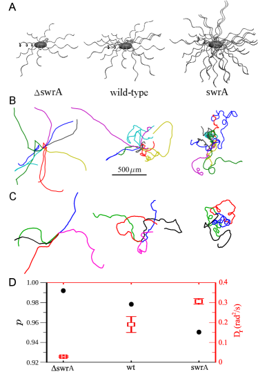
We report and compare the dynamics of three strains of
Bacillus subtilis with different flagellar number
obtained by genetic manipulation (see
Materials and Methods section for details). The
strains , wt (wild-type
NCIB3610) and swrA have ,
and flagella, respectively Guttenplan13
(Fig. 1A). In contrast to the studies on tumbling of
trapped cells Mears14 , here we track the motion
of freely swimming cells. A few typical trajectories
of each strain are shown in Fig. 1B, which consist
of successive active run and tumble phases.
Distinct motility patterns
Strikingly, we find that the curvatures of the trajectories
strongly depend on the flagellar number. The paths are clearly
less curved with decreasing , which may originate either
from the change in their tumbling statistics or from having a
larger persistence length in the run phase. While the influence
of on the run-and-tumble statistics will be discussed
in the next section, here we focus on the latter possibility, i.e. the variations of the persistency in the run phase with changing
. To characterize the running persistency, we note that
each run trajectory comprises a set of recorded positions of the
bacterium. Given these data, one can extract the local direction
of motion and, thus, the directional change along
the trajectory, resulting in the distribution of directional changes
Burov13 . Then, the persistency of the run
phase can be obtained as the Fourier transform of ,
i.e. Shaebani14 ; Sadjadi15 .
Alternatively, the curvature of the trajectories can be characterized
by calculating the rotational mean square displacement , from which one can extract the asymptotic
rotational-diffusion coefficient . The results shown in
Fig. 1D reveal that the directional persistency in the run phase
remarkably depends on the flagellar number; the run paths are
less curved for smaller .
Run-and-tumble statistics
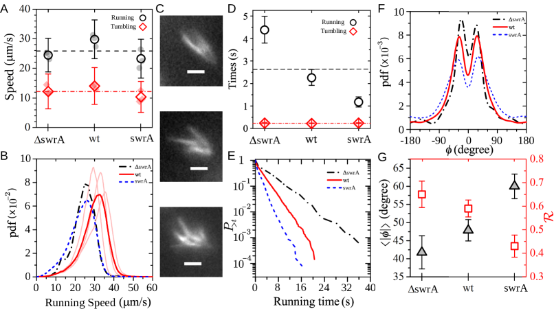
Next, we turn to studying the statistics of run and tumble events. A detailed description of the tumbling detection procedure can be found in Materials and Methods section. According to the results shown in Fig. 2A, the tumbling speed is smaller (but non-negligible) compared to the running speed Turner16 . Moreover, the speeds in both run and tumble phases show no systematic dependence on . A closer look at the probability distributions of the instantaneous speeds (see e.g. the running speed data in Fig. 2B) reveals that even the distributions of a given strain slightly differ from one culture to another due to biological heterogeneity. Thus, there is no significant difference in the speeds of different strains. By approximating the bundle as a single helix Lauga09 ; Kanehl14 ; Rodenborn13 , simple models based on resistive-force theory predict a weak (logarithmic) growth of running speed with , though experiments have shown that hydrodynamic dissipation along the cell body considerably weakens the effect Darnton07 . Additionally, the assumption of a single bundle does not hold in general, according to our observations of fluorescently-stained flagella of swimming bacteria as well as other reports in the literature Valeriani11 ; Hyon12 ; even multiple bundles may form during the active running phase (several examples of a wt strain with multiple bundles are shown in Fig. 2C). The diversity of the locations and interactions between the bundles makes the prediction of the propulsive force and swimming speed highly complex.
To understand whether and to what extent the flagellar number influences the switching frequencies between run and tumble phases, we plot the average excursion times in Fig. 2D. It turns out that the mean tumbling time () and thus the ability of restoring the run phase are not influenced by . Given the mean tumbling time and speed, one finds that the displacement in the tumbling phase is of the order of the cell length (i.e. a few micrometers). However, such a swimmer with the running speed of in an aqueous medium is expected to stop after only and traveling approximately if the pushing force suddenly stops purcell77 . This suggests that the bundles are not fully disrupted in the tumbling phase, thus, the bacterium is partially propelled. The mean running times remarkably depend on the flagellar number. Increasing enhances the probability of switching from running to tumbling. This supports the veto model for bundle disruption Turner00 ; Sneddon12 , which proposes that clockwise rotation of a single or a few flagella (less than ) is sufficient to disrupt the bundle. Figure 2E shows that the tail of the probability distribution of running times is nearly exponential, with a -dependent slope (data censoring for the first and last incomplete periods sharpens the trend even more). We observe similar exponential decays for the tumbling time distributions (not shown), verifying that the switching events between the two states of motility happen spontaneously and can be described by Markovian processes.
Another characteristic of the run-and-tumble dynamics that is affected by the number of flagella is the turning angle between successive run phases. We define the turning angle between two successive runs as the change in the direction of motion from the end of one run to the beginning of the next run (the direction of motion was obtained by a linear fit to four data points for each run phase). The turning-angle distribution obeys left-right symmetry and develops a peak for all strains. However, the peak and, more clearly, the mean of shift towards larger angles with increasing , as can be seen in Figs. 2F and 2G. Similar to the curvature of run trajectories, we can use the Fourier transform of , i.e. , as a measure of the directional change between consecutive run phases. ranges between and , with , , and denoting , , and turning, respectively. Indeed, is correlated with the tumbling time and speed saragosti12 as well as the strength of the torque exerted on the cell body during the re-formation of the bundle Darnton07 . Since the speed and excursion time in the tumbling phase are not considerably affected by , we attribute the reduction of with increasing to experiencing a larger torque during bundle re-formation at higher flagellar numbers Darnton07 .
We also checked that the direction of motion does not vary significantly when switching from running to tumbling. This fact and the relatively high speed in the tumbling phase once again indicate that the bundles are not fully disrupted in the tumbling phase. As a result, one expects that the tumbling is not a purely random motion and should exhibit directional persistency. To verify this idea we assign a dimensionless number to each tumbling segment of the trajectory, obtained by dividing the end-to-end distance by the total length of the trajectory segment. The probability distribution shown in Fig. 3A develops a sharp peak at , indicating that most of the tumbling trajectories are nearly straight during the relatively short periods of tumbling.
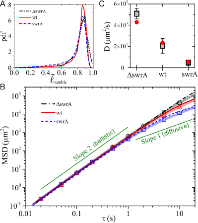
Finally, we measure the mean square displacement
for different strains. According to our findings, the strains
with lower flagellar number have less-curved run trajectories,
switch less frequently from run to tumble phase, and experience
a smaller turning angle between successive run phases. Consequently,
the crossover from a persistent motion to the asymptotic diffusive
dynamics is expected to occur at longer times for smaller values of
, which is confirmed by the experimental results shown
in Fig. 3B. The asymptotic diffusion coefficient is also
a decreasing function of (see Fig. 3C). These results
indicate that for increasing the efficiency of long-distance
transport, it is more beneficial to have less flagella. The
strains with more flagella spend a larger fraction of time
in tumbling state and have a shorter persistence length when
swimming. We note that the trends (as a function of )
discussed in this section are robust against variations of
tumbling detection thresholds.
Model
Based on our findings, we develop a stochastic coarse-grained model for a random walk with spontaneous switchings between two states of motility. We first validate the approach by comparing the analytical predictions with experimental results. Then the model enables us to better understand the influence of the flagellar number on bacterial motility, by numerically exploring the phase space of the run-and-tumble statistical parameters and identifying the isolated role of each influential factor. Moreover, our simulations based on the two-state model evidence for a nonmonotonic dependence of the mean first-passage time on .
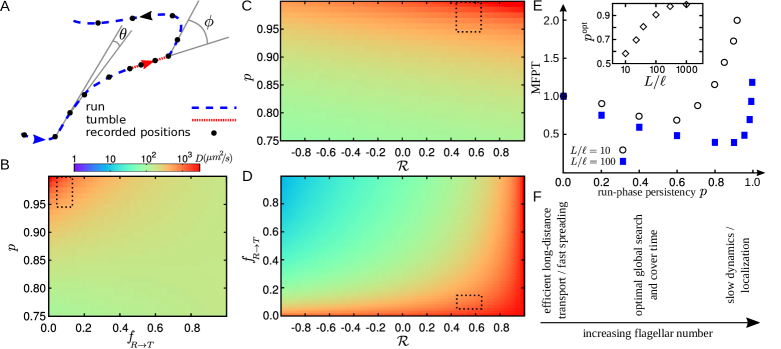
Stochastic two-state models have been widely employed to describe altering phases of motion of e.g. swimmers and cytoskeletal motor proteins, or the locomotive patterns in other systems Theves13 ; Taktikos13 ; Hafner16 ; Pinkoviezky13 ; Shaebani17 ; Kong15 ; Watari10 . Aiming at obtaining analytical insight by including only the most prominent characteristics of the bacterial motility pattern into the model, we introduce a random walk approach in which the walker experiences two states of motility: (i) running with the mean speed and persistency , and (ii) tumbling with the mean speed . The switching probabilities from run to tumble state and vice versa are supposed to be asymmetric and denoted by and , respectively. Assuming constant switching probabilities results in exponential distributions for the residence times in each state, which is in agreement with our experimental observations. In the run state, a persistent random walk in the continuous space is considered which is characterized by its speed and the distribution of directional changes along the run trajectory. Thus, the directional persistence quantifies the curvature of the run trajectories. Based on the results shown in Fig. 3A, the tumbling trajectories are approximated by straight lines along the last direction of motion in the run phase (see Fig. 4A). The last key property is the turning angle between two successive run phases. Using the turning-angle distribution , we calculate to quantify the directional change between consecutive run phases. Denoting the time spacing between consecutive frames by , we describe the process in discrete time by introducing the probability densities and to find the walker at position along the direction at time in the run and tumble states, respectively. The dynamical evolution is defined by the following set of master equations
| (1) |
By the two terms on the right-hand side of each equation, we consider the possibilities of being at each of the two states in the previous step. On the right-hand side of the second equation, the possibilities of moving along a straight line or switching from running to tumbling are considered. For simplicity we assume that the direction of motion does not change when switching from run to tumble state. By means of a Fourier-z-transform technique Sadjadi08 ; Sadjadi11 , we obtain exact analytical expressions for temporal evolution of arbitrary moments of displacement (see Materials and Methods section for the details of the method and the explicit lengthy expression of MSD).
| Table 1: Parameter values extracted from experiments. |
| strain | ||||||
|---|---|---|---|---|---|---|
| 24.5 | 12.1 | 4.39 | 0.244 | 0.99 | 0.65 | |
| wt | 29.8 | 14.0 | 2.27 | 0.224 | 0.98 | 0.59 |
| swrA | 23.3 | 10.3 | 1.18 | 0.240 | 0.95 | 0.43 |
The parameter values extracted from experiments are given in Table 1 (for the calculation of we chose ). Using these data as the input of the model, the time evolution of the mean square displacement can be predicted via Eq. 13 (plotted in Fig. 3B for different strains). We also performed extensive Monte Carlo simulations of a stochastic process with the same parameter values. The results agree perfectly with the analytical predictions. The agreement with experiments is also strikingly good; Despite all the simplifications, the model recovers the MSDs of all strains over the whole range of time. By introducing and , we obtain the following expression for the asymptotic diffusion coefficient
| (2) |
The measured diffusion coefficients for different strains are in satisfactory agreement with the analytical predictions, as shown in Fig. 3C. Equation (2) reduces to the asymptotic diffusion coefficient of a single-state persistent random walker for and Tierno16 .
While it is extremely difficult to systematically vary the flagellar number in experiments, we can numerically explore the full phase space of the influential parameters. Among the key elements in determining the transport properties of the Bacillus subtilis, the speeds and , and the switching probability do not vary significantly with . Thus, we fix them at their average experimental values, which allows us to reduce the degrees of freedom to three sensitive parameters, i.e. , the directional persistency , and the switching probability . Figures 4B-4D show 2D profiles of in the (, , ) phase space, revealing that varies by several orders of magnitude by varying these key parameters. However, limiting the parameter values to the accessible range in experiments with Bacillus subtilis reveals that here and the long-distance transport are mainly affected by the variation of the run-phase persistency . For example, by fixing the two other degrees of freedom at their average experimental values and calculating the variations of the asymptotic diffusion coefficient when the third parameter varies within the accessible range in our experiments, we get and for varying , , and , respectively. More generally, by changing the flagellar number of other types of bacteria one may deal with other regions of the 6-fold phase space.
Our analytical approach allows us to extract further information about the transport properties, such as the crossover time to asymptotic diffusive regime as a function of the key parameters. The crossover time can be estimated by balancing the linear terms in time and the nonlinear contribution in the MSD equation. We find that also varies by several orders of magnitude in the (, , ) phase space (not shown). Note that due to the interplay between the key parameters, multiple transitions between different types of anomalous diffusive dynamics occur on varying time scales in general Hafner16 .
Finally, we study the mean first-passage time (MFPT) of a two-state
persistent random walk in confinement by means of extensive Monte
Carlo simulations. The MFPT is defined as the mean time taken by the
random walker to reach a particular position in the system for the
first time. For simplicity, random walk on a cubic lattice with
periodic boundary conditions is considered, and all run-and-tumble
parameters except (i.e. the most sensitive factor upon varying
) are fixed at their mean experimental values. The walker
is supposed to search for hidden targets during its both states of
motility. The results reveal that the MFPT is a nonmonotonic function
of and admits a minimum (Fig. 4E). From the monotonic
dependence of on (Fig. 1D), one concludes
that the search efficiency is optimized at intermediate values of
. Denoting the size of the confinement with , and the
reaction range of the searcher with , the efficient persistency
grows with increasing (inset of Fig. 4E); thus,
the optimal number of flagella shifts towards
smaller values with increasing the effective system size.
DISCUSSION
Our investigation of the dynamics of Bacillus subtilis revealed that the strains with lower flagellar number have higher persistency and longer excursion time in the run phase and, additionally, their abrupt directional change when switching back to the run phase is smaller; Consequently, having too few flagella enhances the efficiency of long-distance transport (reflected e.g. in their higher translational diffusion coefficient), and causes fast spreading. Increasing the number of flagella considerably increases the probability of switching from running to tumbling as well as the curvature of the trajectories in the run phase; Moreover, switching from tumbling to running is accompanied by a larger directional change when the bacterium has more flagella. As a result, the overall orientation changes more frequently if there are too many flagella. This leads to slow dynamics and can be beneficial for the processes which require localization, such as the formation of biofilms.
However, the flagellar number of the wild-type Bacillus
subtilis falls in the middle of the range, where the search
efficiency could be optimized according to our simulation
results (see Fig. 4F). Intermittent random walks are known
to be beneficial for search efficiency in confined geometries
Benichou06 ; Chabaud15 ; Rupprecht16 . For example, when
slow diffusion periods are interrupted by fast relocating
ballistic flights, it has been shown that there exists an
optimal ratio between the excursion times in the two states
which leads to a global minimum of the search time
Benichou06 . The optimal choice varies with the system
size and the reaction range of the searcher. In addition, it
has been recently shown for a single-state persistent random
walk, that the mean first-passage time to find a target
admits a minimum as a function of the persistency Tejedor12 .
In a general two-state random walk with tumbling and persistent
running phases (such as the motion of Bacillus subtilis),
one expects that further complexity to determine the optimal
search time arises due to the interplay between the directional
persistency of the run phase, the switching probabilities
and between run and tumble
(which determine the ratio between the excursion times in the
two states), the size of the confinement, and the characteristics
of the bacterial chemotaxis system (esp. the reaction range).
However, from our experimental findings we expect that changing
the flagellar number of Bacillus subtilis influences the
dynamics and, thus, the search time mainly via affecting the
run-phase persistency . If the number of flagella of wild-type
Bacillus subtilis has been solely evolved towards optimizing
the search efficiency, then our simulation results suggest that
it should be most appropriate to live in environments with a
typical size of the order of a few hundreds larger than the
reaction radius of the bacterium (e.g. an environment size
of less than a millimeter for a reaction radius comparable
to the typical size of Bacillus subtilis i.e. a few
microns). Our results thus provide a new insight into the nature
of bacterial motility, which goes beyond the mechanochemical
description of chemotaxis and mechanisms of flagellar bundle
formation and disruption.
MATERIALS AND METHODS
Genetic manipulation of bacteria
Allelic replacement was performed using the pMAD-system
according to [43]. All strains originated from the recently described
NCIB3610 Bacillus subtilis strain harbouring a point mutation
in the gene [44]. Briefly, the gene
encoding flagellin was amplified including up- and downstream flanking
regions and cloned into the pMAD plasmid. The T209 to C mutation was
obtained by quick-change mutagenesis. NCIB3610 was transformed and positive clones selected according
to [43]. Exchange of the native gene to the was confirmed by sequencing and light
microscopy. The strain is referred to as wild-type NCIB3610 in terms
of the flagellar number in the text. We are grateful to Daniel Kearns
for strain DK1693 (, ; mls). This
strain was used as both and
when induced with IPTG.
Sample preparation
The temperature was set to 37∘C for all
steps of the experiment. First, 20 ml of frozen stock Bacillus
subtilis was streaked onto a LB-agar plate. The plate was incubated
for 16 hours and then a few colonies of bacteria from the plate
were stirred in LB and grown over night. The cultures were diluted to
0.1 next morning and grown for two more hours to reach
the early exponential phase within the optical density range of 0.5
and 0.8. After dilution, swrA strain was induced by 1 mM
IPTG solution to synthesize more flagella. The optical density of
the cells in the exponential phase was first adjusted to 0.5 and
then further diluted (15 folds) in a fresh LB that had been
previously purified by 0.4 m syringe filter. Because of strong
sticking of strain to the surface,
PVP-40 (polyvinylpyrrolidones) was added before the experiments [45].
For fluorescent labeling of cells, the dye was prepared by solving
1 mg Alexa Fluor568 C5-maleimide in 200 l DMSO
(Dimethyl sulfoxide). 1 ml of the cell culture in 1
was centrifuged at 8000g for one minute and gently washed three
times in PBS pH 7.4(1X). The pellet was resuspended in 200 l
PBS together with 5 l of the dye solution. The suspension was
mixed and incubated in the dark at room temperature for 20 min
and washed three times again in PBS buffer and re-energized by
half hour outgrowth in LB to observe motile cells.
Fluorescence microscopy samples were prepared by adding 30 l
of labeled cell suspension into a FluoroDish FD35-100 and covering
it by circular cover glass (VWR, diameter 22 mm, thickness NO. 1.5). A Nikon Eclipse Ti microscope together with a Nikon N Plan
Apo 60x, N.A. 1.4 oil immersion objective were used
for fluorescence microscopy. The dye was excited by a mercury
lamp and images were acquired using Hamamatsu ORCA-Flash 4.0
camera with 25 ms exposure time and binning. Tracking
chamber was a superposition of a cover slip (VWR,
mm, thickness NO. 1) on a microscope slide (Carl Roth GmbH,
Karlsruhe, mm) separated by a thin layer of
silicone grease (GE Bayer Silicones Baysilone, medium viscosity)
as spacer. The chambers were quasi-two-dimensional with a height
ranging between 30 and 50 m. The lateral sides were sealed
by silicone grease after filling the cavity with bacterial
suspension. Microscopy was performed with a Nikon Eclipse
TE2000-s microscope and a Nikon 4x, N.A. 0.2 objective in
the dark field mode. For each sample, sequences of images for
2 min with 60 Hz frame rate were recorded using a Point Grey
FL3-U3-88s2cc camera. The experiments were repeated with
three different cultures for each strain.
Trajectory selection and tumbling detection
A triangular smoothing filter was applied to smooth the data.
Circular trajectories in the vicinity of the surface, extremely
short tracks, and abnormal ones belonging to the cells in late
exponential or dividing phase were discarded. More than
cell trajectories were eventually selected for further analysis.

The tumbling detection algorithm was based on identifying the
dramatic changes in speed and magnitude of angular velocity
of the bacterium Theves13 ; Masson12 . First, all
local minima (maxima) of () over time were determined.
Each minimum (maximum) is surrounded by two maxia (minima)
located at and . The depth of the
minimum or the height of the maximum can be
characterized as or , respectively. To identify a tumbling event
two criteria were imposed: (i) If , there can be a tumbling phase around .
The possible tumbling period is limited to those times
around where . (ii) The local maximum of may indicate
a tumbling event if the total directional change during the
time interval exceeds .
The corresponding tumbling period consists of those times
around where . Imposing both conditions to identify a
tumbling event ensures that the reduction of speed is
accompanied by a sudden change in the direction of motion.
See Theves13 ; Masson12 for more details. A sample
trajectory with detected tumbling events is shown in Fig. 5
(see also Suppl. Movie S1).
Analytical approach
To describe the persistent motion of bacteria interrupted
by stochastic tumbling periods, we developed an analytical framework
for a random walker with two states of motility, as described by the
set of master Eqs. 1. In the following, we briefly
explain how arbitrary moments of displacement can be obtained by a
Fourier-z-transform technique. Here, a two-dimensional motion is
considered but extension to three dimensions is straightforward.
The Fourier transform of is defined as
| (3) |
with . An arbitrary moment of displacement can be obtained as
| (4) | ||||
| (5) |
The master Eqs. 1 after Fourier transformation read
| (6) | ||||
| (7) | ||||
where we used the th order Bessel’s function
| (8) |
the Fourier transforms of the turning-angle distribution
| (9) |
and the distribution of the directional change along the run trajectory
| (10) |
The Fourier transform of the probability can be expanded as a Taylor series
| (11) | ||||
and the moments of displacement can be read in terms of the Taylor expansion coefficients. For example,
| (12) |
Thus we expand both sides of the master equations 6 and 7 and collect all terms with the same power in . As a result, coupled recursion relations for the Taylor expansion coefficients of terms with the same power in can be obtained. Next, the time indices on both sides of these equations can be equalized by means of -transform, which enables us to obtain the moments of displacement in the -space. For example, one obtains the following exact expression for the mean square displacement
| (13) | ||||
where
| (14) |
By inverse -transforming the moments of displacement in the -space,
the moments can be obtained as a function of time.
SUPPLEMENTARY MATERIALS
movie S1. A sample bacterial trajectory.
References
- (1) H. C. Berg, E. coli in motion (Springer Verlag, New York), (2004).
- (2) J. Schuhmacher, K. M. Thormann, G. Bange, How bacteria maintain location and number of flagella. FEMS Rev. Microbiol. 39, 812-822 (2015).
- (3) H. C. Berg, R. A. Anderson, Bacteria swim by rotating their flagellar filaments. Nature 245, 380-382 (1973).
- (4) E. Lauga, T. R. Powers, The hydrodynamics of swimming microorganisms. Rep. Prog. Phys. 72, 096601 (2009).
- (5) H. C. Berg, D. A. Brown, Chemotaxis in escherichia coli analysed by three-dimensional tracking. Nature 239, 500-504 (1972).
- (6) O. Benichou, M. Coppey, M. Moreau, P. H. Suet, R. Voituriez, Optimal search strategies for hidden targets. Phys. Rev. Lett. 94, 198101 (2005).
- (7) F. Bartumeus, S. A. Levin, Fractal reorientation clocks: Linking animal behavior to statistical patterns of search. Proc. Natl. Acad. Sci. 105, 19072 (2008).
- (8) O. Benichou, C. Loverdo, M. Moreau, R. Voituriez, Intermittent search strategies. Rev. Mod. Phys. 83, 81-129 (2011).
- (9) G. H. Wadhams, J. P. Armitage, Making sense of it all: bacterial chemotaxis. Nat. Rev. Mol. Cell Biol. 5, 1024-1037 (2004).
- (10) S. Chattopahyay, R. Moldovan, C. Yeung, X. L. Wu, Swimming efficiency of bacterium escherichia coli. Proc. Natl. Acad. Sci. 103, 13712-13717 (2006).
- (11) N. C. Darnton, L. Turner, S. Rojevsky, H. C. Berg, On torque and tumbling in swimming escherichia coli. J. Bacteriol. 89, 1756-1764 (2007).
- (12) P. J. Mears, K. Santosh, C. V. Rao, I. Golding, Y. R. Chemla, Escherichia coli swimming is robust against variations in flagellar number. Elife 3, e01916 (2014).
- (13) L. Turner, W. S. Ryu, H. C. Berg, Real-time imaging of fluorescent flagellar filaments. J. Bacteriol. 182, 2793-2801 (2000).
- (14) E. A. Korobkova, T. Emonet, H. Park, P. Cluzel, Hidden stochastic nature of a single bacterial motor. Phys. Rev. Lett. 96, 058105 (2006).
- (15) S. Terasawa, H. Fukuoka, Y. Inoue, T. Sagawa, H. Takahashi, A. Ishijima, Coordinated reversal of flagellar motors on a single escherichia coli cell. Biophys. J. 100, 2193-2200 (2011).
- (16) B. Hu, Y. Tu, Coordinated switching of bacterial flagellar motors: evidence for direct motor-motor coupling? Phys. Rev. Lett. 110, 158703 (2013).
- (17) J. Hu, M. Yang, G. Gompper, R. G. Winkler, Modelling the mechanics and hydrodynamics of swimming E. coli. Soft Matter 11, 7867-7876 (2015).
- (18) N. Vladimirov, D. Lebiedz, V. Sourjik, Predicted auxiliary navigation mechanism of peritrichously flagellated chemotactic bacteria. PLoS Comput. Biol. 6, e1000717 (2010).
- (19) N. W. Sneddon, W. Pontius, T. Emonet, Stochastic coordination of multiple actuators reduces latency and improves chemotactic response in bacteria. Proc. Natl. Acad. Sci. 109, 805-810 (2012).
- (20) P. Kanehl, T. Ishikawa, Fluid mechanics of swimming bacteria with multiple flagella. Phys. Rev. E 89, 042704 (2014).
- (21) S. Y. Reigh, R. G. Winkler, G. Gompper, Synchronization and bundling of anchored bacterial flagella. Soft Matter 8, 4363-4372 (2012).
- (22) D. B. Kearns, R. Losick, Cell population heterogeneity during growth of Bacillus subtilis. Genes Dev. 19, 3083-3094 (2005).
- (23) S. B. Guttenplan, S. Shaw, D. B. Kearns, The cell biology of peritrichous flagella in Bacillus subtilis. Mol. Microbiol. 87, 211-229 (2013).
- (24) S. Burov et al., Distribution of directional change as a signature of complex dynamics. Proc. Natl. Acad. Sci. 110, 19689-19694 (2013).
- (25) M. R. Shaebani, Z. Sadjadi, I. M. Sokolov, H. Rieger, L. Santen, Anomalous diffusion of self-propelled particles in directed random environments. Phys. Rev. E 90, 030701(R) (2014).
- (26) Z. Sadjadi, M. R. Shaebani, H. Rieger, L. Santen, Persistent random walk approach to anomalous transport of self-propelled particles. Phys. Rev. E 91, 062715 (2015).
- (27) L. Turner, L. Ping, M. Neubauer, H. C. Berg, Visualizing flagella while tracking bacteria. Biophys. J. 111, 630-639 (2016).
- (28) B. Rodenborn, C.-H. Chen, H. L. Swinney, B. Liu, H. P. Zhang, Propulsion of microorganisms by a helical flagellum. Proc. Natl. Acad. Sci. 110, E338-E347 (2013).
- (29) C. Valeriani, M. Li, J. Novosel, J. Arlt, D. Marenduzzo, Colloids in a bacterial bath: simulations and experiments. Soft Matter 7, 5228-5238 (2011).
- (30) Y. Hyon, T. R. Powers, R. Stocker, H. C. Fu, The wiggling trajectories of bacteria. J. Fluid Mech. 705, 58-76 (2012).
- (31) E. M. Purcell, Life at low Reynolds number. Am. J. Phys. 45, 3-11 (1977).
- (32) J. Saragosti, P. Silberzan, A. Buguin, Modeling E. coli tumbles by rotational diffusion: Implications for chemotaxis. PLoS ONE 7, e35412 (2012).
- (33) M. Theves, J. Taktikos, V. Zaburdaev, H. Stark, C. Beta, A bacterial swimmer with two alternating speeds of propagation. Biophys. J. 105, 1915-1924 (2013).
- (34) J. Taktikos, H. Stark, V. Zaburdaev, How the motility pattern of bacteria affects their dispersal and chemotaxis. PLoS ONE 8, e81936 (2013).
- (35) A. E. Hafner, L. Santen, H. Rieger, M. R. Shaebani, Run-and-pause dynamics of cytoskeletal motor proteins. Sci. Rep. 6, 37162 (2016).
- (36) I. Pinkoviezky, N. S. Gov, Transport dynamics of molecular motors that switch between an active and inactive state. Phys. Rev. E 88, 022714 (2013).
- (37) M. R. Shaebani, A. E. Hafner, L. Santen, Geometrical considerations for anomalous transport in complex neuronal dendrites. submitted (2017).
- (38) M. Kong, Y. Wu, G. Liab, R. G. Larson, A bead-spring model for running and tumbling of flagellated swimmers: detailed predictions compared to experimental data for E. coli. Soft Matter 11, 1572-1581 (2015).
- (39) N. Watari, R. G. Larson, The hydrodynamics of a run-and-tumble bacterium propelled by polymorphic helical flagella. Biophys. J. 98, 12-17 (2010).
- (40) Z. Sadjadi, M. F. Miri, M. R. Shaebani, S. Nakhaee, Diffusive transport of light in a two-dimensional disordered packing of disks: Analytical approach to transport mean free path. Phys. Rev. E 78, 031121 (2008).
- (41) Z. Sadjadi, M. F. Miri, Diffusive transport of light in two-dimensional granular materials. Phys. Rev. E 84, 051305 (2011).
- (42) P. Tierno, M. R. Shaebani, Enhanced diffusion and anomalous transport of magnetic colloids driven above a two-state flashing potential. Soft Matter 12, 3398-3405 (2016).
- (43) O. Benichou, C. Loverdo, M. Moreau, R. Voituriez, Two-dimensional intermittent search processes: An alternative to Levy flight strategies. Phys. Rev. E 74, 020102 (2006).
- (44) M. Chabaud et al., Cell migration and antigen capture are antagonistic processes coupled by myosin II in dendritic cells. Nat. Commun. 6, 7526 (2015).
- (45) J. F. Rupprecht, O. Benichou, R. Voituriez, Optimal search strategies of run-and-tumble walks. Phys. Rev. E 94, 012117 (2016).
- (46) V. Tejedor, R. Voituriez, O. Benichou, Optimizing persistent random searches. Phys. Rev. Lett. 108, 088103 (2012).
- (47) M. Arnaud, A. Chastanet, M. Débarbouillé, New vector for efficient allelic replacement in naturally nontransformable, low-GC-content, gram-positive bacteria. Appl. Enviro. Microbiol. 70, 6887-6891 (2004).
- (48) M. A. Konkol, K. M. Blair, D. B. Kearns, Plasmid-Encoded ComI Inhibits Competence in the Ancestral 3610 Strain of Bacillus subtilis. J. Bacteriol. 195, 4085-4093 (2013).
- (49) A. P. Berke, L. Turner, H. C. Berg, E. Lauga, Hydrodynamic attraction of swimming microorganisms by surfaces. Phys. Rev. Lett. 101, 038102 (2008).
- (50) J. B. Masson, G. Voisinnea, J. Wong-Nga, A. Celania, M. Vergassolaa, Noninvasive inference of the molecular chemotactic response using bacterial trajectories. Proc. Natl. Acad. Sci. 109, 1802-1807 (2012).
Acknowledgments: We thank Daniel B. Kearns for providing us with strain DK1693, and Z. Sadjadi for fruitful discussions. Funding: M. R. S. acknowledges support by DFG within SFB 1027 (A7). Author contributions: C. W. and G. B. designed the research. F. A. prepared the strains of the bacterium Bacillus subtilis. C. W., T. J., and J. N. designed and built the experimental setup. J. N. carried out the experiments and analyzed the results. M. R. S. developed the analytical model and performed simulations. All authors contributed to the interpretation of the results. M. R. S. drafted the manuscript. Competing interests: The authors declare that they have no competing interests. Data and materials availability: All data needed to evaluate the conclusions in the paper are present in the paper. Additional data related to this paper may be requested from the authors.