Application of Deep Learning in Fundus Image Processing for Ophthalmic Diagnosis - A Review
Abstract
An overview of the applications of deep learning in ophthalmic diagnosis using retinal fundus images is presented. We also review various retinal image datasets that can be used for deep learning purposes. Applications of deep learning for segmentation of optic disk, blood vessels and retinal layer as well as detection of lesions are reviewed. Recent deep learning models for classification of diseases such as age-related macular degeneration, glaucoma, diabetic macular edema and diabetic retinopathy are also reported.
keywords:
Deep Learning, Ophthalmology, Image Segmentation, Classification, Fundus Photos, Fundus Image Datasets, Retina1 Introduction
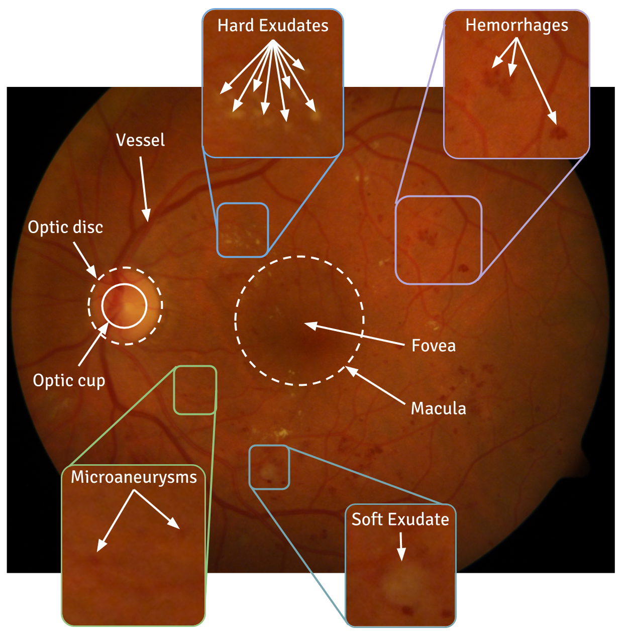
In the United States, more than 40 million people suffer from acute eye related diseases that may lead to complete vision loss if left untreated [1]. Many of these diseases involve the retina. Glaucoma, diabetic retinopathy and age-related macular degeneration are some of the most common retinal diseases. Figure 1 is a fundus photograph of the retina with various structures and disease manifestations.
Glaucoma is one of the major causes of blindness; it is estimated that by 2020 glaucoma will affect almost 80 million people in the world [2]. The two main types of this disease are open-angle glaucoma and angle closure glaucoma. About 90% of the affected people suffer from primary open-angle glaucoma [3]. Traditionally glaucoma is diagnosed by calculating what is called the optic cup to disk ratio . Neuroretinal rim loss, visual fields and retinal nerve fibre layer defects are also some of the measures used by ophthalmologists for diagnosis. Diabetic retinopathy (DR) is another common cause of human vision loss. It is expected that the percentage of diabetic patients worldwide will increase from 2.8% in 2000 to 4.4% in 2030. Diabetes is quite common in persons above the age of 30; uncontrolled diabetes can lead to DR [4]. Early stages of DR are less severe and clincially managed.. It is characterized by various abnormalities in retina such as microaneurysms (MA) and other small lesions caused by rupture of thin retinal capillaries; these are early indicators for DR. Some of the other manifestations include hard exudates, soft exudates or cotton wool spots (CWS), hemorrhages (HEM), neovascularization (NV) and macular edema (ME) (see Figure 1) [5].
Age-related macular degeneration (AMD) is another common vision related problem. It can result in loss of vision in the middle of the visual field in the human eye, and with time there is a complete loss of central vision [6]. In the United States, about 0.4% people from age range 50 to 60 suffer from this disease and around 12% people who are over 80 years old are affected [7]. Health-care in most countries suffers from a low doctor to patient ratio. Due to an overburdened patient-care system, diagnosis and proper treatment becomes error-prone and time-intensive. On the other hand, sufficient amount of data are generated everyday in various health clinics and hospitals, but it is rarely utilized for computer aided diagnostics (CAD) applications and not available publicly. [5]. During the past few years, artificial intelligence algorithms have been used in classifying different types of data including images.In retinal image analysis, the traditional CAD system architectures takes several predefined templates and kernels to compare with manually annotated and segmented parts of these images. Deep learning models are extremely powerful architectures to find patterns between different nonlinear combinations of different types of data. It derives relevant necessary representations from the data without the requirement of manual feature extraction. In recent years, deep learning algorithms are replacing most of the traditional machine learning algorithms and in most of cases outperforming the traditional classifiers. General details of the different deep learning architectures like Alexnet [8], VGG [9], Sparse Autoencoder [10] can be found in [11]. This review focuses on the application of different deep learning architectures and algorithms for retinal fundus image processing especially for segmentation and classification problems. Table 1 gives an overview of existing fundus image datasets which are commonly used in deep learning models. Section 2 reviews various applications of deep learning for detection and diagnosis of ophthalmic diseases from retinal fundus images. Section 3 discusses several future research directions and critical insights.
2 Application in Retinal Image Processing Techniques
| Dataset Name | Images | Usage | Camera | Availability |
| ACHIKO-K [12] | 258 manually annotated images, 114 Glaucoma, 144 Normal | Glaucoma detection | Available Online | |
| AREDS [13] | Approx. 206,500 images | AMD detection | Upon Request | |
| CHASE [14] | 28 images | Blood vessel segmentation | Available Online | |
| CLEOPATRA [15] | 298 images | OD segmentation | Not Available Publicly | |
| DIARETDB1 [16] | 88 images,84 DR and 4 normal | DR detection | Fundus Camera FOV 50° | Available Online |
| DIARETDB0 [17] | 130 images, 20 normal and 110 DR | DR detection | Available Online | |
| DRIONS-DB [18] | 110 images, 23.1% Chronic Glaucoma and 76.9% Eye Hypertensio | Glaucoma detection | Available Online | |
| DRISHTI-GS [19] | 101 images | Glaucoma detection | Fundus Camera with FOV 30° | Available Online |
| DRIVE [20] | 40 images,33 normal and 7 mild DR | Vessel segmentation | Canon CR5 non-mydriatic 3CCD camera with FOV 45° | Avaliable Online |
| Kaggle/ EyePACS [21] | 35126 images | DR detection | Available on Registration | |
| HRF [22] | 45 images,15 images each of healthy, DR, glaucomatous patients | Glaucoma detection | Canon CR-1 fundus camera with FOV 45° | Available Online |
| KORA [23] | images from 2,840 patients | AMD detection | Available Online | |
| SEED [24] | 235 images,43 glaucoma and 192 normal | Glaucoma | Not available online | |
| STARE [25] | 400 images,blood vessel annotation on 40 images | Vessel segmentation | TRV50 fundus camera with FOV 35° | Available Online |
| MESSIDOR [26] | 1200 images | OD segmentation,Lesion detection | Color Video 3CCD camera with FOV 45° | Available Online MESSIDOR-2 Upon Request |
| e-optha [27] | 47 images with exudates, 35 without. 233 normal images and 148 MA images | Lesion detection | Available Online | |
| ONHSD [28] | 99 images | OD,ON segmentation | Canon CR6 45MMNf with FOV 45° | Available Online |
| ORIGA [29] | 650 retinal images | Glaucoma detection | Not Available Online | |
| RIGA [30] | 760 retinal fundus images | Glaucoma detection | Available Online | |
| RIM-ONE [31] | 783 images | OD segmentation | Nidek AFC-210 Can EOS 5D Mark II | Not Available Publicly |
| REFUGE [32] | 1200 annotated images | Glaucoma detection | Available Online | |
| Retinopathy Online Challenge [33] | 100 fundus images | Lesion detection | Topcon NW 100, Topcon NW 200, Canon CR5-45NM | Available on Registration |
To the best of our knowledge, the very first application of computer-aided methods to clinical ophthalmology was by Goldbaum et al. in 1994 [34]. The authors concluded that a neural network could be trained and modelled as efficiently as a trained reader for glaucoma visual field interpretation. Another early application was the use of a neural network to predict astigmatism after cataract surgery [35].
Segmentation is an important step for automatic cropping of the region of interest for further processing. An image may possess some unwanted distortions which hamper proper processing. Noise can be present in the images and the illumination may not be uniform across the image. Hence for proper visualization, different parts of an image should be segmented. Over the last few years different deep learning approaches combining with various methodologies were reported to solve segmentation problems.
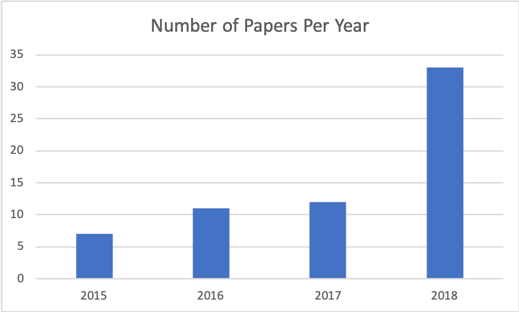
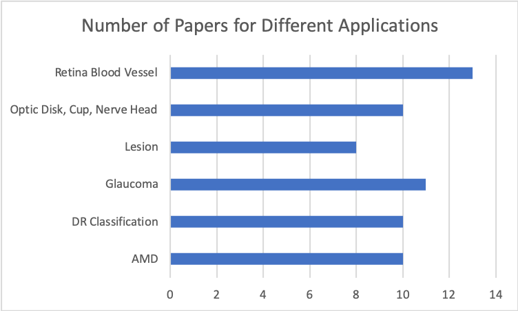
In this review, we will discuss recent articles where different deep learning architectures have been implemented for ophthalmic applications with fundus images. Figure 2 shows yearwise trends of published literature and also number of papers for different application areas. It can be seen that number of published papers on deep learning for fundus imaging for ophthalmic diagnosis has increased significantly starting from 2014. In this review, published papers upto December 2018 have been reviewed. Papers were collected through search queries on google scholar with various keywords like deep learning, ophthalmology, image segmentation, classification, fundus photos, image datasets (e.g.- MESSIDOR, DRIVE, STARE, EYEPACS, RIGA etc), retina. Different performance measures like accuracy (Acc) , sensitivity (SN), specificity (SP), area under curve (AUC), F1 score, DICE Score are mentioned for different application areas. Please refer to [36] for details on the performance indicators discussed herein.
2.1 Fundus Image Applications
2.1.1 Optic Disc(OD), Cup(OC) and Nerve Head Segmentation(ONH)
The first implementation of deep learning architecture in OD segmentation was proposed by Lim et al. [37] in 2015. During this time (2015) CNN had already been successfully implemented in various biomedical segmentation problems [38] [39]. The authors developed CNN for calculating cup-to-disc ratio as a measure of the presence of glaucoma to overcome the need for hand-crafted feature extraction methods of shallow machine learning algorithms. Since this publication, there have been significant advances in deep learning architectures. Maninis et al. [40] experimented on fundus images to segment both blood vessels and OD together using the VGG model [9] with a smaller modification of layers. Feng et al. [41] performed both OD and exudates segmentation. A fully convolutional neural network (FCN) of U-Net architecture, very popular for biomedical image segmentation problems [42], was modified by replacing convolutional layers with residual blocks, (inspired by He et al. [43]) and used to build a unified architecture. Sevastopolsky [44] also used U-Net architecture(with reduced number of filters in each convolutional layer) to segment both OD and OC decreasing both time and space complexity. In [44] the authors segmented both OD and OC separately. Edupuganti et al. [45] implemented one shot segmentation pipeline for segmenting OD and OC for glaucoma analysis.ImageNet(http://www.image-net.org/) was used for initialization of the FCN encoder. Utilizing the concept of unified segmentation architecture like [40] or [41] Al-Bander et al. [46] proposed deep learning based segmentation architecture for both OD and fovea together. Recently Mitra et al. [47] reported some drawbacks of Al-Bander et al. [46], as Al-Bander et al. used grayscale images which resulted in some data loss. Their proposed architecture in [46] utilized Dropout layers at different stages which arbitrarily dropped neurons resulting in further data loss. To address and overcome these shortcomings, [47] used batch-normalization in CNN for OD detection. More recently Liu et al. [48] used fundus images to implement deep learning based segmentation architectures to segment glaucomatous OD. A previously trained model with ImageNet database was used and the output layer was replaced by a new output layer with 2 nodes for 2 different classes- normal and glaucoma. In contrast with the previous studies, this work gathered a larger amount of data from different sources with different image qualities and resolutions. Hence this model can be considered as more robust than most of the other works. Sun et al. [49] employed a faster R-CNN architecture as a deep object detection architecture to segment OD from fundus. Ghassabi et al. [50] introduced a consolidated approach of ONH and cup segmentation for glaucoma assessment which is effective even when there are non-obvious neuroretinal rim, peripapillary atrophy and low intensities of the optic cup as it gave a better performance (as shown by the overlapping error).
Reference Architecture Dataset Acc SN SP AUC F1 Score DICE Score Overlapping Error Lim et al. 3-class CNN MESSIDOR, SEED-DB .847 Maninis et al. CNN DRIU DRIVE, STARE, DRIONS-DB, RIM-ONE .822, .831, .971, .959 Feng et al. FCN DRIONS-DB 93.12% 99.56% 0.9093 Sevastopolsky U-Net with lesser filters DRIONS-DB, RIM-ONE .94, .95 Edupuganti et al. VGG16 FCN Drishti-GS .967 Al-Bander et al. MESSIDOR 96.89% Mitra et al. CNN MESSIDOR, EyePACS 99.05%, 98.78% 99.14%, 98.17% Sun et al. Faster R-CNN ORIGA Dataset 93.1% Liu et al. ResNet50 FCN 3 centres from Sydney, HRF, RIM-ONE 91.6% 86.7% 96.5% .97 Ghassabi et al. WTA Neural Network, SOM Neural Network Stein Eye Institute, Labbafi Nedjad hospital of Iran, RIMONE, DIARETDBO 9.6%(ONH Seg.) & 25.1%(Cup Seg.) Tan et al. 10-layer CNN CLEOPATRA-DB 87.58%-exudates 71.58%-dark lesions
2.1.2 Lesion Segmentation and Detection
Lesion detection is an important step for DR screening. Different deep learning based studies on lesion detection and segmentation are discussed below.
Haloi [51] was the first to implement deep neural network to detect MA for DR screening. He used a 5 layer pixel based deep neural network to detect MA. Shan et al. [52] found biological cell nuclei detection and MA detection problems quite similar and employed stacked sparse autoencoder (SSAE) proposed in a nuclei detection problem [53]. Classification was done for MA and non-MA patches. Image patches were passed through the SSAE to obtain features and a softmax layer was used to classify the labels. The previous studies mainly addressed detection of MA, but in DR screening bigger hemorrhages are also important. Grinven et al.[54] proposed a methodology to detect hemorrhages in retinal fundus images by classifying the lesions. CNN was implemented and a selective sampling algorithm was introduced to dynamically select misclassified training samples. It was found to decrease the time of epochs and also to enhance the AUC as compared for the CNN with no selective sampling. All previous studies [51] [52] [54] tried to detect different lesions separately, Tan et al. [55] applied a 10 layer CNN to automatically segment exudates, MA and hemorrhages using a single framework. Orlando et al. [56] also worked on detection of both MA and HE together using 3 different data-sets combining hand-crafted feature and deep features learned from CNN. Previously there were very few studies analyzing the effectiveness of combining two feature methods [57]. Deep learned feature vectors using 4 convolutional layers and 1 fully connected layer from CNN(trained using LeNet architecture) were created by combining handcrafted features drawn from the green channel of the normalized and equalized image. Lam et al. [58] used a deep learning architecture to detect the presence of five classes of red lesions i.e. normal, microaneurysms, hemorrhages, exudates, retinal neovascularization using EyePACS. The CNN was trained with 1050 images using GoogleNet architecture [11]. Patches were extracted with varying shapes and sizes according to the size of the lesions. For testing, a sliding window was introduced to make a full scan over the whole image by the CNN to give a multiclass outcome probability. Son et al. [59] proposed a cost-effective method to localize lesions which improved precision during training by using regional annotation of findings. Badar et al. [60] used an encoder-decoder based FCN architecture calculating pixel-wise segmentation of multi-class retinal pathologies (exudates, hemorrhages & cotton wool spots) and achieved state of the art results. Khojasteh et al. [61] introduced an innovative framework and architecture for CNN by inserting a pre-processing layer for recognition of HE and MAs. Chudzik et al [62] presented a segmentation method which utilized similar a combination of CNN and codebook structure.
Reference Architecture Dataset Acc SN SP AUC Haloi 5 layer CNN MESSIDOR, ROC 96% 97% 96% .982, .98 Shan et al. Transfer Learning (SSAE) DIARETDB 91.38% .916 Grinven et al. CNN using OxfordNet MESSIDOR, EyePACS 91.9%, 83.7% 91.8%, 85.1% .972, .895 Tan et al. 10 layer CNN CLEOPATRA 87.58%, 71.58% Orlando et al. CNN using LeNet architecture e-optha, MESSIDOR .8812, .8932 Lam et al. CNN using GoogleNet3 EyePACS, e-optha 98% .95 Son et al. CNN with residual, reduction, avg. pooling, atrous pyramid pooling layers Seoul National University Bundang Hospital 0.9895 Badar et al. Encoder-Decoder based FCN MESSIDOR 97.86% 80.93% 98.54% Khojasteh et al. CNN with preprocessing after 1st conv layer DIARETDB1 90.0% Chudzik et al. FCNN & Auxiliary Codebook E-Optha MA & E-Optha EX 0.8666 0.9998 0.982
2.1.3 Retinal Blood Vessel Segmentation
Retinal blood vessels are important for different eye disease diagnosis. In this section we will discuss different results on vessel segmentation from retinal fundus images. In one of the first studies in this domain, Maji et al. [63] used a hybrid of random forest and deep neural network (DNN) for blood vessel segmentation. The DNN performed unsupervised learning of vessel dictionaries using sparse trained denoising auto-encoders (DAE). It was followed by supervised learning of random forest on the DNN response. However this method could not outperform the conventional approaches. Around the same time Liskowski et al. [64] proposed a deep learning based blood vessel segmentation framework of retinal fundus images datasets. Images were standardized by subtracting the mean from every patch and dividing it by the standard deviation to avoid contrast and brightness fluctuations in the image pixels. It outperformed many existing approaches. In another pioneering work Melinscak et al. [65] used a deep neural network, inspired by a similar problem of segmenting neuronal membranes [66] using DNN as pixel classifier. To improve the performance proposed in [63] Maji et al. [67] used an ensemble of 12 CNNs to segment retinal blood vessels. The networks were trained individually on the dataset of 60,000 randomly chosen 3x31x31 patches. During inference, the responses were averaged to form the final segmentation. RMSProp was used as the optimizer and a minibatch size of 200 was used. Fu et al. [68] found several disadvantages in [65] as it used pixel based approach and hence Fu et al. proposed a fully CNN architecture based on image-to-image training system. Multi-scale and multi-level CNN was used and combined with conditional random field (CRF) to model the long-range interactions between pixels. Leopold et al. [69] investigated use of CNN to segment blood vessels using ADAM parameter optimization. The green channel of each image was used for classifying vessels and non-vessels image pixels. The model gave the probability maps of every pixel to classify between vessel and non-vessel. Gabor filters were used to smooth and finalize the decision. Zhang et al. [70] applied U-Net which was also used in other works for OD segmentation [41] [44]. The authors proposed a modified U-Net based architecture to segment blood vessels from fundus images. By adding some additional labels on boundary areas the problem was converted into a multi-class task. Stochastic gradient descent (SGD) was used to optimize model parameters. Oliveira et al. [71] implemented deep learning architecture for blood vessel segmentation. Previous deep learning architectures only processed raw data but here, initially, stationary wavelet transform was applied to each training image to keep multi-resolution information. A fully convolutional network was used to generate feature maps. Stochastic Gradient Descent with Nesterov momentum was implemented during training to decrease the cross-entropy loss function. The final probability maps for all of the image patches were merged and averaged to get a final value and thresholding was done to get the ultimate unique segmentation. Liu et al. [72] used densely Connected CNN to segment blood vessels in fundus images. A 17 layer architecture was used and the layer number X got input from the output of the previous X-1 layers and thus used the back layers of the network as features of the front layer. Similar to [68] Hu et al. [73] proposed an image-to-image deep learning vessel detection model using the CNN combined with conditional random field (CRF). Main contribution of this work was to combine features from each of the convolutional layers and to incorporate class-balanced cross-entropy loss to improve detection accuracy. VGG-16 model was used. Lepetit-Aimon et al [74] introduced the LRFFCN which did better than the U-Net [42]in retinal artery and vein classification and manifested high sensitivity in comparison to other state of the art algorithms to segment vessels. Chudzik et al. [75] gave a two stage architecture combining visual codebook framework with CNN.
Reference Architecture Dataset Acc SN SP AUC Maji et al. RF and DNN DRIVE 93.27% .9195 Liskowski et al. CNN DRIVE, STARE, CHASE_DB 97% .99 Maji et al. ConvNet ensemble DRIVE 94.7% .9283 Fu et al. Multi-scale and Multi-level CNN DRIVE, STARE, CHASE_DB1 95.23%, 95.85%, 94.89% Leopold et al. FCN using RETSEG13 DRIVE 94.78% 68.23% 98.01% .9707 Zhang et al. CNN(U-Net) DRIVE, STARE, CHASE_DB1 95.04%, 97.12%, 97.7% 87.23%, 76.73%, 76.7% 96.18%, 99.01%, 99.09% .9799, .9882, .99 Oliveira et al. CNN DRIVE, STARE, CHASE_DB1 95.76%, 96.94%, 96.53% 80.39%, 83.15%, 77.79% 98.04%, 98.58%, 98.64% .9821, .9905, .9855 Liu et al. Densely Connected CNN DRIVE 95% Lepetit-Aimon et al. FCNN with large receptive field MESSIDOR, STARE, DRIVE 95.9% Chudzik et al. CNN DRIVE, STARE 0.7881, 0.8269 0.9741, 0.9804 0.9646, 0.9837
2.1.4 AMD Classification
Recent results on AMD disease classification from fundus images are discussed below.
One of the very first publications in this domain was done by Burlina et al. [76] where they used OverFeat features from DCNN (pretrained in ImageNet database) and used Support Vector Machine to classify between early and intermediate stages of AMD. Later Burlina et al. [77] used a completely data-driven approach using deep CNN (DCNN-A) to perform a binary classification between early-stage AMD and advanced stage AMD using the same AREDS database implemented on AlexNet model [11]. This method was compared with the previous methods combining both deep features and transfer learning.Based upon the work of [76] Horta et al. [78] reported a hybrid method employing deep image features and random forest to combine different patient non-visual data e.g. lifestyle, cataract, demographics with the image for AMD classification. To extract deep image features, the CNN (pre-trained with 1.2 million image data) was used. The deep features combined with the non-medical, non-visual information of the patients were used to train a Random Forest Classifier to perform binary classification for higher severity AMD and lower severity of AMD. The combined features were found to achieve higher accuracy than individual feature set. Govindaiah et al.[79] reported an extended study of [80] with a modified deeper VGG16 architecture. The macula was chosen as a Region of Interest and images were resized to a common reference level. For comparison with the VGG16, a 50 layer Keras implementation of residual neural network was used. Matsuba et al. [81] published a new approach for detecting AMD disease from ultra wide-range Optos ophthalmoscope color fundus images. Three convolutional layers, with ReLU unit and max-pooling layers were used to perform this experiment on pre-processed fundus images. The accuracy of DCNN using images was compared with human grading by six ophthalmologists. Tan et al. [82] used a 14 layer CNN to detect AMD. Three fully-connected layers, 4 max-pooling layers, and 7 convolutional layers were implemented in this work. Adam optimization [83] was used for tuning the CNN model’s parameters.Grassmann et al. [84] proposed a deep learning based classification architecture to predict the severity of AMD. In this study, an ensemble of several convolutional neural networks was used to classify among 13 different classes of AMD [85]. Mainly four different steps can be found in this methodology. Six different neural networks (AlexNet, GoogLeNet, VGG, ResNet, Inception-v3, 1-ResNet-v2 ) were used independently to train the model. With the result obtained from each of the individual neural networks, a random forest ensemble model was developed. Govindaiah et al. [86] used an ensemble network consisting of state of the art network architectures thereby reaching a satisfactory performance level in AMD classification.
Reference Architecture Dataset Acc SN SP AUC Burlina et al. Deep Features with SVM AREDS 95% 96.4% 95.6% Burlina et al. DCNN using AlexNet AREDS 91.6% .96 Horta et al. DCNN AREDS 79.04% 66.34% 88.95% .8476 Govindaiah et al. VGG16 AREDS dataset 92.5% Matsuba et al. DCNN Tsukazaki Hospital 99.76% Tan et al. CNN Kasturba Medical College 95.45% 96.43% 93.45% Grassmann et al. Ensemble (AlexNet, GoogleNet, VGG, ResNet, Inception-v3, 1-ResNet-v2) AREDS and KORA 94.3% 84.2% Govindaiah et al. Ensemble Network (Inception-ResNet-V2 & Xception) AREDS 86.13%
2.1.5 Glaucoma Classification
One of the early publications in glaucoma classification using deep learning was by Chen et al.[87]. They implemented a CNN with dropout and data augmentation. A six layers deep CNN with 4 convolutional layers of progressively decreasing filter size (11, 5, 3, 3) followed by 2 dense layers was used. Improving their previous work, [87] Chen et al. [88] presented a model using Contextualized CNN (C-CNN) architecture. It combined the output of convolutional layers of multiple CNN to a final dense layer to obtain the softmax probabilities. The 5 C-CNN model which was a concatenation of outputs of last convolutional layers of 5 CNNs each of depth 6 (5 convolutional layers + 1 MLP). Asoaka et al. [89] used a 3 layer deep Feed-forward Neural Network (FNN) on a private dataset of 171 Glaucoma images. Chakravarty [90] was first to propose a method for joint segmentation of OD, OC and glaucoma prediction. In this method CNN feature sharing for different tasks ensured better learning and over-fitting prevention. The parts of the model that were shared with U-net contained 8 times fewer number of CNN filters than the conventional U-net. It used an encoder network to downsample the feature and then a decoder network to restore the image size. Two different convolutional layers were applied on the decoder network’s output for OC and OD segmentation. The OC and OD segmentation masks were merged into separate channels and CNN was applied to it. The outputs of the CNN and encoder output were combined and fed to a single neuron to predict glaucoma. With a lower number of parameters this method achieved comparable performance with existing architecture e.g. [91]. Zhixi et al. [92] used the Inception-v3 architecture to detect glaucomatous optic neuropathy. Here researchers graded the images by trained ophthalmologists before applying the algorithm. Local space average color subtraction was applied in pre-processing to accommodate for varying illumination. Chai et al.[93] presented a framework on a dataset of fundus images obtained from various hospitals by incorporating both domain knowledge and features learned from a deep learning model. This method was also used in other applications [56] The disk image provided local CNN features, the whole image provided global CNN features whereas domain knowledge features were obtained from diagnostic reports. It used a total of 25 features including 3 numerical features: intraocular pressure, age, and visual acuity as well as 22 binary features such as swollen eye, headache, blurred vision and failing visual acuity. The disk and whole images were fed to two separate CNN while domain knowledge features were fed to a third branch consisting of a fully connected neural network. These three branches were concatenated by a merge layer followed by two dense layers and a logistic regression classifier. Perdomo et al. [94] used curriculum learning [95] in DCNN’s to achieve better results using a reduced set of training examples. Pal et al. [96] introduced the G-EyeNet architecture which proved to be more robust given its results on low quality images.
Reference Architecture Dataset Acc SN SP AUC F-Score Chen et al. 6 layer CNN ORIGA, SCES .831, .887 Asoaka et al. 3 layer FNN Private: 171 .926 Chakravarty et al. Multi-task CNN REFUGE .9456 Zhixi et al. Inception-v3 Private:48000+ 95.6% 92.0% .986 Chai et al. MB-NN Private: 2554 91.51% Chen et al. C-CNN ORIGA, SCES .838, .898 Perdomo et al. DCNN RIM-ONE-v1, RIM-ONE-v3, DRISHTI-GS1 89.4%(RIM-ONE-v1) 0.82 (DRISHTI-GS) Pal et al. CAE with CNN classifier DRIONS-DB 0.923
2.1.6 Diabetic Retinopathy Classification
In this section different applications of deep learning algorithms for diabetic retinopathy detection are described briefly.
Abràmoff et al [97] described DR detection using a device called IDx-DR X2.1. Here retinal images were used in a CNN based on AlexNet to classify different types of DR. The main classes of diseases were referable DR (rDR), vision-threatning DR (vtDR) and proliferative DR (pDR). The CNN-based architectures were designed to characterize and detect optic disc, fovea and lesion characteristics. Using a retinal fundus image dataset consisting of 70000 images, Colas et al. [98] proposed a DR grading method. There were 4 different classes of DR images in the dataset- no DR, mild DR, moderate DR and acute DR. Gulshan et al. [99] used deep learning algorithms to identify the presence of diabetic retinopathy. Five different types of DR and the presence of macular edema were graded by expert clinicians. Inception v-3 model was used, stochastic gradient descent method for optimization was used and batch normalization was done with a pre-trained model with ImageNet data. Gargeya et al.[100] reported a deep learning architecture to classify between normal and DR fundus images and also reported heatmap visualization of the result. Using the principle of deep residual learning, the CNN model was built to learn deep discriminative features for detecting DR. From average pooling layer of CNN, 1024 features were obtained. Metadata features related to 3 metadata variables i.e. pixel height, pixel width and field of view of the image were appended to form a final feature vector with 1027 features. A second level tree-based gradient boosting classifier was designed. Quellec et al. [101] discussed a method to detect referable DR as well as lesions with ConvNet architecure using o_O Solution [21].Unlike the previous studies this method attempted to classify between normal and DR on both image level and pixel level. This proposed model was mainly based on visualization methods of CNN. Heatmap generation modifications were proposed for this purpose to jointly improve the quality of DR and lesion detection. Takahashi et al. [102] graded different stages of the presence of DR using GoogleNet architecture. Unlike in other previously published literature the authors graded the images manually on their own to test the accuracy of the methodology. The model was designed using two different ways, first with manual staging of three color photographs (AI1) and second with manual staging of only one color photograph (AI2). From the GoogleNet model- 5 top layers were deleted, the crop size was expanded and the batch size was reduced. 20 fold cross-validation was used and for comparison AI1 was also trained with ResNet model. García et al. [103] applied different architectures of CNN for DR detection. As a pre-processing step, images were subtracted from color mean and rescaled to 256x256. Data augmentation by flipping the images was done to increase the robustness. In this work several neural network architectures using various learning rates and different number of layers were used to compare different architectures to calculate the highest accuracy among all. Lin et al. [104] used entropy images instead of original fundus images and showed that the feature maps are generated faster and competently.
Reference Architecture Dataset Acc SN SP AUC Abràmoff et al. CNN AlexNet Messidor-2 96.8% 87% Colas et al. CNN EyePACS 96.2% 66.6% .946 Gulshan et al. CNN Incetionv-3 EyePACS-1, Messidor-2 90.3%, 87% 98.1%, 98.5% .991, .99 Gargeya et al. CNN Deep Residual Learning EyePACS, MESSIDOR e-Optha2 94% 98% .97 Quellec et al. ConvNet Kaggle, e-optha, DiaretDB1 .954, .949, .955 Takahashi et al. CNN GoogleNet, ResNet 9939 images 80% Garcia et al. CNN AlexNet EyePACS 83.68% Lin et al. CNN Kaggle 86.10% 73.24% 93.81% 0.92
3 Conclusion and Future Research
This review addressed different applications of deep learning methodologies in ophthalmic diagnosis.
Application Reference Method Dataset Acc SN SP AUC F1 Score OD Segmentation Maninis et al. [40] CNN DRIU DRIVE, STARE .822, .831 Soares et al. [105] Wavelets DRIVE, STARE .762, .774 Lesion Detection Haloi et al. [51] 5-layer CNN Messidor 96% 97% 96% .988 Antal et al. [106] Ensemble Model Messidor 90% 90% 91% .989 Retinal Vessel Segmentation Leopold et al. [69] FCN using RETSEG13 DRIVE 94.78% Staal et al. [20] kNN Classifier DRIVE 94.22% AMD Classification Burlina et al. [76] AREDS Deep Features with SVM 95% 96.4% 95.6% Kankanahalli et al. [107] AREDS SURF features with random forest 91.8% 91.3% 92.3% Glaucoma Classification Perdomo et al. [94] DCNN RIM-ONE-v1, RIM-ONE-v3, DRISHTI-GS1 89.4% Gajbhiye et al. [108] KNN 89% DR Classification Gargeya et al. [100] CNN Deep Residual Learning EyePACS, MESSIDOR e-Optha2 94% 98% .97 Roychowdhury et al. [109] kNN (DREAM) MESSIDOR 98.88% 48.72%
Table 8 gives a brief overview of state-of-the-art deep learning approach and traditional imethods for computer-aided diagnosis.It can be noticed that in most of the cases deep learning methods outperformed traditional methodologies.
The previous reviews published in this domain were very clinical or focused on traditional machine learning algorithms or emphasized a particular disease or focused on hardware implementation of artificial intelligence in ophthalmic diagnosis [110][111][112][113][114]. None of them dealt with detailed reviews of different state-of-the art deep learning algorithms used in ophthalmic diagnosis with retinal fundus images. Hence, to the best of our knowledge this is the first review article of deep learning algorithms and performance outcomes for different architectures for ophthalmic diagnosis using retinal fundus images.Deep learning applications in retinal images are quite useful and effective. They reduce the need of manual feature extraction as the methodologies are mainly data-driven. Convolutional neural networks are the most widely used architecture for classification, detection or segmentation of different parts of fundu images. Ensemble, FCN, ResNet and AE based architectures were commonly used in these studies.
However, there are still some limitations which need to be addressed. Some of these and also some possible solutions are discussed below:
-
•
Unlike computer vision problems, large datasets are not available. Also there is a scarcity of manual annotation of data. Deep learning equated large amounts of data since the model mainly learns from the inherent pattern of the data. Hence this is a major problem in this field.Generative models proposed by Goodfellow et al. can be an important and useful solution to mitigate this problem. This is a very state-of-the art area of this research and very few efforts have been made so far [115] [116] to explore the possibilities of generative modelling to synthesize new fundus images with annotations and with proper clinical relevance. Generative Adversarial Network, Variational Auto-encoders are some very popular architecture for image generation. Successful application of these can be used to generate large amounts of clinically relevant synthetic data. It will not only help to increase amount of available data but also it will help to avoid the privacy issues.
-
•
A major problem is the unavailability of standardized KPIs (Key Performance Indicators) for measuring the performance of a particular model. Different researchers use different indices to measure their work. Due to this variablity one cannot easily compare different deep learning architectures for a given disease state. For example, in lesion detection, Lam et al. [58] achieved an accuracy of 98% which is higher than most of the other state-of-the-art methods, whereas in terms of AUC Haloi[51] achieved 0.982 which is higher than other reported AUC. Leopold et al. [36] took this into consideration and also suggested more generalized metrics such as G-mean and MCC to measure a model’s effectiveness.
-
•
Due to the difference in camera settings there is a possibility of domain shift problem. In most of the literature, training and test data come from same image distribution. But in real life this is not always the case. Hence this domain shift can cause a major damage in real life application if not taken care of beforehand. Transfer learning has been used for different applications in this area [45] [77][117][118]. Domain Adaptation is a sub-domain of Transfer Learning where data for both training and testing are extracted from different distributions. In real world, it is not always possible to get test data and training data from the same distribution. Hence the model should be robust enough to deal with data from a different distribution for test purpose. Often it is found that accuracy decreases due to this domain shift problem. More emphasis should be given to deep domain adaptation approaches in order to create robust models which can be implemented for real world ophthalmic diagnosis. Wang et al. [119] have discussed different deep domain adaptation algorithms which can be used to address this problem. A recent paper explored adversarial domain adaptation technique to segment blood vessels of STARE dataset with a model trained on DRIVE dataset and it outperformed other works in terms of F score [120]. In the context of ophthalmic diagnosis it can be an important and necessary direction for future research.
4 Acknowledgement
This research was supported by a Discovery Grant from NSERC, Canada to Vasudevan Lakshminarayanan.
5 Conflict of Interest
The authors declare no conflict of interest.
References
- [1] J P Whitcher, M Srinivasan, and M P Upadhyay. Corneal blindness: a global perspective. Bulletin of the World Health Organization, 79:214–221, 2001.
- [2] C Costagliola, R Dell’Omo, M R Romano, M Rinaldi, L Zeppa, and F Parmeggiani. Pharmacotherapy of intraocular pressure: part i. parasympathomimetic, sympathomimetic and sympatholytics. Expert Opinion on Pharmacotherapy, 10(16):2663–2677, 2009.
- [3] MT Nicolela and JR Vianna. Optic Nerve: Clinical Examination, in Pearls of Glaucoma Management. Springer, Berlin, Heidelberg. pages 17–26, 2016.
- [4] AS Krolewski, JH Warram, LI Rand, AR Christlieb, EJ Busick, and CR Kahn. Risk of proliferative diabetic retinopathy in juvenile-onset type i diabetes: a 40-yr follow-up study. Diabetes Care, 9(5):443–452, 1986.
- [5] H.A. Leopold, J.S. Zelek, and V. Lakshminarayanan. chapter Deep Learning Methods for Retinal Image Analysis in Signal Processing and Machine Learning for Biomedical Big Data. eds. Sejdić, E and Falk, TH., pages 329–365. CRC Press, 2018.
- [6] RD Jager, WF Mieler, and JW Miller. Age-related macular degeneration. New England Journal of Medicine, 358(24):2606–2617, 2008.
- [7] DS Friedman, BJ O’Colmain, Bz Munoz, SC Tomany, De Jong PT Nemesure B Mitchell P Kempen J McCarty, C, et al. Prevalence of age-related macular degeneration in the united states. Arch Ophthalmol, 122(4):564–572, 2004.
- [8] A Krizhevsky, I Sutskever, and GE Hinton. Imagenet classification with deep convolutional neural networks. In Advances in neural information processing systems, pages 1097–1105, 2012.
- [9] Kaiming He, Xiangyu Zhang, Shaoqing Ren, and Jian Sun. Deep residual learning for image recognition. In Proceedings of the IEEE conference on computer vision and pattern recognition, pages 770–778, 2016.
- [10] Andrew Ng et al. Sparse autoencoder. CS294A Lecture notes, 72(2011):1–19, 2011.
- [11] Y Guo, Y Liu, A Oerlemans, S Lao, S Wu, and MS Lew. Deep learning for visual understanding: A review. Neurocomputing, 187:27–48, 2016.
- [12] Z Zhang, J Liu, F Yin, B Lee, DW Wong, and KR Sung. Achiko-k: Database of fundus images from glaucoma patients. In 2013 IEEE 8th Conference on Industrial Electronics and Applications (ICIEA), pages 228–231, 2013.
- [13] TE Clemons, EY Chew, SB Bressler, and W McBee. Age-related eye disease study research, g. national eye institute visual function questionnaire in the age-related eye disease study (areds). Arch Ophthalmology, 121(2):211–7, 2003.
- [14] CG Owen, AR Rudnicka, R Mullen, SA Barman, D Monekosso, PH Whincup, J Ng, and C Paterson. Measuring retinal vessel tortuosity in 10-year-old children: validation of the computer-assisted image analysis of the retina (caiar) program. Investigative Ophthalmology & Visual Science, 50(5):2004–2010, 2009.
- [15] S Sivaprasad, G Arden, AT Prevost, R Crosby-Nwaobi, H Holmes, J Kelly, C Murphy, G Rubin, J Vasconcelos, and P Hykin. A multicentre phase iii randomised controlled single-masked clinical trial evaluating the cl inical e fficacy and safety of light-masks at p reventing dark-a daptation in the tr eatment of ea rly diabetic macular oedema (cleopatra): study protocol for a randomised controlled trial. Trials, 15(1):458, 2014.
- [16] T Kauppi, V Kalesnykiene, J Kamarainen, L Lensu, I Sorri, A Raninen, R Voutilainen, H Uusitalo, H Kälviäinen, and J Pietilä. The diaretdb1 diabetic retinopathy database and evaluation protocol. In BMVC, volume 1, pages 1–10, 2007.
- [17] T Kauppi, V Kalesnykiene, J Kamarainen, L Lensu, I Sorri, H Uusitalo, H Kälviäinen, and J Pietilä. Diaretdb0: Evaluation database and methodology for diabetic retinopathy algorithms. Machine Vision and Pattern Recognition Research Group, Lappeenranta University of Technology, Finland, 73, 2006.
- [18] EJ Carmona, M Rincón, J García-Feijoó, and JM Martínez-de-la Casa. Identification of the optic nerve head with genetic algorithms. Artificial Intelligence in Medicine, 43(3):243–259, 2008.
- [19] J Sivaswamy, SR Krishnadas, GD Joshi, M Jain, and AUS Tabish. Drishti-gs: Retinal image dataset for optic nerve head (onh) segmentation. In 2014 IEEE 11th International Symposium on Biomedical Imaging (ISBI), pages 53–56, 2014.
- [20] J Staal, MD Abràmoff, M Niemeijer, MA Viergever, and B Van Ginneken. Ridge-based vessel segmentation in color images of the retina. IEEE Transactions on Medical Imaging, 23(4):501–509, 2004.
- [21] Kaggle:https://www.kaggle.com/c/diabetic-retinopathy-detection/discussion/15617, accessed- 01/09/2019.
- [22] A Budai, R Bock, A Maier, J Hornegger, and G Michelson. Robust vessel segmentation in fundus images. International Journal of Biomedical Imaging, vol. 2013,ID 154860 pages 11, 2013.
- [23] C Brandl, V Breinlich, K Stark, S Enzinger, M Aßenmacher, and M et al. Olden. Features of age-related macular degeneration in the general adults and their dependency on age, sex, and smoking: results from the german kora study. Plos One, 11(11):e0167181, 2016.
- [24] Y Zheng, C-Y Cheng, E L Lamoureux, PPC Chiang, AR Anuar, JJ Wang, P Mitchell, S Saw, and T Wong. How much eye care services do asian populations need? projection from the singapore epidemiology of eye disease (seed) study. Investigative Ophthalmology & Visual Science, 54(3):2171–2177, 2013.
- [25] AD Hoover, Va Ko, and M Goldbaum. Locating blood vessels in retinal images by piecewise threshold probing of a matched filter response. IEEE Transactions on Medical Imaging, 19(3):203–210, 2000.
- [26] Etienne Decencière, Xiwei Zhang, Guy Cazuguel, Bruno Lay, Béatrice Cochener, Caroline Trone, Philippe Gain, Richard Ordonez, Pascale Massin, Ali Erginay, et al. Feedback on a publicly distributed image database: the messidor database. Image Analysis & Stereology, 33(3):231–234, 2014.
- [27] E Decencière, G Cazuguel, X Zhang, G Thibault, J-C Klein, F Meyer, B Marcotegui, G Quellec, M Lamard, R Danno, et al. Teleophta: Machine learning and image processing methods for teleophthalmology. Innovation and Research in Biomedical Engineering, 34(2):196–203, 2013.
- [28] J Lowell, A Hunter, D Steel, A Basu, R Ryder, E Fletcher, L Kennedy, et al. Optic nerve head segmentation. IEEE Transactions on Medical Imaging, 23(2):256–264, 2004.
- [29] Z Zhang, FS Yin, J Liu, WK Wong, NM Tan, BH Lee, J Cheng, and TY Wong. Origa-light: An online retinal fundus image database for glaucoma analysis and research. In Engineering in Medicine and Biology Society (EMBC), 2010 Annual International Conference of the IEEE, pages 3065–3068, 2010.
- [30] A Almazroa, S Alodhayb, E Osman, E Ramadan, M Hummadi, M Dlaim, MR Alkatee, and V Lakshminarayanan. Retinal fundus images for glaucoma analysis: the riga dataset. In Medical Imaging 2018: Imaging Informatics for Healthcare, Research, and Applications, volume 10579, page 105790. SPIE, 2018.
- [31] F Fumero, S Alayón, JL Sanchez, J Sigut, and M Gonzalez-Hernandez. Rim-one: An open retinal image database for optic nerve evaluation. In 2011 24th International Symposium on Computer-based Medical Systems (CBMS), pages 1–6. IEEE, 2011.
- [32] Refuge. http://refuge.grand-challenge.org. 5th miccai workshop on ophthalmic medical image analysis (omia).
- [33] M Niemeijer, B Van G, M Cree, A Mizutani, G Quellec, CI Sánchez, B Zhang, R Hornero, M Lamard, C Muramatsu, et al. Retinopathy online challenge: automatic detection of microaneurysms in digital color fundus photographs. IEEE Transactions on Medical Imaging, 29(1):185–195, 2010.
- [34] MH Goldbaum, PA Sample, H White, B Colt, P Raphaelian, RD Fechtner, and RN Weinreb. Interpretation of automated perimetry for glaucoma by neural network. Investigative Ophthalmology & Visual Science, 35(9):3362–3373, 1994.
- [35] ML Severns, V. Lakshminarayanan, and P. Smith. Predicting astigmatism after cataract surgery using a neural network. Visual Optics/Noninvasive Assessment of the Visual System Technical Digest,Optical Society of America, Washington D.C., 3:34–37, 1993.
- [36] HA Leopold, J Orchard, JS Zelek, and V Lakshminarayanan. Pixelbnn: Augmenting the pixelcnn with batch normalization and the presentation of a fast architecture for retinal vessel segmentation. Journal of Imaging, 5(2), 26., 2019.
- [37] G Lim, Y Cheng, W Hsu, and ML Lee. Integrated optic disc and cup segmentation with deep learning. In Tools with Artificial Intelligence (ICTAI), 2015 IEEE 27th International Conference on, pages 162–169, 2015.
- [38] D Ciresan, A Giusti, LM Gambardella, and J Schmidhuber. Deep neural networks segment neuronal membranes in electron microscopy images. In Advances in Neural Information Processing Systems, pages 2843–2851, 2012.
- [39] DC Cireşan, A Giusti, LM Gambardella, and J Schmidhuber. Mitosis detection in breast cancer histology images with deep neural networks. In In: International Conference on Medical Image Computing and Computer-assisted Intervention eds. Mori K., Sakuma I., Sato Y., Barillot C., Navab N., volume 8150, pages 411–418. Springer, 2013.
- [40] K Maninis, J Pont-Tuset, P Arbeláez, and L Van Gool. Deep retinal image understanding. In International Conference on Medical Image Computing and Computer-Assisted Intervention eds. Ourselin S., Joskowicz L., Sabuncu M., Unal G., Wells W., volume 9901, pages 140–148. Springer, 2016.
- [41] Z Feng, J Yang, L Yao, Y Qiao, Q Yu, and X Xu. Deep retinal image segmentation: A fcn-based architecture with short and long skip connections for retinal image segmentation. In International Conference on Neural Information Processing eds. Liu D., Xie S., Li Y., Zhao D., El-Alfy ES., volume 10637, pages 713–722. Springer, 2017.
- [42] O Ronneberger, P Fischer, and T Brox. U-net: Convolutional networks for biomedical image segmentation. In International Conference on Medical image computing and computer-assisted intervention eds. Navab N., Hornegger J., Wells W., Frangi A., pages 234–241. Springer, 2015.
- [43] K He, X Zhang, S Ren, and J Sun. Deep residual learning for image recognition. In The IEEE Conference on Computer Vision and Pattern Recognition, pages 770–778, 2016.
- [44] A Sevastopolsky. Optic disc and cup segmentation methods for glaucoma detection with modification of u-net convolutional neural network. Pattern Recognition and Image Analysis, 27(3):618–624, 2017.
- [45] VG Edupuganti, A Chawla, and A Kale. Automatic optic disk and cup segmentation of fundus images using deep learning. In 2018 25th IEEE International Conference on Image Processing (ICIP), pages 2227–2231, 2018.
- [46] B Al-Bander, W Al-Nuaimy, B M Williams, and Y Zheng. Multiscale sequential convolutional neural networks for simultaneous detection of fovea and optic disc. Biomedical Signal Processing and Control, 40:91–101, 2018.
- [47] A Mitra, PSh Banerjee, S Roy, S Roy, and SK Setua. The region of interest localization for glaucoma analysis from retinal fundus image using deep learning. Computer Methods and Programs in Biomedicine, 165:25–35, 2018.
- [48] S Liu, SL Graham, A Schulz, M Kalloniatis, B Zangerl, W Cai, Y Gao, B Chua, H Arvind, J Grigg, et al. A deep learning-based algorithm identifies glaucomatous discs using monoscopic fundus photographs. Ophthalmology Glaucoma, 1(1):15–22, 2018.
- [49] X Sun, Y Xu, W Zhao, T You, and J Liu. Optic disc segmentation from retinal fundus images via deep object detection networks. In 2018 40th Annual International Conference of the IEEE Engineering in Medicine and Biology Society (EMBC), pages 5954–5957, 2018.
- [50] Z Ghassabi, J Shanbehzadeh, and K Nouri-Mahdavi. A unified optic nerve head and optic cup segmentation using unsupervised neural networks for glaucoma screening. In 2018 40th Annual International Conference of the IEEE Engineering in Medicine and Biology Society (EMBC), pages 5942–5945, 2018.
- [51] M Haloi. Improved microaneurysm detection using deep neural networks. arXiv preprint arXiv:1505.04424, 2015.
- [52] J Shan and L Li. A deep learning method for microaneurysm detection in fundus images. In 2016 IEEE First International Conference on Connected Health: Applications, Systems and Engineering Technologies (CHASE), pages 357–358, 2016.
- [53] J Xu, L Xiang, Q Liu, H Gilmore, J Wu, J Tang, and A Madabhushi. Stacked sparse autoencoder (ssae) for nuclei detection on breast cancer histopathology images. IEEE Transactions on Medical Imaging, 35(1):119–130, 2016.
- [54] MJJP van Grinsven, B van Ginneken, CB Hoyng, T Theelen, and CI Sánchez. Fast convolutional neural network training using selective data sampling: application to hemorrhage detection in color fundus images. IEEE Transactions on Medical Imaging, 35(5):1273–1284, 2016.
- [55] JH Tan, H Fujita, S Sivaprasad, SV Bhandary, AK Rao, KC Chua, and UR Acharya. Automated segmentation of exudates, haemorrhages, microaneurysms using single convolutional neural network. Information Sciences, 420:66–76, 2017.
- [56] JI Orlando, E Prokofyeva, M del Fresno, and MB Blaschko. An ensemble deep learning based approach for red lesion detection in fundus images. Computer Methods and Programs in Biomedicine, 153:115–127, 2018.
- [57] R Annunziata and E Trucco. Accelerating convolutional sparse coding for curvilinear structures segmentation by refining scird-ts filter banks. IEEE Transactions on Medical Imaging, 35(11):2381–2392, 2016.
- [58] C Lam, C Yu, L Huang, and D Rubin. Retinal lesion detection with deep learning using image patches. Investigative Ophthalmology & Visual Science, 59(1):590–596, 2018.
- [59] J Son, W Bae, S Kim, SJ Park, and K Jung. Classification of findings with localized lesions in fundoscopic images using a regionally guided cnn. in: Stoyanov d. et al. (eds) computational pathology and ophthalmic medical image analysis,omia 2018, compay 2018. lecture notes in computer science, vol 11039. springer, cham.
- [60] M Badar, M Shahzad, and MM Fraz. Simultaneous segmentation of multiple retinal pathologies using fully convolutional deep neural network. in: Nixon m., mahmoodi s., zwiggelaar r. (eds) medical image understanding and analysis. miua 2018. communications in computer and information science, vol 894 pp. 313-324, springer, cham.
- [61] P Khojasteh, B Aliahmad, SP Arjunan, and DK Kumar. Introducing a novel layer in convolutional neural network for automatic identification of diabetic retinopathy. In 2018 40th Annual International Conference of the IEEE Engineering in Medicine and Biology Society (EMBC), pages 5938–5941, 2018.
- [62] P Chudzik, B Al-Diri, F Calivá, G Ometto, and A Hunter. Exudates segmentation using fully convolutional neural network and auxiliary codebook. In 2018 40th Annual International Conference of the IEEE Engineering in Medicine and Biology Society (EMBC), pages 770–773, 2018.
- [63] D Maji, A Santara, S Ghosh, D Sheet, and P Mitra. Deep neural network and random forest hybrid architecture for learning to detect retinal vessels in fundus images. In Engineering in Medicine and Biology Society (EMBC), 2015 37th Annual International Conference of the IEEE, pages 3029–3032, 2015.
- [64] P Liskowski and K Krawiec. Segmenting retinal blood vessels with deep neural networks. IEEE Transactions on Medical Imaging, vol. 35(11):2369–2380, 2016.
- [65] M Melinščak, P Prentašić, and S Lončarić. Retinal vessel segmentation using deep neural networks. In VISAPP 2015 (10th International Conference on Computer Vision Theory and Applications),vol 1, 2015.
- [66] D Cireşan, U Meier, and J Schmidhuber. Multi-column deep neural networks for image classification. arXiv preprint arXiv:1202.2745, 2012.
- [67] D Maji, A Santara, P Mitra, and D Sheet. Ensemble of deep convolutional neural networks for learning to detect retinal vessels in fundus images. arXiv preprint arXiv:1603.04833, 2016.
- [68] H Fu, Y Xu, DWK Wong, and J Liu. Retinal vessel segmentation via deep learning network and fully-connected conditional random fields. In Biomedical Imaging (ISBI), 2016 IEEE 13th International Symposium on, pages 698–701, 2016.
- [69] HA Leopold, J Orchard, J Zelek, and V Lakshminarayanan. Segmentation and feature extraction of retinal vascular morphology. In Medical Imaging 2017: Image Processing, volume 10133, pages 101–330. SPIE, 2017.
- [70] Y Zhang and A Chung. Deep supervision with additional labels for retinal vessel segmentation task. arXiv preprint arXiv:1806.02132, 2018.
- [71] AFM Oliveira, SRM Pereira, and CAB Silva. Retinal vessel segmentation based on fully convolutional neural networks. Expert Systems with Applications, 112:229–242, 2018.
- [72] Z Liu, Y Zhang, P Liu, Y Zhang, Y Luo, Y Du, Y Peng, and P Li. Retinal vessel segmentation using densely connected convolution neural network with colorful fundus images. Journal of Medical Imaging and Health Informatics, 8(6):1300–1307, 2018.
- [73] J Mo, L Zhang, and Y Feng. Exudate-based diabetic macular edema recognition in retinal images using cascaded deep residual networks. Neurocomputing, 290:161–171, 2018.
- [74] G Lepetit-Aimon, R Duval, and F Cheriet. Large receptive field fully convolutional network for semantic segmentation of retinal vasculature in fundus images. in: Stoyanov d. et al. (eds) computational pathology and ophthalmic medical image analysis. omia 2018, compay 2018. lecture notes in computer science, vol 11039. pp 201-209 springer, cham.
- [75] P Chudzik, B Al-Diri, F Calivá, and A Hunter. Discern: Generative framework for vessel segmentation using convolutional neural network and visual codebook. In 2018 40th Annual International Conference of the IEEE Engineering in Medicine and Biology Society (EMBC), pages 5934–5937, 2018.
- [76] P Burlina, DE Freund, N Joshi, Y Wolfson, and NM Bressler. Detection of age-related macular degeneration via deep learning. In Biomedical Imaging (ISBI), 2016 IEEE 13th International Symposium on, pages 184–188, 2016.
- [77] PM Burlina, N Joshi, M Pekala, KD Pacheco, DE Freund, and NM Bressler. Automated grading of age-related macular degeneration from color fundus images using deep convolutional neural networks. JAMA Ophthalmology, 135(11):1170–1176, 2017.
- [78] A Horta, N Joshi, M Pekala, KD Pacheco, J Kong, N Bressler, DE Freund, and P Burlina. A hybrid approach for incorporating deep visual features and side channel information with applications to amd detection. In Machine Learning and Applications (ICMLA), 2017 16th IEEE International Conference on, pages 716–720, 2017.
- [79] A Govindaiah, MdA Hussain, RT Smith, and A Bhuiyan. Deep convolutional neural network based screening and assessment of age-related macular degeneration from fundus images. In Biomedical Imaging (ISBI 2018), 2018 IEEE 15th International Symposium on, pages 1525–1528. IEEE, 2018.
- [80] P Burlina, KD Pacheco, N Joshi, DE Freund, and NM Bressler. Comparing humans and deep learning performance for grading amd: A study in using universal deep features and transfer learning for automated amd analysis. Computers in Biology and Medicine, 82:80–86, 2017.
- [81] S Matsuba, H Tabuchi, H Ohsugi, H Enno, N Ishitobi, H Masumoto, and Y Kiuchi. Accuracy of ultra-wide-field fundus ophthalmoscopy-assisted deep learning, a machine-learning technology, for detecting age-related macular degeneration. International Ophthalmology, 39(6):1269–1275, Jun 2019.
- [82] JH Tan, SV Bhandary, S Sivaprasad, Y Hagiwara, A Bagchi, U Raghavendra, AK Rao, B Raju, NS Shetty, A Gertych, et al. Age-related macular degeneration detection using deep convolutional neural network. Future Generation Computer Systems, 87:127–135, 2018.
- [83] DP Kingma and J Ba. Adam: A method for stochastic optimization. arXiv preprint arXiv:1412.6980, 2014.
- [84] Mengelkamp J. Brandl C. Harsch S. Zimmermann M.E. Linkohr B. Peters A. Heid I.M. Palm C. Grassmann, F. and B.H. Weber. A deep learning algorithm for prediction of age-related eye disease study severity scale for age-related macular degeneration from color fundus photography. Ophthalmology, 125:1410–1420, 2018.
- [85] G Ying, MG Maguire, J Alexander, RW Martin, and AN Antoszyk. Description of the age-related eye disease study 9-step severity scale applied to participants in the complications of age-related macular degeneration prevention trial. Archives of Ophthalmology, 127(9):1147–1151, 2009.
- [86] A Govindaiah, RT Smith, and A Bhuiyan. A new and improved method for automated screening of age-related macular degeneration using ensemble deep neural networks. In 2018 40th Annual International Conference of the IEEE Engineering in Medicine and Biology Society (EMBC), pages 702–705, 2018.
- [87] X Chen, Y Xu, DWK Wong, TY Wong, and J Liu. Glaucoma detection based on deep convolutional neural network. In Engineering in Medicine and Biology Society (EMBC), 2015 37th Annual International Conference of the IEEE, pages 715–718, 2015.
- [88] X Chen, Y Xu, S Yan, DWK Wong, TY Wong, J. In: Stoyanov D. et al. (eds) Computational Pathology Liu, and vol 11039. pp 669-677 Springer Cham Ophthalmic Medical Image Analysis. OMIA 2018, COMPAY 2018. Lecture Notes in Computer Science. Automatic feature learning for glaucoma detection based on deep learning.
- [89] R Asaoka, H Murata, A Iwase, and M Araie. Detecting preperimetric glaucoma with standard automated perimetry using a deep learning classifier. Ophthalmology, 123(9):1974–1980, 2016.
- [90] A Chakravarty and J Sivswamy. A deep learning based joint segmentation and classification framework for glaucoma assesment in retinal color fundus images. arXiv preprint arXiv:1808.01355, 2018.
- [91] H Fu, J Cheng, Y Xu, C Zhang, DWK Wong, J Liu, and X Cao. Disc-aware ensemble network for glaucoma screening from fundus image. IEEE Transactions on Medical Imaging, 37:2493–2501, 2018.
- [92] L Zhixi, Y He, S Keel, W Meng, RT Chang, and M He. Efficacy of a deep learning system for detecting glaucomatous optic neuropathy based on color fundus photographs. Ophthalmology, 125(8):1199–1206, Aug 2018.
- [93] Y Chai, H Liu, and J Xu. Glaucoma diagnosis based on both hidden features and domain knowledge through deep learning models. Knowledge-Based Systems, 161:147–156, 2018.
- [94] O Perdomo, V Andrearczyk, F Meriaudeau, H Müller, and FA González. Glaucoma diagnosis from eye fundus images based on deep morphometric feature estimation. In Computational Pathology and Ophthalmic Medical Image Analysis, pages 319–327. Springer, 2018.
- [95] Y Bengio, J Louradour, R Collobert, and J Weston. Curriculum learning. In Proceedings of the 26th annual international conference on machine learning, pages 41–48. ACM, 2009.
- [96] A Pal, MR Moorthy, and A Shahina. G-eyenet: A convolutional autoencoding classifier framework for the detection of glaucoma from retinal fundus images. In 2018 25th IEEE International Conference on Image Processing (ICIP), pages 2775–2779, 2018.
- [97] MD Abràmoff, Y Lou, A Erginay, W Clarida, R Amelon, JC Folk, and M Niemeijer. Improved automated detection of diabetic retinopathy on a publicly available dataset through integration of deep learning. Investigative Ophthalmology & Visual Science, 57(13):5200–5206, 2016.
- [98] E Colas, A Besse, A Orgogozo, B Schmauch, N Meric, and E Besse. Deep learning approach for diabetic retinopathy screening. Acta Ophthalmologica, 94, 2016.
- [99] Coram M et al. Gulshan V, Peng L. Development and validation of a deep learning algorithm for detection of diabetic retinopathy in retinal fundus photographs. Journal of American Medical Association, 316(22):2402–2410, 2016.
- [100] R Gargeya and T Leng. Automated identification of diabetic retinopathy using deep learning. Ophthalmology, 124(7):962–969, 2017.
- [101] G Quellec, K Charrière, Y Boudi, B Cochener, and M Lamard. Deep image mining for diabetic retinopathy screening. Medical Image Analysis, 39:178–193, 2017.
- [102] H Takahashi, H Tampo, Y Arai, Y Inoue, and H Kawashima. Applying artificial intelligence to disease staging: Deep learning for improved staging of diabetic retinopathy. Plos One, 12(6):e0179790, 2017.
- [103] G García, J Gallardo, A Mauricio, J López, and C Del Carpio. Detection of diabetic retinopathy based on a convolutional neural network using retinal fundus images. In International Conference on Artificial Neural Networks ed. Lintas A., Rovetta S., Verschure P., Villa A., volume 11071, pages 635–642. Springer, 2017.
- [104] G Lin, M Chen, C Yeh, Y Lin, H Kuo, M Lin, M Chen, SD Lin, Y Gao, A Ran, et al. Transforming retinal photographs to entropy images in deep learning to improve automated detection for diabetic retinopathy. Journal of ophthalmology, 2018, 2018.
- [105] J Soares, J Leandro, R Cesar, H Jelinek, and M Cree. Retinal vessel segmentation using the 2-d gabor wavelet and supervised classification. IEEE Transactions on medical Imaging, 25(9):1214–1222, 2006.
- [106] B Antal and A Hajdu. An ensemble-based system for automatic screening of diabetic retinopathy. Knowledge-based systems, 60:20–27, 2014.
- [107] Srihari Kankanahalli, Philippe M. Burlina, Yulia Wolfson, David E. Freund, and Neil M. Bressler. Automated Classification of Severity of Age-Related Macular Degeneration from Fundus PhotographsAutomated AMD Classification. Investigative Ophthalmology & Visual Science, 54(3):1789–1796, 03 2013.
- [108] GO Gajbhiye and AN Kamthane. Automatic classification of glaucomatous images using wavelet and moment feature. In 2015 annual IEEE India conference (INDICON), pages 1–5. IEEE, 2015.
- [109] S Roychowdhury, DD Koozekanani, and KK Parhi. Dream: diabetic retinopathy analysis using machine learning. IEEE journal of biomedical and health informatics, 18(5):1717–1728, 2013.
- [110] Z Zhang, R Srivastava, H Liu, X Chen, L Duan, DW K Wong, CK Kwoh, TY Wong, and J Liu. A survey on computer aided diagnosis for ocular diseases. BMC Medical Informatics and Decision Making, 14(1):80, 2014.
- [111] P Teikari, RP Najjar, L Schmetterer, and D Milea. Embedded deep learning in ophthalmology: Making ophthalmic imaging smarter. arXiv preprint arXiv:1810.05874, 2018.
- [112] E Rahimy. Deep learning applications in ophthalmology. Current Opinion in Ophthalmology, 29(3):254–260, 2018.
- [113] DT Hogarty, DA Mackey, and AW Hewitt. Current state and future prospects of artificial intelligence in ophthalmology: a review. Clinical & Experimental Ophthalmology, 47(1):128–139, 2019.
- [114] N Salamat, MS Missen, and A Rashid. Diabetic retinopathy techniques in retinal images: a review. Artificial intelligence in medicine, 2018.
- [115] A Diaz-Pinto, An Colomer, V Naranjo, S Morales, Y Xu, and AF Frangi. Retinal image synthesis and semi-supervised learning for glaucoma assessment. IEEE transactions on medical imaging, 2019.
- [116] P Costa, A Galdran, MI Meyer, MD Abràmoff, M Niemeijer, AM Mendonça, and A Campilho. Towards adversarial retinal image synthesis. arXiv preprint arXiv:1701.08974, 2017.
- [117] DS Kermany, M Goldbaum, W Cai, CCS Valentim, H Liang, SL Baxter, A McKeown, G Yang, X Wu, F Yan, et al. Identifying medical diagnoses and treatable diseases by image-based deep learning. Cell, 172(5):1122–1131, 2018.
- [118] GCY Chan, A Muhammad, SAA Shah, TB Tang, C Lu, and F Meriaudeau. Transfer learning for diabetic macular edema (dme) detection on optical coherence tomography (oct) images. In Signal and Image Processing Applications (ICSIPA), 2017 IEEE International Conference on, pages 493–496, 2017.
- [119] M Wang and W Deng. Deep visual domain adaptation: A survey. Neurocomputing, 312:135–153, 2018.
- [120] M Javanmardi and T Tasdizen. Domain adaptation for biomedical image segmentation using adversarial training. In 2018 IEEE 15th International Symposium on Biomedical Imaging (ISBI 2018), pages 554–558, 2018.