Two-Photon Absorption and Saturable Absorption of Mid-IR in Graphene
Abstract
We report on the response of graphene to high intensity mid-IR radiation and show that graphene exhibits saturable absorption and significant two-photon absorption in the spectral region from 1.55 µm to 3.50 µm (0.35 eV to 0.80 eV). We find that the effective modulation depth of multilayer graphene is limited by two-photon absorption which will affect its performance as a laser mode-locking element. The measured saturation intensities of femtosecond pulses were found to depend on the third power of photon energy when we combined our results with others reported in literature, while those of longer pulses were found to have a square root dependence.
keywords:
Graphene, saturable absorption, two-photon absorption, saturation intensity, modulation depth, mid-IR.University of Adelaide] Department of Physics and Institute of Photonics & Advanced Sensing, University of Adelaide, Adelaide University of Tokyo] Research Center for Advanced Science and Technology, University of Tokyo, Tokyo University of Adelaide] Department of Physics and Institute of Photonics & Advanced Sensing, University of Adelaide, Adelaide \abbreviationsIR,mid-IR,near-IR,CVD,SA,2PA,PMMA,TLS,FTIR
1 Introduction
The optical and electronic properties of graphene are important for a number of applications including solar cells, photodetectors, light-emitting devices, and touch screens 1. Graphene has also been used extensively as a saturable absorber to mode-lock ultrafast lasers because it has broadband saturable absorption (SA), large modulation depth, and ultrafast relaxation time 2, 3, 4, 5, 6, 7, 8, 9. Graphene mode-locked fiber lasers were first demonstrated in the telecommunication band 1.5 µm (0.8 eV) 2, 4, 5 and subpicosecond pulses were achieved. The longest wavelength demonstrated in a graphene mode-locked laser is 2.8 µm (0.4 eV) in which 42 ps pulses were achieved 9. There is currently an urgent need to understand the mid-IR saturation properties of graphene to facilitate growing demand for mode-locked mid-IR laser sources in fields such as molecular spectroscopy and medicine 10, 11.
Graphene has a variety of interesting electronic properties including linear dispersion near the Dirac points, zero bandgap, and charge carriers that have speed and mimic relativistic particles with zero rest mass 12. The electronic band structure can be described using a tight-binding Hamiltonian 13. Pauli blocking at high intensity combined with ultrafast response times and linear dispersion make graphene an ideal broadband saturable absorber for passive mode-locking 1, 14, 15. Compared to traditional semiconductor saturable absorber mirrors (SESAMs) and single-walled carbon nanotubes, graphene has an extremely broad wavelength response, high modulation depth and low saturation intensity 2. The modulation depth can be extended by stacking multiple graphene layers 16, 17.
The saturation intensity, , has been suggested to have a wavelength dependence given by the empirical relationship , where and are expressed in units of and respectively 8. The empirical fit was made to saturation intensities measured using wavelengths between 780 nm and 1560 nm (0.8 eV to 1.6 eV). However, this relationship did not hold for some extremely small reported values of 2, 3. We present a revised empirical fit to measured with wavelengths between 435 nm and 3.5 µm (0.4 eV to 2.9 eV).
Two-photon absorption (2PA) has been observed in the spectral region between 435 nm and 1100 nm (1.1 eV to 2.9 eV) 18, 19. The strength of 2PA is greater in stacked layers than single layer graphene due to the increased number of energy bands caused by interlayer coupling and thus a greater number of possible electronic transitions 18. The effective modulation depth of graphene is limited by 2PA which can be detrimental to passive mode-locking 20.
In this work, we study the saturation behavior of trilayer graphene mounted on \ceCaF2 using the well known z-scan technique to measure intensity dependent transmission 21, 22. We use a 100 fs tunable laser source at a range of wavelengths, ranging from 1.55 µm (0.80 eV), where measured saturation intensity is compared with values in literature 2, 5, 8, up to 3.5 µm (0.35 eV), where recent advances have been made in fiber lasers 23, 24, 25, 26, 27, 28, 29. We show that 2PA limits the effective modulation depth, as well as the slope of the nonlinear transmission curve, at mid-IR wavelengths. We also show that saturation intensities of femtosecond pulses follow the empirical relation where is the photon energy, while for longer pulses, . To the best of our knowledge, this is the first reported measurement of the transmission of high-intensity radiation through graphene using a single sample and a fixed pulse duration over a wide spectral region in the near to mid-IR.
The SA and 2PA processes are described by Eqn. 1 where is the intensity and is the depth into the material. The absorption parameters , , and are the saturable, non-saturable, and 2PA parameters respectively. The non-saturable coefficient is included because saturable absorbers are generally imperfect and do not saturate absorption down to zero 30, 31.
| (1) |
We make no assumptions about the change in intensity within the sample and treat the trilayer thickness as dimensionless and unity such that . The transmission is then described by Eqn. 2,
| (2) |
where is the transmission through the substrate. Note that in this expression, the units of are inverse intensity and the SA parameters and are dimensionless.
2 Experiment
The intensity dependent transmission of mid-IR radiation through graphene was measured using the z-scan technique. Graphenea (Spain) produced the graphene using chemical vapor deposition (CVD) and transferred three monolayers separately onto the face of a 25 mm diameter, 5 mm thick \ceCaF2 window (Thorlabs WG51050). The tunable light source was an optical parametric amplifier (Light Conversion TOPAS-C) pumped by an 800 nm regenerative amplifier system (Spectra Physics Spitfire Pro XP). The pulse duration was 100 fs full width at half maximum and the repetition rate was 1 kHz. The intensity dependent transmission was measured at six wavelengths - 1550 nm, 2000 nm, 2500 nm, 2800 nm, 3200 nm, and 3500 nm.
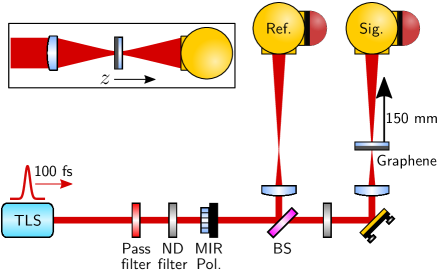
The experiment setup is illustrated in Figure 1. The beam was split into a reference path and signal path using a beamsplitter (BS, Thorlabs BSW511). Each beam was focussed using a \ceCaF2 plano convex lens ( mm). The spot radius at the focal point was between 65 µm and 78 µm depending on the wavelength. Each detection system included a gold integrating sphere (Newport 819D-GL-4) and PbSe detector (Thorlabs PDA20H-C), suitable for the 1.5 µm to 4.8 µm spectral region. The integrating spheres were implemented to minimize the effects of beam jitter. The beams that entered each integrating sphere were matched in size and closely matched in power to ensure comparable detector response.
At each sample position, , the pulse energies of the signal and reference were measured simultaneously, both with and without the sample in place. The sample position was controlled by a motorized translation stage and motorized flip mount. In this way, the time between measurements was minimized, reducing effects from fluctuations in the source power. See Supporting Information for more detail on the z-scan procedure.
The quality of the graphene sample was assessed using Raman spectroscopy and scanning Raman microscopy. The Raman spectra of the CVD single layer graphene used to produce the trilayer sample, as provided by the supplier (Graphenea, Spain), are presented in Figure 2a. These single layers were stacked on a \ceCaF2 substrate to form the trilayer graphene used in this experiment. The most prominent features in the Raman spectra of graphene are the G band near and G′ band near . The large intensity of the G′ band relative to the G band is explained by a triple resonance process that can occur due to graphene’s linear dispersion 32. The D band arises from the breathing modes of the hexagonal carbon rings and requires the presence of a defect for its activation 33.
Raman maps were measured at 1 mm and 1 µm spacings after the experiment was completed to analyze the degrees of uniformity and disorder in the graphene sample. The laser excitation wavelength used for the Raman maps was 532 nm (2.33 eV) with a spot size. The ratio of the integrated G band to G′ band over the area of the sample is shown by the color map in Figure 2b. The variations may be explained by local changes in stacking orientation which affect the degree of coupling between layers 34 and variations in distance between layers. Although the quality of the graphene is high, it is not crystalline. The orientation of each layer is random and an inhomogeneous residue of polymethyl methacrylate (PMMA), resulting from the wet-transfer of CVD graphene, may exist between the layers. The higher ratio of exhibits the Raman signature of a strong coupling between the layers while the lower ratio resembles single layer graphene. The ratio was averaged over all Raman map locations indicating that no significant disorder was introduced since the graphene was produced 33.
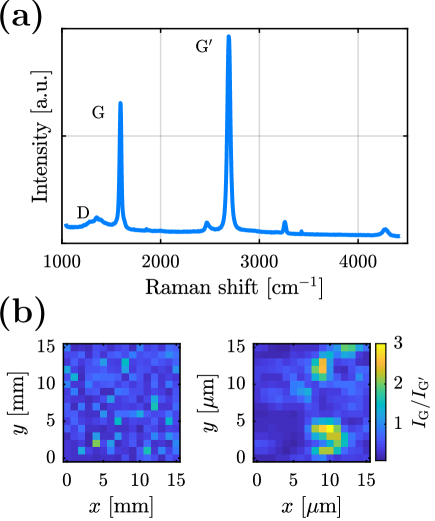
The Raman maps show that large variations in the degree of coupling between graphene layers occur on the scale of several . As the smallest spot radius used in the z-scan transmission measurements was , any effects from variations in layer coupling are averaged over the relatively large beam area and are thus not likely to change with beam location. See Supporting Information for Fourier transform infrared (FTIR) spectra of the graphene sample.
3 Results and Discussion
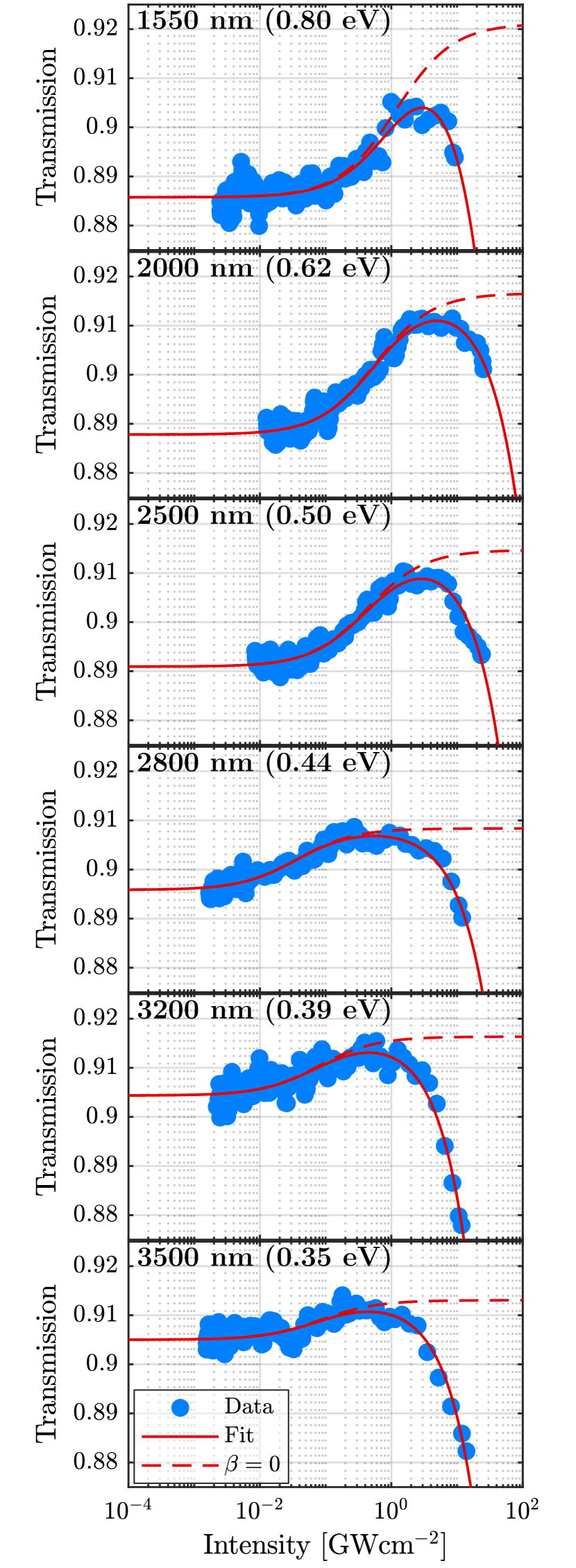
The transmission of femtosecond pulses at each wavelength were measured at each position and converted to a function of intensity . The transmission data were then fitted to Eqn. 2 and are presented in Figure 3. The data at all wavelengths show an increase in transmission with intensity that is consistent with SA. At intensities above , the dependence of 2PA dominates the effects of SA and the transmission rolls off. At wavelengths 2.8 µm (0.44 eV), 3.2 µm (0.39 eV), and 3.5 µm (0.35 eV), the transmission reduces to well below unsaturated values. Similar observations have been made in the near-IR regime with bilayer graphene 18 as well as SESAMs 20. The highest peak intensities incident on the graphene were limited to below damage thresholds determined by experiment at each wavelength. The femtosecond laser induced damage threshold tests were performed on a sacrificial sample of single layer graphene mounted on \ceCaF2. See Supporting Information for listed damage thresholds.
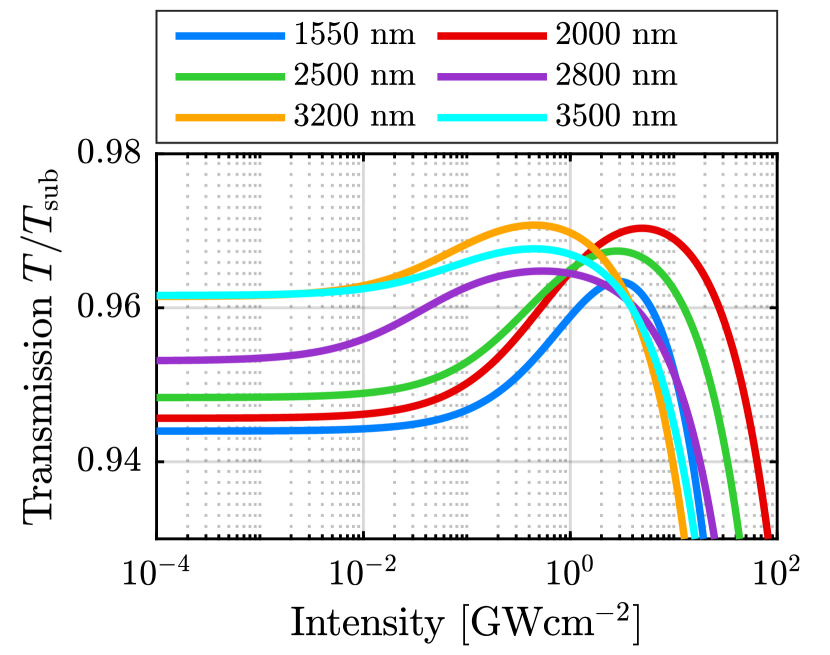
The transmission functions fitted to Eqn. 2 were normalized to the transmission of the \ceCaF2 substrate at each wavelength and are presented in Figure 4. There are several interesting features displayed here. Firstly, the low intensity transmission increases with wavelength, which is also observed in the FTIR spectra presented in the Supporting Information. This is in agreement with some reports in literature 7, 35, however it does contradict other reports of a completely flat absorption spectrum 36, 37 and may be due to an interaction between the graphene and the \ceCaF2 substrate. Secondly, the saturation intensity decreases with wavelength with the exception of 2.8 µm (0.44 eV) where is the lowest of all wavelengths. Thirdly, the modulation depth is highest at 2.0 µm (0.62 eV) and lowest at 3.5 µm (0.35 eV). The effective modulation depth and slope of the nonlinear transmission curve are reduced by 2PA.
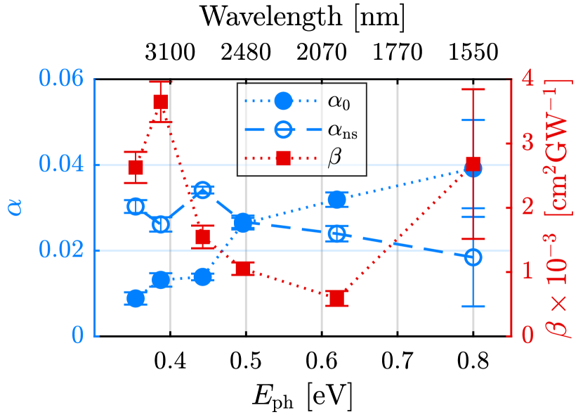
The fitted absorption parameters for SA and 2PA are shown graphically in Figure 5. Resonant features in 2PA are observed with a peak at around 3200 nm (0.39 eV) which may be explained by interlayer coupling. This peak location agrees with quantum perturbation theory used for the case of AB stacked bilayer graphene 18. There is insufficient data to resolve a possible second peak below 2000 nm (above 0.62 eV) that may exist due to three layer coupling. The sum of the SA parameters decreases with wavelength which corresponds to the transmission increase in the low intensity regime. See Supporting Information for listed values of the fitted absorption parameters , , and .
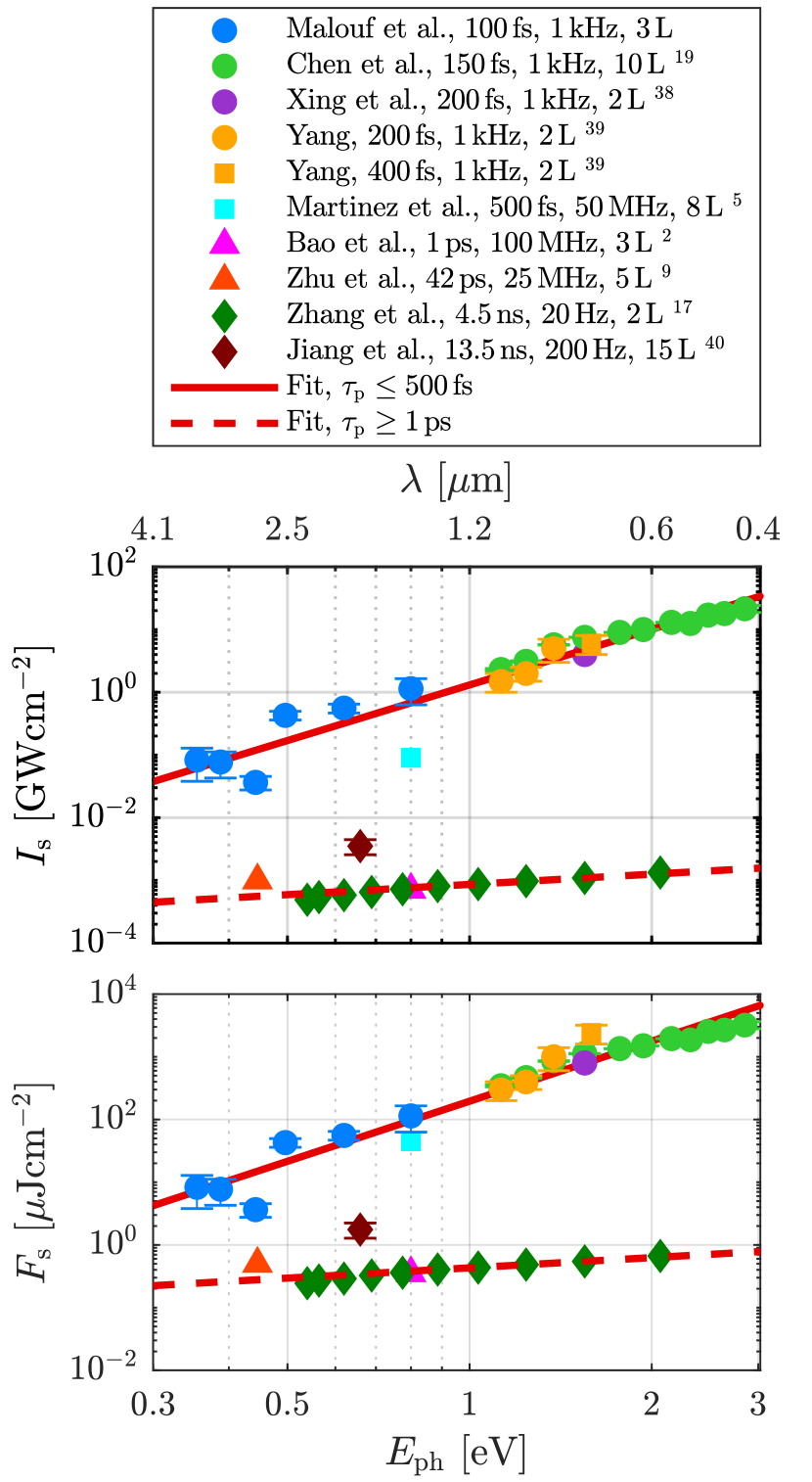
The measured saturation intensities, , and saturation fluences, , are presented in Figure 6 as functions of photon energy, (See Supporting Information for the listed values), and compared with measurements published in literature 19, 38, 18, 39, 5, 2, 9, 17. In each case, graphene sheets were produced by either mechanical exfoliation, epitaxy, or CVD and the direction of incident radiation was perpendicular to the graphene plane. Cases for graphene flakes suspended in a solution or polymer were excluded from the analysis because the angle of incidence is not uniform and the incident beam is more likely to interact with flake edges where disorder is high. One reported measurement of for graphene produced by spin coating is also included in Figure 6 for comparison, since the graphene was mounted on a flat glass substrate and the radiation was at normal incidence 40.
The relationship between and is affected by incident pulse duration, , relative to the carrier lifetime. The relaxation of photogenerated carriers in graphene is described by two distinct time scales, and , where may be attributed to carrier-carrier intraband scattering while may be explained by carrier-phonon intraband scattering or electron-hole recombination 41, 37, 2. The fractional amplitudes of the biexponential fits are for time constant and for 41, which result in a mean lifetime of . The saturation intensity data in Figure 6 are divided into two pulse duration regimes, and , where the boundary between the two regimes is comparable with the mean carrier lifetime. Each set of data were fitted to and , where and are the fit parameters. The saturation intensity of graphene produced by spin coating 40 is significantly higher than the case for CVD graphene 17 when measured with similar photon energy and pulse duration, which is likely due to high disorder as a result of the spin-coating process. Therefore, and for the spin-coated graphene were excluded from the fits.
In the short pulse regime, the parameters are and while the parameters are and . In the long pulse regime, , and the fit parameters are , , , and . That is, the empirical fits suggest that and when incident pulse durations are below the mean carrier lifetime. In the case of longer pulses, and .
4 Conclusion
We have characterized the response of trilayer graphene to high intensity radiation between 1.55 µm and 3.50 µm (from 0.35 eV to 0.80 eV). We have shown that multilayer graphene exhibits SA and 2PA in response to 100 fs pulses. Resonant features in 2PA were observed over the spectral region measured, however more data is required to resolve these features. The 2PA limits the effective modulation depth and can be detrimental to mode-locking ultrafast lasers. Saturation intensities of femtosecond pulses are shown empirically to be proportional to the third power of photon energy, while those of longer pulses are shown to have a square root dependence.
{acknowledgement}
The authors thank Tak W. Kee and Patrick Tapping for the femtosecond laser facilities and Jason Gascooke for performing Raman measurements. The authors are grateful to Elizaveta Klantsataya for useful discussions. The authors acknowledge the expertise, equipment, and support provided by the Australian National Fabrication Facility (ANFF) at Flinders University. This research was supported by the Australian Research Council through ARC LIEF Grant LE098974 and the South Australian Government Premier’s Research and Industry Fund (PRIF).
Z-scan procedure, transmission measurements, absorption and saturation intensity values, method of beam profile measurement, damage thresholds, and FTIR spectra.
References
- Bonaccorso et al. 2010 Bonaccorso, F.; Sun, Z.; Hasan, T.; Ferrari, A. Graphene photonics and optoelectronics. Nat. Photonics 2010, 4, 611
- Bao et al. 2009 Bao, Q.; Zhang, H.; Wang, Y.; Ni, Z.; Yan, Y.; Shen, Z. X.; Loh, K. P.; Tang, D. Y. Atomic-layer graphene as a saturable absorber for ultrafast pulsed lasers. Adv. Funct. Mater. 2009, 19, 3077–3083
- Tan et al. 2010 Tan, W. D.; Su, C. Y.; Knize, R. J.; Xie, G. Q.; Li, L. J.; Tang, D. Y. Mode locking of ceramic Nd:yttrium aluminum garnet with graphene as a saturable absorber. Appl. Phys. Lett. 2010, 96, 031106
- Sun et al. 2010 Sun, Z.; Hasan, T.; Torrisi, F.; Popa, D.; Privitera, G.; Wang, F.; Bonaccorso, F.; Basko, D. M.; Ferrari, A. C. Graphene mode-locked ultrafast laser. ACS Nano 2010, 4, 803–810
- Martinez et al. 2011 Martinez, A.; Fuse, K.; Yamashita, S. Mechanical exfoliation of graphene for the passive mode-locking of fiber lasers. Appl. Phys. Lett. 2011, 99, 121107
- Ugolotti et al. 2012 Ugolotti, E.; Schmidt, A.; Petrov, V.; Kim, J. W.; Yeom, D.-I.; Rotermund, F.; Bae, S.; Hong, B. H.; Agnesi, A.; Fiebig, C.; Erbert, G.; Mateos, X.; Aguiló, M.; Diaz, F.; Griebner, U. Graphene mode-locked femtosecond Yb:KLuW laser. Appl. Phys. Lett. 2012, 101, 161112
- Cizmeciyan et al. 2013 Cizmeciyan, M. N.; Kim, J. W.; Bae, S.; Hong, B. H.; Rotermund, F.; Sennaroglu, A. Graphene mode-locked femtosecond Cr:ZnSe laser at 2500 nm. Opt. Lett. 2013, 38, 341–343
- Yamashita et al. 2014 Yamashita, S.; Martinez, A.; Xu, B. Short pulse fiber lasers mode-locked by carbon nanotubes and graphene. Opt. Fiber Technol. 2014, 20, 702–713
- Zhu et al. 2016 Zhu, G.; Zhu, X.; Wang, F.; Xu, S.; Li, Y.; Guo, X.; Balakrishnan, K.; Norwood, R. A.; Peyghambarian, N. Graphene mode-locked fiber laser at 2.8 µm. IEEE Photonics Technol. Lett. 2016, 28, 7–10
- Vainio and Halonen 2016 Vainio, M.; Halonen, L. Mid-infrared optical parametric oscillators and frequency combs for molecular spectroscopy. Phys. Chem. Chem. Phys. 2016, 18, 4266–4294
- Serebryakov et al. 2010 Serebryakov, V.; Boĭko, E.; Petrishchev, N.; Yan, A. Medical applications of mid-IR lasers. Problems and prospects. J. Opt. Technol. 2010, 77, 6–17
- Novoselov et al. 2005 Novoselov, K. S.; Geim, A. K.; Morozov, S. V.; Jiang, D.; Katsnelson, M. I.; Grigorieva, I. V.; Dubonos, S. V.; Firsov, A. A. Two-dimensional gas of massless Dirac fermions in graphene. Nature 2005, 438, 197–200
- Wallace 1947 Wallace, P. R. The band theory of graphite. Phys. Rev. 1947, 71, 622–634
- Vasko 2010 Vasko, F. T. Saturation of interband absorption in graphene. Phys. Rev. B 2010, 82, 245422
- Marini et al. 2017 Marini, A.; Cox, J. D.; García de Abajo, F. J. Theory of graphene saturable absorption. Phys. Rev. B 2017, 95, 125408
- Sobon et al. 2015 Sobon, G.; Sotor, J.; Pasternak, I.; Krajewska, A.; Strupinski, W.; Abramski, K. M. Multilayer graphene-based saturable absorbers with scalable modulation depth for mode-locked Er- and Tm-doped fiber lasers. Opt. Mater. Express 2015, 5, 2884–2894
- Zhang et al. 2015 Zhang, F.; Han, S.; Liu, Y.; Wang, Z.; Xu, X. Dependence of the saturable absorption of graphene upon excitation photon energy. Applied Physics Letters 2015, 106, 091102
- Yang et al. 2011 Yang, H.; Feng, X.; Wang, Q.; Huang, H.; Chen, W.; Wee, A. T. S.; Ji, W. Giant two-photon absorption in bilayer graphene. Nano Lett. 2011, 11, 2622–2627, PMID: 21650165
- Chen et al. 2015 Chen, W.; Wang, Y.; Ji, W. Two-photon absorption in graphene enhanced by the excitonic fano resonance. J. Phys. Chem. C 2015, 119, 16954–16961
- Grange et al. 2005 Grange, R.; Haiml, M.; Paschotta, R.; Spühler, G.; Krainer, L.; Golling, M.; Ostinelli, O.; Keller, U. New regime of inverse saturable absorption for self-stabilizing passively mode-locked lasers. Appl. Phys. B: Lasers Opt. 2005, 80, 151–158
- Sheik-Bahae et al. 1990 Sheik-Bahae, M.; Said, A. A.; Wei, T. H.; Hagan, D. J.; Stryland, E. W. V. Sensitive measurement of optical nonlinearities using a single beam. IEEE J. Quantum Electron. 1990, 26, 760–769
- Chapple et al. 1997 Chapple, P. B.; Staromlynska, J.; Hermann, J. A.; Mckay, T. J.; Mcduff, R. G. Single-beam z-scan: measurement techniques and analysis. J. Nonlinear Opt. Phys. Mater. 1997, 06, 251–293
- Henderson-Sapir et al. 2017 Henderson-Sapir, O.; Malouf, A.; Bawden, N.; Munch, J.; Jackson, S. D.; Ottaway, D. J. Recent advances in 3.5 µm erbium-doped mid-infrared fiber lasers. IEEE J. Sel. Top. Quantum Electron. 2017, 23, 1–9
- Henderson-Sapir et al. 2016 Henderson-Sapir, O.; Jackson, S. D.; Ottaway, D. J. Versatile and widely tunable mid-infrared erbium doped ZBLAN fiber laser. Opt Lett 2016, 41, 1676–9
- Fortin et al. 2016 Fortin, V.; Maes, F.; Bernier, M.; Bah, S. T.; D’Auteuil, M.; Vallee, R. Watt-level erbium-doped all-fiber laser at 3.44 µm. Opt. Lett. 2016, 41, 559–62
- Jobin et al. 2018 Jobin, F.; Fortin, V.; Maes, F.; Bernier, M.; Vallée, R. Gain-switched fiber laser at 3.55 µm. Opt. Lett. 2018, 43, 1770–1773
- Yang et al. 2018 Yang, J.; Zhong, H.; Zhang, S.; Tang, Y.; Fan, D. Cascade-gain-switching for generating 3.5-µm nanosecond pulses from monolithic fiber lasers. IEEE Photonics J. 2018, 10, 1–12
- Qin et al. 2018 Qin, Z.; Hai, T.; Xie, G.; Ma, J.; Yuan, P.; Qian, L.; Li, L.; Zhao, L.; Shen, D. Black phosphorus Q-switched and mode-locked Er:ZBLAN fiber lasers at 3.5 µm. 2018; arXiv:1802.00710v1
- Malouf et al. 2016 Malouf, A.; Henderson-Sapir, O.; Gorjan, M.; Ottaway, D. J. Numerical modeling of 3.5 µm dual-wavelength pumped erbium-doped mid-infrared fiber lasers. IEEE J. Quantum Electron. 2016, 52, 1–12
- Boyd 2008 Boyd, R. Nonlinear Optics; Academic Press: Amsterdam Boston, 2008
- Keller et al. 1996 Keller, U.; Weingarten, K. J.; Kartner, F. X.; Kopf, D.; Braun, B.; Jung, I. D.; Fluck, R.; Honninger, C.; Matuschek, N.; der Au, J. A. Semiconductor saturable absorber mirrors (SESAM’s) for femtosecond to nanosecond pulse generation in solid-state lasers. IEEE J. Sel. Top. Quantum Electron. 1996, 2, 435–453
- Malard et al. 2009 Malard, L.; Pimenta, M.; Dresselhaus, G.; Dresselhaus, M. Raman spectroscopy in graphene. Phys. Rep. 2009, 473, 51 – 87
- Pollard et al. 2014 Pollard, A. J.; Brennan, B.; Stec, H.; Tyler, B. J.; Seah, M. P.; Gilmore, I. S.; Roy, D. Quantitative characterization of defect size in graphene using Raman spectroscopy. Appl. Phys. Lett. 2014, 105, 253107
- Kim et al. 2012 Kim, K.; Coh, S.; Tan, L. Z.; Regan, W.; Yuk, J. M.; Chatterjee, E.; Crommie, M. F.; Cohen, M. L.; Louie, S. G.; Zettl, A. Raman spectroscopy study of rotated double-layer graphene: misorientation-angle dependence of electronic structure. Phys. Rev. Lett. 2012, 108, 246103
- Hu et al. 2017 Hu, H.; Liao, B.; Guo, X.; Hu, D.-B.; Qiao, X.; Liu, N.; Liu, R.; Chen, K.; Bai, B.; Yang, X.; Dai, Q. Large-scale suspended graphene used as a transparent substrate for infrared spectroscopy. Small 2017, 13
- Nair et al. 2008 Nair, R. R.; Blake, P.; Grigorenko, A. N.; Novoselov, K. S.; Booth, T. J.; Stauber, T.; Peres, N. M. R.; Geim, A. K. Fine structure constant defines visual transparency of graphene. Science 2008, 320, 1308–1308
- Dawlaty et al. 2008 Dawlaty, J. M.; Shivaraman, S.; Chandrashekhar, M.; Rana, F.; Spencer, M. G. Measurement of ultrafast carrier dynamics in epitaxial graphene. Appl. Phys. Lett. 2008, 92, 042116
- Xing et al. 2010 Xing, G.; Guo, H.; Zhang, X.; Sum, T. C.; Huan, C. H. A. The physics of ultrafast saturable absorption in graphene. Opt. Express 2010, 18, 4564–4573
- Yang 2012 Yang, H. Saturable Absorption and Two-Photon Absorption in Graphene. Ph.D. thesis, National University of Singapore, 2012
- Jiang et al. 2018 Jiang, X.; Gross, S.; Withford, M. J.; Zhang, H.; Yeom, D.-I.; Rotermund, F.; Fuerbach, A. Low-dimensional nanomaterial saturable absorbers for ultrashort-pulsed waveguide lasers. Opt. Mater. Express 2018, 8, 3055–3071
- Shang et al. 2012 Shang, J.; Yan, S.; Cong, C.; Tan, H.-S.; Yu, T.; Gurzadyan, G. G. Probing near Dirac point electron-phonon interaction in graphene. Opt. Mater. Express 2012, 2, 1713–1722
![[Uncaptioned image]](/html/1810.12981/assets/x7.png)