Topological Hall effect in thin films of Mn1.5PtSn
Abstract
Spin chirality in metallic materials with non-coplanar magnetic order can give rise to a Berry phase induced topological Hall effect. Here, we report the observation of a large topological Hall effect in high-quality films of Mn1.5PtSn that were grown by means of magnetron sputtering on MgO(001). The topological Hall resistivity is present up to T below the spin reorientation transition temperature, K. We find, that the maximum topological Hall resistivity is of comparable magnitude as the anomalous Hall resistivity. Owing to the size, the topological Hall effect is directly evident prior to the customarily performed subtraction of magnetometry data. Further, we underline the robustness of the topological Hall effect in Mn2-xPtSn by extracting the effect for multiple stoichiometries (x = 0.5, 0.25, 0.1) and film thicknesses (t = 104, 52, 35 nm) with maximum topological Hall resistivities between cm and cm at 150 K.
I Introduction
Topological magnetic structures have become of great interest recently, attributed to the emergent transport phenomena associated with the magnetic texture Fert et al. (2013). One of these phenomena is the transverse Hall current, that arises from the interplay of magnetic order and intrinsic band structure or scattering Nagaosa et al. (2010). Experimentally, the measured Hall resistivity can be separated into the ordinary Hall effect (OHE) Hall (1879) dependent on the external field () and the anomalous Hall effect (AHE) which scales with the saturation magnetization. The modern understanding of the AHE ascribes the effect to scattering mechanisms Karplus and Luttinger (1954); Smit (1958); Berger (1970) and the intrinsic momentum space Berry curvature Fang et al. (2003). However, recently an additional Hall-type contribution was proposed that scales neither with the magnetization () nor with the externally applied field, termed the topological Hall effect (THE) Ye et al. (1999); Bruno et al. (2004). This THE has been proposed to originate from a finite scalar spin chirality Taguchi et al. (2001), skyrmions Ye et al. (1999), and Weyl points Kübler and Felser (2014). The prior two are connected through the magnetic texture and the latter is connected to the momentum space dispersion. Here, we focus on the magnetic texture induced THE which has become of great interest in Heusler compounds due to their tunability Manna et al. (2018); Wollmann et al. (2017).
There are two limiting cases for the stabilization of magnetic textures: the scalar spin chirality and the skyrmionic lattice Rößler et al. (2006), which originate from a competition of exchange, e.g. Heisenberg and Dzyaloshinki-Moriya interaction Dzyaloshinsky (1958); Moriya (1960), with anisotropy and external fields. In the limit of discrete spins, there is a finite scalar spin chirality caused by three non-coplanar spins that subtend a finite cone angle and give rise to the momentum space dependent THE Taguchi et al. (2001); Sürgers et al. (2014). In the adiabatic limit, the spin chirality is taken to be continuous as the integer winding of the real space Berry curvature Ye et al. (1999); Bruno et al. (2004); Neubauer et al. (2009). As electrons couple to such spin textures, they acquire a finite Berry phase acting as a magnetic field. This in turn results in an additional contribution to the Hall effect Berry (1984).
The THE has been observed in a variety of materials including the B20 compounds Neubauer et al. (2009); Kanazawa et al. (2011); Huang and Chien (2012), perovskites Nakamura et al. (2018); Vistoli et al. (2018) and Heusler compounds Rana et al. (2016); Liu et al. (2018); Li et al. (2018). The Heusler compounds are of particular interest, owing to the recent discovery of antiskyrmions in Mn1.4Pt0.9Pd0.1Sn, a new type of topological texture due to the symmetry Nayak et al. (2017). The ferrimagnetic Mn2YZ (Y being a transition metal and Z a main-group element) inverse Heusler compounds that crystallize in a non-centrosymmetric structure with symmetry are promising candidates to realize such spin textures through competing interactions of the magnetic sublattices and magnetocrystalline anisotropy caused by tetragonal distortion Meshcheriakova et al. (2014). In thin films the presence of geometric constraints can additionally stabilize the desired spin textures in a wider field and temperature range Butenko et al. (2010). Recently, the THE was observed in single crystal thin films of Mn2RhSn Rana et al. (2016) as well as in bulk Mn2PtSn Liu et al. (2018) below a spin reorientation transition temperature () Meshcheriakova et al. (2014), and in polycrystalline Mn2PtSn Li et al. (2018) films for all temperatures below the Curie temperature. Conversely, the work of Jin et al. shows no topological Hall signal or in epitaxially grown films of Mn2PtSn Jin et al. (2018).
In this paper, we focus on Mn1.5PtSn thin films, with the closest stoichiometry relation to the antiskyrmion compound Mn1.4Pt0.9Pd0.1Sn. We demonstrate the presence of a THE below a spin reorientation transition temperature and up to high fields, evident prior to the customarily performed subtraction of magnetometry data. Further, we point out the robustness of the THE in Mn2-xPtSn by comparing different compositions and film thicknesses, as well as previously reported results on Mn2PtSn films.
II Experimental Details
High-quality Mn2-xPtSn films were grown on single crystal MgO (001) substrates in a BESTEC UHV magnetron sputtering system. Mn, Pt and Sn were deposited from 2” targets using DC magnetron co-sputtering. The stoichiometry was controlled by adjusting the power of the magnetrons. The deposition was performed in confocal geometry with a target to substrate distance of 200 mm. Prior to deposition, the chamber was evacuated to a base pressure below mbar, while during deposition a process gas pressure of mbar (Ar, 15 sccm) was maintained. The films were deposited at and post-annealed for 30 min at the same temperature in order to improve the chemical ordering. The annealed films were capped at room temperature with 3 nm Al, in order to prevent oxidization.
The film compositions were confirmed using energy-dispersive x-ray (EDX) microscopy. The film surface topography was analyzed by atomic force microscopy (AFM) on an Asylum Research MFP-3D Origin by Oxford Instruments. Structural characterization was carried out using x-ray diffractometry (XRD) with Cu-K 1 radiation ( Å) on a PANalytical X’Pert PRO system. The film thickness () was determined by x-ray reflectivity (XRR) measurements.
Magnetization measurements were performed on a vibrating sample magnetometer (MPMS 3, Quantum Design). In order to infer the magnetization of the films, we subtracted the diamagnetic substrate contribution as well as a low-temperature paramagnetic contribution from the raw data. Here, the paramagnetic contribution can be attributed to impurities in the MgO substrate. The diamagnetic susceptibility () of MgO was determined from reference measurements. The paramagnetic contribution was fitted and subtracted from the raw data using the Brillouin function.
Four-probe and five-probe measurements were performed to obtain the resistivity along the longitudinal direction and the Hall resistivity, respectively. Therefore, an in-plane current, , was applied along a film stripe with a width of (-direction). Voltages were recorded simultaneously along the current direction (), with a lead distance of (-direction), as well as perpendicular to the current direction (), with a lead distance of . The magnetic field was applied along the out-of-plane () direction (MgO [001]). In order to obtain a clean resistivity, , the raw resistivity, , was symmetrized by averaging at positive and negative fields with respect to the field sweep directions. To obtain a clean Hall resistivity, , the raw Hall resistivity, , was antisymmetrized by averaging the difference of at positive and negative fields with respect to the field sweep directions.
III Results and Discussion
III.1 Structural Characterization
In the following, we discuss the properties of a Mn1.5PtSn thin film in detail, since it has the closest stoichiometry relation to the antiskyrmion compound Mn1.4Pt0.9Pd0.1Sn. In Fig. 1(a) we show the x-ray reflectivity, together with AFM analysis, confirming the smoothness of the film with a r.m.s roughness of 0.3 nm in the obtained 5 m 5 m scan. The Kiessig fringes, reaching beyond the measurement range, are further evidence of a high-quality surface as well as a high-quality substrate to film interface. A thickness of 104.7 nm and a roughness of less than 0.5 nm is inferred from XRR fitting.
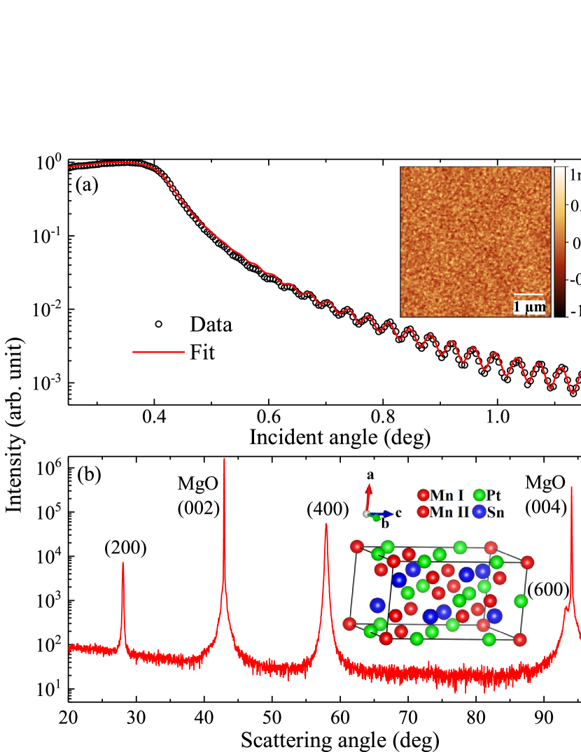
Furthermore, we use x-ray diffraction radial scans () as shown in Fig. 1(b) and Fig. S1 (see the Supplemental Material Sup ) to determine the crystal structure of our film. The symmetric radial scans in Fig. 1(b) confirm epitaxial growth since only the (00) series of Bragg peaks, attributed to the Mn1.5PtSn film, can be observed. The full-width at half-maximum of the (400) out-of-plane rocking curve of 1.147∘ verifies high crystallinity. Additionally, more than 10 asymmetric Bragg peaks (Fig. S1) can be indexed using a unit cell similar to bulk Mn1.4PtSn Nayak et al. (2017). Analogous to the bulk structure, we describe our unit cell by the space group (#122), which is derived from the inverse tetragonal Heusler structure. This is supported by the observation of a systematic absence of Bragg peaks corresponding to this crystal symmetry (Fig. S1). By modeling the peak intensities we find that Mn atoms occupy the and () positions, while the Pt and Sn atoms occupy the () and () positions, respectively. A detailed analysis of the peak positions shows that the film geometry stabilizes the axis in the film plane, slightly breaking the equivalence of the and parameters, reflected in the lattice parameters Å Å, Å Å and Å Å.
From the {112} pole figure and the comparison of the corresponding azimuthal scan as well as the splitting of high-angle peaks (see Fig. S1 and S2 in the Supplemental Material Sup ) we conclude that two orientations of the axis, along [] and [110] of the MgO substrate are present. For the two lattice directions within the film plane, this corresponds to a lattice mismatch of 2.5% and 6.5%, respectively.
III.2 Magnetometry and Magnetotransport Properties
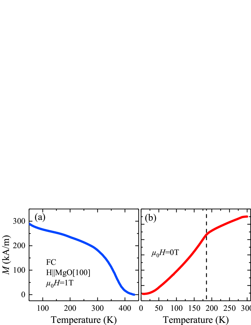
Figure 2(a) depicts the temperature dependence of the magnetization at 1 T with a single transition at 400 K representing the Curie temperature for the 104 nm thick Mn1.5PtSn film. A spin reorientation is not clearly evident for this field. Figure 2(b) shows the temperature dependence of the longitudinal resistivity. In analogy to the case of Mn2RhSn Meshcheriakova et al. (2014), a change in the slope at K marks a transition from a collinear () into a non-collinear () magnetic structure following spin reorientation of one Mn sub-lattice. A similar feature was also observed in related compounds Liu et al. (2018); Nayak et al. (2017); Li et al. (2018).
The out-of-plane magnetization for the 104 nm thick Mn1.5PtSn film is shown for 10 K, 150 K, and 300 K at magnetic fields up to 7 T in Fig. 3(a). The loops are reminiscent of hard-axis behavior with a small coercive field. We attribute this to the tetragonal axis lying in the film plane. Here, the saturation magnetization is 415 kA/m, 550 kA/m and 590 kA/m at 300 K, 150 K, and 10 K, respectively, which is comparable to determined for the bulk material Nayak et al. (2017). The saturation field is estimated to be about 1.2 T at 300 K, increasing to about 3.5 T at 10 K.
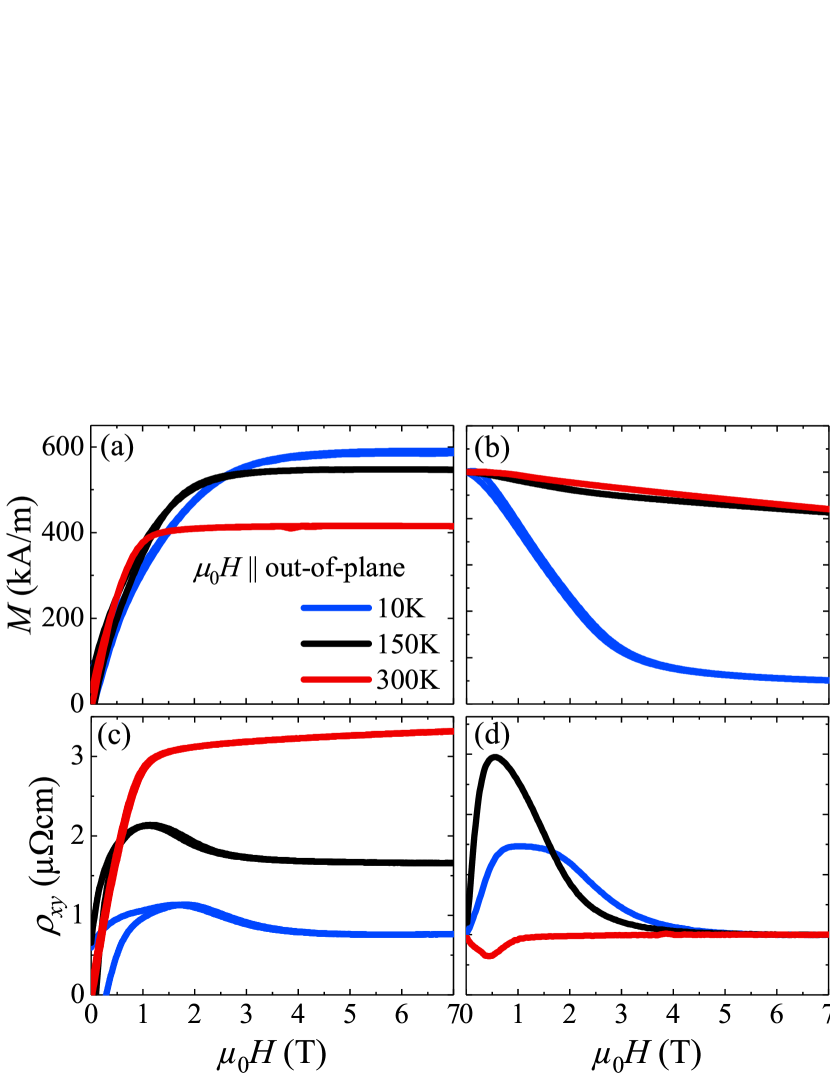
The magneto-resistance (MR) in Fig. 3(b), recorded with the applied along the out-of-plane direction, is depicted as the ratio . The MR is negative for all temperatures and is composed of two parts: First, a steep part leveling off around 4 T and visible at 10 K. This likely originates from the alignment of the spins in the non-coplanar phase and scales with the magnetometry data (Fig. 3(a)). Second, a linear field dependent part which does not saturate at 7 T. Furthermore, the absolute value of the MR ratio at 7 T clearly decreases with increasing temperature.
The Hall resistivity at 300 K in Fig. 3(c) resembles (Fig. 3(a)) with a steep increase at low fields and and a linear behavior at high fields. Those two regimes can be attributed to the AHE and the OHE, respectively. Below , at 150 K and 10 K, an additional non-linear part appears up to approximately 4 T. Here, does not trace , which is reminiscent of the THE. The three different contributions can be summarized as:
| (1) |
where corresponds to the OHE scaling linearly with applied field (H), is the AHE scaling with the magnetization component perpendicular to the film and represents the THE.
The AHE can arise from intrinsic and/or extrinsic mechanisms scaling with different powers of the resistivity Nagaosa et al. (2010). Therefor, we write , with corresponding to intrinsic and side-jump scattering and corresponding to skew scattering. In an independent analysis, we determined from the scaling relation that the underlying mechanism is of primarily of intrinsic origin with (see Fig. S3 in the Supplemental Material Sup ). The zero-field conductivity , supports the notion that the intrinsic and side jump mechanisms dominate Miyasato et al. (2007); Nagaosa et al. (2010). Therefore, we focus on the skew scattering independent contributions in our evaluation in the following, taking .
In order to quantify the different contributions to the field dependant Hall resistivity, we follow the customarily performed separation process Kanazawa et al. (2011). Therefore, we take into account that only the AHE and OHE contribute to the Hall resistivity once the magnetization is saturated at high fields. Hence, and can be obtained through a linear fit to our transport data taken at high magnetic fields, using the resistivity and the (separately measured) magnetization as . Finally, we can calculate the topological Hall resistivity as
| (2) |
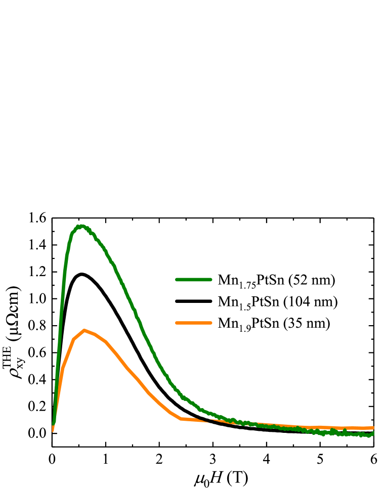
As evident from Fig. 3(d) the THE in the 104 nm thick Mn1.5PtSn film can be observed up to fields of T with a maximum topological Hall resistivity cm at 150 K. From an anolagous analysis in films of Mn1.75PtSn (52 nm) and Mn1.9PtSn (35 nm) we obtained cm and cm, respectively at 150 K (Fig. 4). Our data shows that a large THE is present in a wide range of stoichiometries, underlining the robustness of the effect. This is in agreement with the presence of a (weaker) THE, previously reported in bulk Mn2PtSn Liu et al. (2018) and polycrystalline Mn2PtSn films () Li et al. (2018). Notably, in single crystalline Mn2PtSn films () Jin et al. (2018) with the axis in the plane, no and no THE were observed. We therefore propose that the contradicting observations (presence or absence of the THE in seemingly similar thin films) might be attributed to the different crystal structures and crystal orientations relative to the applied field.
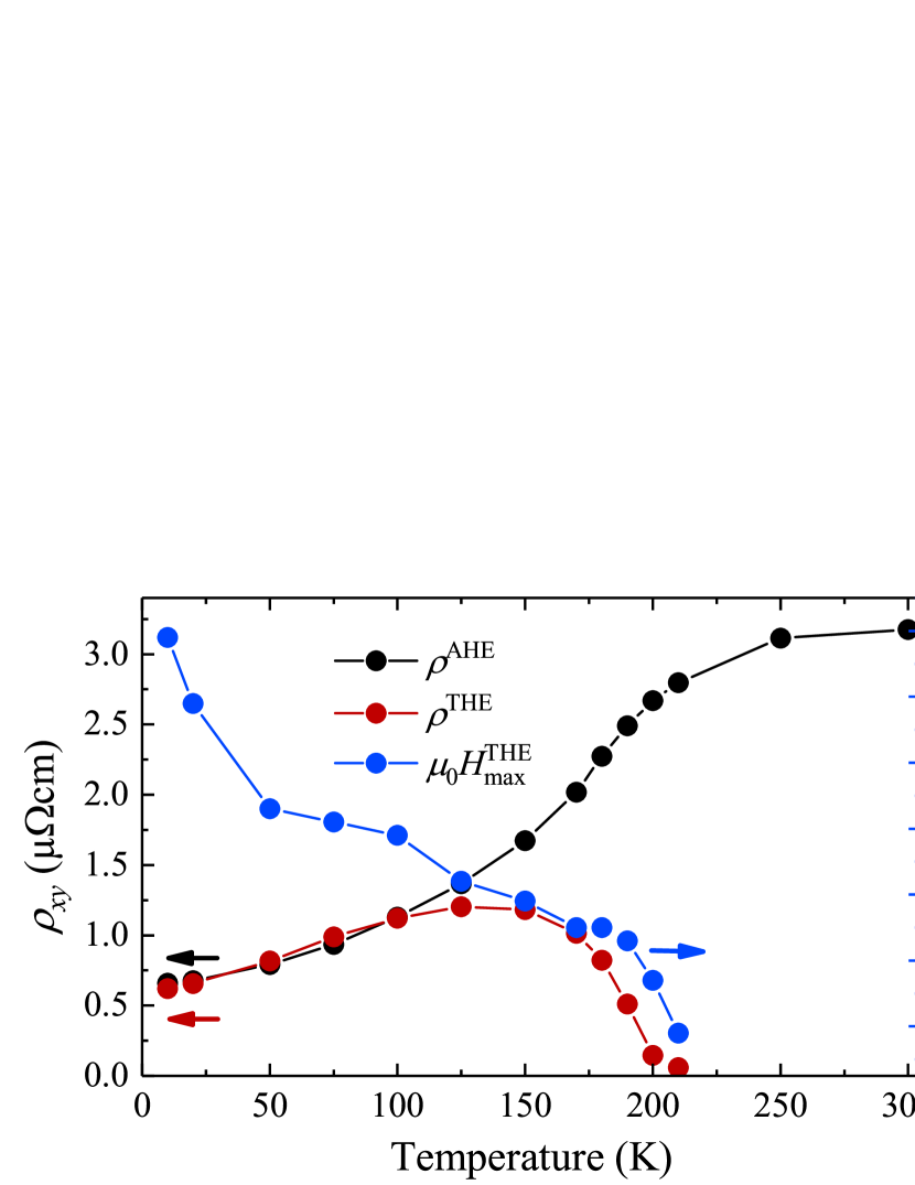
Figure 5 summarizes the evolution of , and the field at which THE reaches its maximum, with temperature. decreases continuously with temperature, having the largest slope around K. The THE appears below , and thus must be connected with a non-coplanar spin texture at finite fields, with peaking at 150 K. Interestingly, and have the same magnitude between 100 K and 10 K, suggesting that a similar microscopic mechanisms is responsible for both effects. The field at which the maximal topological Hall resistivity is observed increases continuously with decreasing temperature following the same trend as the saturation field in the magnetization (Fig. 3(a)).
Since magnetization experiments in films are challenging, the employed extraction procedure is highly susceptible to small misalignments in sample mounting or temperature differences between the transport and magnetometry measurements. This can result in significant errors of the THE values or even mimic non-existent effects. It is therefore unclear whether the THE signature at low fields and above (Fig. 3(d)) is genuine or attributable to the THE extraction process Ishizuka and Nagaosa (2018). However, our findings would agree with the presence of antiskyrmions above in Mn1.4Pt0.9Pd0.1Sn Nayak et al. (2017).
In contrast to the majority of reports on the THE in conjunction with the AHE, we find that in Mn1.5PtSn thin films the size of the THE is of the same magnitude as the corresponding AHE. Typically, the AHE by far surpasses the THE Neubauer et al. (2009); Huang and Chien (2012); Rana et al. (2016); Spencer et al. (2018). Nevertheless, similar behavior as in Mn1.5PtSn was also presented in the non-collinear metallic Mn5Si3 and the correlated oxide charge-transfer insulator (Ca,Ce)MnO3 Sürgers et al. (2014); Vistoli et al. (2018). Interestingly, one can also find a few examples where the THE appears in conjunction with a vanishing AHE, such as the Weyl semimetal GdPtBi and the helimagnetic metal MnGe Suzuki et al. (2016); Kanazawa et al. (2011). Thus, the dependence of the underlying mechanism (i.e. skyrmions/bubbles, Weyl points or non-coplanar magnetic structure) in the respective material system (e.g. thin film, bulk or multilayer) determines the relation of the THE to the AHE, which can range over several orders of magnitude. The physics regarding the relation of the THE to the AHE have not been completely explored or understood, where in our films we clearly observe a difference in the relation that depends on the spin reorientation transition temperature.
IV Conclusion
In this work, we report a non-trivial behavior of the Hall response in Mn1.5PtSn thin films (space group ) identified as the THE. The signature is clearly evident even prior to the customarily performed subtraction of magnetometry data. The THE is present up to a spin reorientation transition temperature, K, and a field of T. The same magnitude of and below 100 K implies a similar microscopic mechanism for the AHE and THE. While we focused on a 104 nm thick Mn1.5PtSn film, similar experiments in different Mn2-xPtSn films show that the THE is robust over various stoichiometries and thicknesses, reaching up to cm at 150 K. All together, Mn2-xPtSn is an interesting compound for the understanding and application of transport phenomena in topological magnetic structures.
acknowledgement
The authors acknowledge funding by the Deutsche Forschungsgemeinschaft (DFG, German Research Foundation) under SPP 2137 (Project number 403502666), ERC Advanced Grant 742068 “TOPMAT” and EU FET Open RIA Grant No. 766566 grant (ASPIN). P. S. acknowledges financial support by the International Max Planck Research School for Chemistry and Physics of Quantum Materials (IMPRS-CPQM).
References
- Fert et al. (2013) A. Fert, V. Cros, and J. Sampaio, Nature Nanotechnology 8, 152 (2013).
- Nagaosa et al. (2010) N. Nagaosa, J. Sinova, S. Onoda, A. H. MacDonald, and N. P. Ong, Rev. Mod. Phys. 82, 1539 (2010).
- Hall (1879) E. H. Hall, American Journal of Mathematics 2, 287 (1879).
- Karplus and Luttinger (1954) R. Karplus and J. M. Luttinger, Physical Review 95, 1154 (1954).
- Smit (1958) J. Smit, Physica 24, 39 (1958).
- Berger (1970) L. Berger, Physical Review B 2, 4559 (1970).
- Fang et al. (2003) Z. Fang, N. Nagaosa, K. S. Takahashi, A. Asamitsu, R. Mathieu, T. Ogasawara, H. Yamada, M. Kawasaki, Y. Tokura, and K. Terakura, Science 302, 92 (2003).
- Ye et al. (1999) J. Ye, Y. B. Kim, A. J. Millis, B. I. Shraiman, P. Majumdar, and Z. Tečanović, Physical Review Letters 83, 3737 (1999).
- Bruno et al. (2004) P. Bruno, V. K. Dugaev, and M. Taillefumier, Physical Review Letters 93, 096806 (2004).
- Taguchi et al. (2001) Y. Taguchi, Y. Oohara, H. Yoshizawa, N. Nagaosa, and Y. Tokura, Science 291, 2573 (2001).
- Kübler and Felser (2014) J. Kübler and C. Felser, EPL (Europhysics Letters) 108, 67001 (2014).
- Manna et al. (2018) K. Manna, Y. Sun, L. Muechler, J. Kübler, and C. Felser, Nature Reviews Materials 3, 244 (2018).
- Wollmann et al. (2017) L. Wollmann, A. K. Nayak, S. S. P. Parkin, and C. Felser, Annual Review of Materials Research 47, 247 (2017).
- Rößler et al. (2006) U. K. Rößler, A. N. Bogdanov, and C. Pfleiderer, Nature 442, 797 (2006).
- Dzyaloshinsky (1958) I. Dzyaloshinsky, Journal of Physics and Chemistry of Solids 4, 241 (1958).
- Moriya (1960) T. Moriya, Physical Review 120, 91 (1960).
- Sürgers et al. (2014) C. Sürgers, G. Fischer, P. Winkel, and H. Löhneysen, Nature Communications 5, 3400 (2014).
- Neubauer et al. (2009) A. Neubauer, C. Pfleiderer, B. Binz, A. Rosch, R. Ritz, P. G. Niklowitz, and P. Böni, Physical Review Letters 102, 186602 (2009).
- Berry (1984) M. V. Berry, Proceedings of the Royal Society of London. A. Mathematical and Physical Sciences 392, 45 (1984).
- Kanazawa et al. (2011) N. Kanazawa, Y. Onose, T. Arima, D. Okuyama, K. Ohoyama, S. Wakimoto, K. Kakurai, S. Ishiwata, and Y. Tokura, Physical Review Letters 106, 156603 (2011).
- Huang and Chien (2012) S. X. Huang and C. L. Chien, Physical Review Letters 108, 267201 (2012).
- Nakamura et al. (2018) M. Nakamura, D. Morikawa, X. Yu, F. Kagawa, T. Arima, Y. Tokura, and M. Kawasaki, Journal of the Physical Society of Japan 87, 074704 (2018).
- Vistoli et al. (2018) L. Vistoli, W. Wang, A. Sander, Q. Zhu, B. Casals, R. Cichelero, A. Barthélémy, S. Fusil, G. Herranz, S. Valencia, R. Abrudan, E. Weschke, K. Nakazawa, H. Kohno, J. Santamaria, W. Wu, V. Garcia, and M. Bibes, Nature Physics (2018), 10.1038/s41567-018-0307-5.
- Rana et al. (2016) K. G. Rana, O. Meshcheriakova, J. Kübler, B. Ernst, J. Karel, R. Hillebrand, E. Pippel, P. Werner, A. K. Nayak, C. Felser, and S. S. P. Parkin, New Journal of Physics 18, 085007 (2016).
- Liu et al. (2018) Z. H. Liu, A. Burigu, Y. J. Zhang, H. M. Jafri, X. Q. Ma, E. Liu, W. H. Wang, and G. H. Wu, Scripta Materialia 143, 122 (2018).
- Li et al. (2018) Y. Li, B. Ding, X. Wang, H. Zhang, W. Wang, and Z. Liu, Applied Physics Letters 113, 062406 (2018).
- Nayak et al. (2017) A. K. Nayak, V. Kumar, T. Ma, P. Werner, E. Pippel, R. Sahoo, F. Damay, U. K. Rößler, C. Felser, and S. S. P. Parkin, Nature 548, 561 (2017).
- Meshcheriakova et al. (2014) O. Meshcheriakova, S. Chadov, A. K. Nayak, U. K. Rößler, J. Kübler, G. André, A. A. Tsirlin, J. Kiss, S. Hausdorf, A. Kalache, W. Schnelle, M. Nicklas, and C. Felser, Physical Review Letters 113, 087203 (2014).
- Butenko et al. (2010) A. B. Butenko, A. A. Leonov, U. K. Rößler, and A. N. Bogdanov, Physical Review B 82, 052403 (2010).
- Jin et al. (2018) Y. Jin, S. Valloppilly, P. Kharel, J. Waybright, P. Lukashev, X. Z. Li, and D. J. Sellmyer, Journal of Applied Physics 124, 103903 (2018).
- (31) See Supplemental Material at for additional x-ray diffraction radial scans, pole figure, -scan, power law scaling and topological Hall data of Mn1.75PtSn.
- Miyasato et al. (2007) T. Miyasato, N. Abe, T. Fujii, A. Asamitsu, S. Onoda, Y. Onose, N. Nagaosa, and Y. Tokura, Physical Review Letters 99, 086602 (2007).
- Ishizuka and Nagaosa (2018) H. Ishizuka and N. Nagaosa, Science Advances 4 (2018).
- Spencer et al. (2018) C. S. Spencer, J. Gayles, N. A. Porter, S. Sugimoto, Z. Aslam, C. J. Kinane, T. R. Charlton, F. Freimuth, S. Chadov, S. Langridge, J. Sinova, C. Felser, S. Blügel, Y. Mokrousov, and C. H. Marrows, Physical Review B 97, 214406 (2018).
- Suzuki et al. (2016) T. Suzuki, R. Chisnell, A. Devarakonda, Y. T. Liu, W. Feng, D. Xiao, J. W. Lynn, and J. G. Checkelsky, Nature Physics 12, 1119 (2016).