Tunable amplification and cooling of a diamond resonator with a microscope
Abstract
Controlling the dynamics of mechanical resonators is central to quantum science and metrology applications. Optomechanical control of diamond resonators is attractive owing to diamond’s excellent physical properties and its ability to host electronic spins that can be coherently coupled to mechanical motion. Using a confocal microscope, we demonstrate tunable amplification and damping of a diamond nanomechanical resonator’s motion. Observation of both normal mode cooling from room temperature to 80K, and amplification into self–oscillations with of optical power is observed via waveguide optomechanical readout. This system is promising for quantum spin-optomechanics, as it is predicted to enable optical control of stress-spin coupling with rates of 1 MHz (100 THz) to ground (excited) states of diamond nitrogen vacancy centers.
I Introduction
The interaction between light and mechanical systems underlies breakthroughs in physics ranging from optical tweezers [1] to gravitational wave detection [2]. Nanoscale systems harnessing this interaction have led to advances in quantum nanomechanics [3, 4, 5, 6, 7, 8, 9], sensing [10, 11, 12, 13], and nonlinear optics [14, 15, 16, 17, 18]. An essential ingredient to many of these demonstrations is dynamic optomechanical back action, which allows energy exchange between optical and mechanical domains [19, 20]. Controlling diamond nanomechanical systems via optomechanical back action is of growing interest, fueled by diamond’s exceptional properties [21], and by demonstrations of diamond spin manipulation using piezoelectronically driven mechanical resonators [22, 23, 24, 25, 26, 24, 27, 28, 29, 30, 31]. Controlling resonator motion optomechanically provides a path towards photon-phonon-spin coupling and technologies ranging from spin-spin entanglement [32, 33] to quantum transduction [34]. Back action can also enhance the performance of diamond resonators used for sensing [35, 36, 37]. In this article, we show that optomechanical back action acting on modes of a diamond resonator vibrating in or out of plane can be selectively be created and controlled by adjusting the focal position of a microscope that commonly serves as an optical interface with diamond colour centres. Using this technique, we cool a diamond nanomechanical resonator, as well as amplify its motion sufficiently for mechanical control of diamond spins via their predicted coupling to phonons.
Optomechanical damping and amplification, for example by optical gradient [38], radiation pressure [39, 40, 4], or photothermal [41, 42, 43, 36, 44, 45] forces typically relies on feedback from a cavity [19, 20], waveguide coupler [45], or external optoelectronics [46]. Inspired in part by optical tweezers, here we introduce a system that operates in an optical intensity gradient dominated regime of optomechanics and does not does not require a cavity or coupling to optical resonances [44]. The dynamic optomechanical back action is photothermal in nature, and is tuned through translation of a microscope focus, allowing both the strength and the sign of the optomechanical damping to be adjusted. This system, which adds confocal microscopy to our previously demonstrated waveguide optomechanical experiment [45], provides a combination of tunable optomechanical actuation and sensitive optomechanical readout. It allows normal–mode cooling of a diamond mechanical resonator from room temperature to , and excitation of nanomechanical self–oscillations whose stress field is predicted to allow control of nitrogen vacancy (NV) center spins [22]. These self–oscillations are observed for continuous wave excitation from a 532 nm laser with power as low as . Unlike previous demonstrations of photothermal backaction [41, 42, 43, 36, 44], neither an external cavity nor a wavelength tunable laser is required to adjust the backaction between damping and anti–damping regimes. Furthermore, we leverage its sensitivity to the microscope field gradient to selectively excite vertical as well as horizontal modes of the nanobeam, the latter of which can be difficult to probe using conventional optical interferometry measurements of nanomechanical devices [47] as their motion primarily induces intensity rather than phase changes on reflected light.
II Device and experimental setup
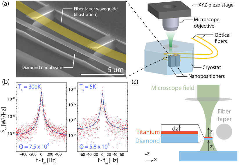
The optomechanical system studied here, illustrated in Fig. 1(a), consists of a diamond nanobeam (dimensions ) illuminated by a green (532 nm) laser input to an objective (Sumitomo long working distance, 0.55 NA) mounted on a three–axis stage. The nanobeam is fabricated from single crystal diamond (Element Six, optical grade, 3 3 mm2 area, polished by Delaware Diamond Knives) using undercut etching [45], and its top surface is coated with titanium ( nm thickness, deposited using electron beam evaporation), which enhances photothermal effects discussed below. The nanobeam is suspended above the diamond substrate, as shown in Fig. 1(a).
In the results presented below, we show that translating the microscope controls the dynamics of the nanobeam’s motion. These dynamics are monitored using an optical fiber taper waveguide (diameter ) [48] evanescently coupled to the nanobeam, as illustrated in Fig. 1(a). The fiber taper and the diamond sample are mounted in a closed cycle cryostat (Montana Instruments) operating in high vacuum over temperatures from 5K to 300K, and are aligned using nanopositioners (Attocube). Nanobeam motion is monitored with up to sensitivity by detecting fluctuations in the coupling between the fiber taper and the nanobeam, as described in Ref. [45]. Nanobeam resonance dynamics are measured from the power spectral density of the photodetected transmission of a source through the fiber taper.
Characterization of the fundamental nanobeam vertical mechanical resonance () in absence of the microscope field is shown in Fig. 1(b), which plots over the frequency () range spanning resonance frequency , in high vacuum ( Torr) at 300K and 5K operating temperatures. The peak in is thermally driven motion of , whose dynamics are determined by dissipation rate where is mechanical quality factor. Fitting with a thermomechanical noise spectrum [49] we find and at 300K and 5K, respectively. The fiber taper input power is sufficiently low (a few W) so that it does not affect the dynamics.
III Tunable optomechanical backaction
Turning on the microscope field introduces optomechanical back action that can be analyzed using the geometry in Fig. 1(c). The field intensity in the nanobeam depends on both the nanobeam’s height above the substrate, , and its distance to the microscope focal plane, , and can be approximated as . Here describes the dependence of . The etalon enhancement factor describes interference between reflections from the etched diamond surface below the nanobeam, the titanium coated nanobeam, and the incident field, which will combine to create a standing wave pattern. In this simplified model implicitly accounts for geometry related local field corrections, for example local optical resonances of the nanobeam and the effect of the titanium layer, and we have assumed that changes in from nanobeam motion are sufficiently small that the etalon contribution can be treated as a separable scaling factor. Vertical nanobeam displacement modifies and by , respectively, which in turn changes . This optomechanical feedback, when combined with a lag between the nanobeam position and forces proportional to , amplifies or damps mechanical motion.
The dominant optical microscope forces on the nanobeam were found to be photothermal [42, 36], whose optomechanical damping is given by
| (1) |
where , and are intrinsic values in absence of the microscope field. This model follows and modifies that previously analyzed in [45] in absence of a microscope field. Unlike in [45], the field from the fiber taper does not sufficiently heat the nanobeam to induce any backaction. Instead, the nanobeam is deflected by power absorbed from the microscope field. The nanobeam deflection for absorbed power is determined by photothermal coupling coefficient (units of m/W), and depends on the nanobeam’s geometry and internal compressive stress [45]. The titanium layer increases absorption cross-section , but is not generally necessary to observe dynamic back action [45]. A non-instantaneous thermal response time is required for optomechanical heating or cooling. Finite element (COMSOL) simulations predict , accounting for the reduced thermal conductivity of nanostructured diamond [50].
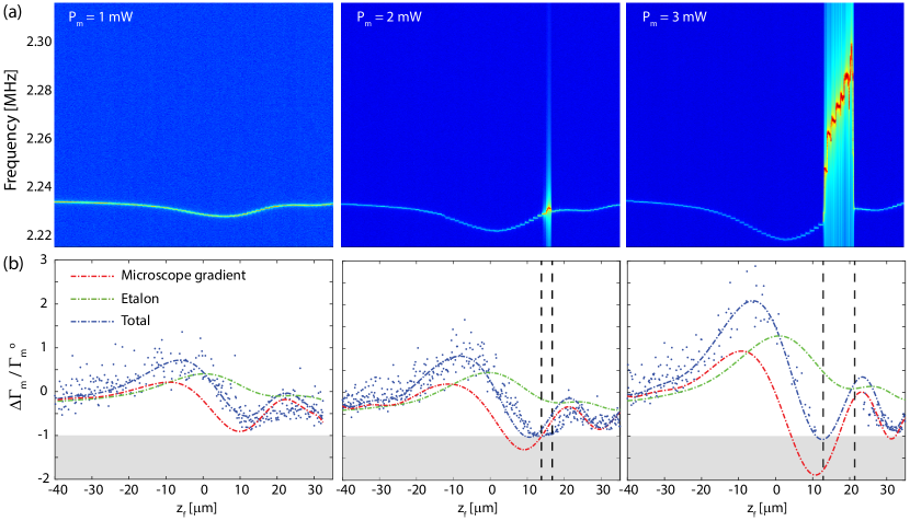
The gradient of the microscope intensity plays a critical role in determining whether , and as a result , is positive or negative. This is in contrast to cavity optomechanics, where back action is dominated by whose sign is independent of external optics. To study the microscope back action, the objective was aligned with the center of the nanobeam and scanned vertically ( steps, 2.9 s/step) while monitoring . Figure 2(a) shows this measurement at room temperature for microscope powers and 3 mW. The mechanical frequency decreases as the microscope is focused on the nanobeam, consistent with optical heating of a compressively stressed device [45]. follows a profile reminiscent of the microscope laser intensity’s dependence, providing a measure of the profile that can be input to the model in Eq. (1), as discussed below. The asymmetry and oscillations in are related to aberrations from the cryostat window [51]. In general, is also affected by dynamic photothermal, and dynamic and static optical gradient force effects. However, they are predicted to be smaller than the observed [45].
The influence of the microscope on the nanobeam dynamics is revealed dramatically in Fig. 2(a) near , where for and 3 mW the peak value of increases, indicating nanomechanical self-oscillation, and shifts due to nonlinear nanomechanical effects related to large amplitude motion [45]. To analyze this quantitatively, the measured is plotted in Fig. 2(b), showing that motion is either damped () or amplified () depending on the microscope focus: the sign of changes as the focus is scanned from above to below the nanobeam. Near , , the nanobeam enters a regime of self-oscillation, in agreement with the increase in peak amplitude.
This behavior illustrates a key feature of this system: the dependence of the sign of on the microscope intensity gradient. By fitting the data in Figs. 2(b) with the model from Eq. (1), the relative contribution from the microscope gradient and the etalon were extracted. This requires expanding the intensity gradient,
| (2) |
and inferring and from to within a proportionality constant. In addition to this constant, the fit requires a fitting parameter that governs the relative contributions of the intensity gradient and the etalon terms in Eq. (2). Contributions from these two terms are shown in Fig. 2(b), showing that in our experiment the microscope gradient is the dominant factor while the smaller etalon contribution damps mechanical motion and shifts the zero of . The imperfect fits reveal the approximate nature of the model. For example, it is possible that the microscope position and the etalon response, which in general is a standing wave pattern, are not entirely separable. In future, detailed numerical simulations of the microscope field and its interaction with the nanobeam and the surrounding diamond structure would provide additional insight into optimization of the strength of the gradient contribution and minimization of the etalon contribution.
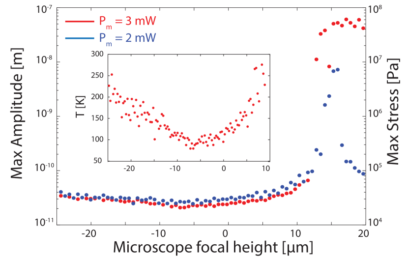
The amplification and damping is further analyzed in Fig. 3, which plots the RMS amplitude for varying and , extracted from the area under normalized by the thermomechanical vibration amplitude in absence of the microscope field [52]. When , the self-oscillations reach close to 100 nm, three orders of magnitude greater than the nanobeam’s intrinsic thermal motion. In contrast, when the microscope position is set to , the thermal motion of the nanobeam is damped, cooling the resonance to from the sample temperature , as shown in the inset to Fig. 3. This inference of temperature from resonance area was found to be consistent with predicted from in Fig. 2(b) [20].
IV Cryogenic operation
IV.1 Low power self-oscillations and cooling
The impact of back action is increased in cryogenic conditions where the intrinsic mechanical dissipation of the diamond resonator is reduced. The improvement in device performance at low temperature is illustrated in Fig. 1(b), which shows that increases by an order of magnitude when the sample temperature is lowered from 300 K to 5 K. As a result, for a given optomechanical backaction , which is nominally independent of temperature and , the relative change in mechanical dissipation, , increases. This lowers the power required for optomechanical self-oscillation () or cooling to a desired .
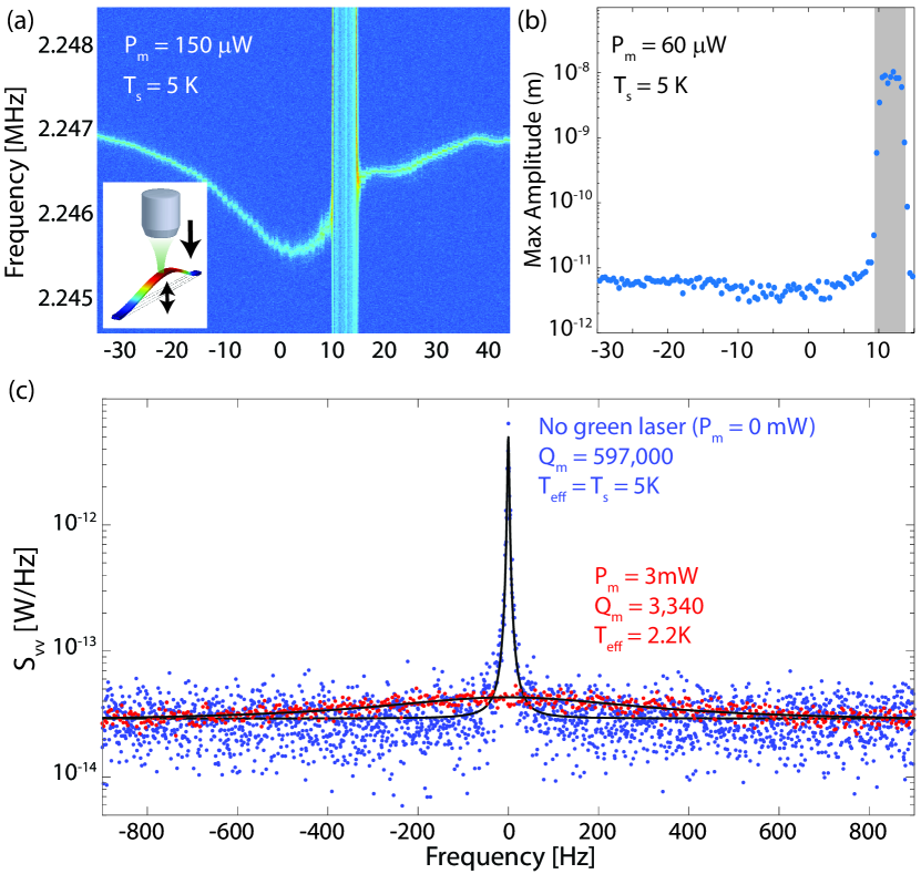
Figures 4(a) and 4(b) illustrate this effect by showing and the mechanical vibration amplitude, respectively, at , for varying . In these measurements the microscope power was reduced to and , respectively. Despite the order of magnitude lower compared to the room temperature measurements in Figs. 2 and 3, self-oscillations with comparable amplitude are observed.
A tantalizing prospect given the nanobeam’s increase in at cryogenic temperature is optomechanical cooling: for the mode is naively expected at from the observed at room temperature in Fig. 2(b). Figure 4(c) compares of this mode at with and without the 3 mW microscope field turned on. With the field on and the focus optimized to maximize damping, is inferred from the resonance linewidth. Although this is a two orders of magnitude increase in linewidth, the corresponding measured area under was only reduced by a factor of 2.4 by the microscope field, resulting in .
The discrepancy between the large broadening of the resonance and the comparatively modest change in can arise from several sources. At the specific heat of diamond is four orders of magnitude smaller than at room temperature. As a result, the microscope field can more easily increase the bath temperature of the nanobeam, counteracting cooling via optomechanical damping. Additional linewidth broadening could arise from fluctuations of the mechanical resonance frequency induced by the microscope field. Comparing the resonance linewidth for at in Fig. 4(c) with the corresponding maximum room temperature linewidth in Fig. 2(b), we see infer if damping rate is assumed to be proportional to the linewidth. Such an enhancement in damping at low temperature requires that photothermal coupling increases in cryogenic conditions, for example due to changes in the nanobeam’s compressive stress [45]. However, measurements discussed below reveal that other mechanisms can contribute significantly to linewidth broadening at cryogenic temperatures.
IV.2 In-plane mode excitation and line broadening
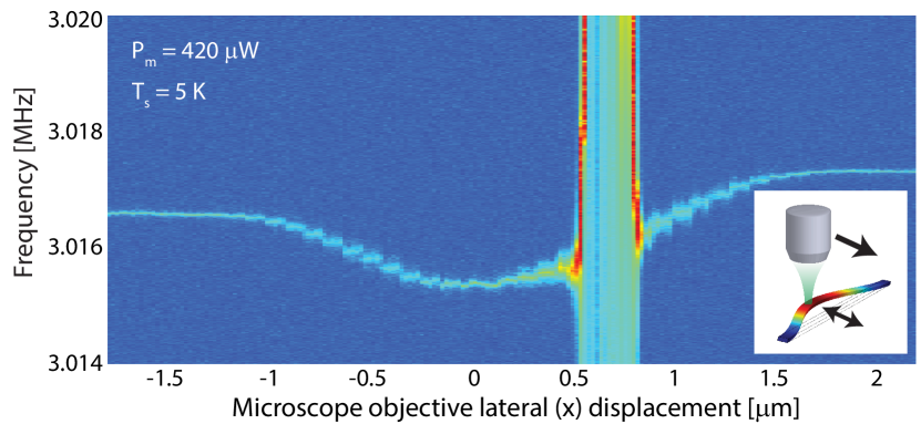
The device’s higher at cryogenic temperatures also enabled excitation of in-plane nanobeam motion. This is shown in Fig. 5, which plots of the in-plane fundamental resonance near as a function of lateral () displacement of the objective for . The scan length is smaller than the scans owing to the microscope’s tight lateral focus in comparison to its depth of focus. Self-oscillation occurs near , while for negative damping is observed. This asymmetric optomechanical response indicates that the nanobeam deflects laterally in a fixed direction when heated, independent of whether the focus is on the right or left side of the nanobeam.
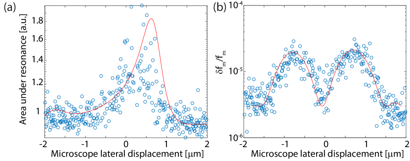
Further analysis of the dependence of the resonance dynamics on the microscope field confirm that mechanisms in addition to optomechanical backaction broaden the mechanical lineshape. Figure 6(a) plots the resonance area () for varying microscope displacement along with set below the threshold for self-oscillation. This clearly illustrates the asymmetric response of the optomechanical damping as a function of , and is consistent with the self-oscillation data in Fig. 5. In contrast, the measured linewidth, plotted in Fig. 6(b), varies symmetrically with . Broadening is maximized when the nanobeam is positioned adjacent to the microscope focus, where the lateral gradient of the microscope field intensity is strong. This broadening occurs even when the nanobeam motion is being amplified. These observations suggest that displacements of the nanobeam relative to the microscope focus are causing spectral diffusion of .
To better understand the dependence of the mode area () and the measured linewidth () on microscope position, we fit the dependent data in Fig. 6 with:
| (3) | ||||
| (4) |
where fitting parameter describes the increase in from the microscope field, and parameter describes the strength of the photothermal optomechanical backaction and its effect on . Fitting parameter describes a contribution to spectral broadening resulting from the nanobeam’s overlap with the gradient of the microscope field intensity, for example that manifest due to variations in the position of the focal spot relative to the nanobeam. The fits in Fig. 6 are created by assuming that is directly proportional to the measured , similar to the room temperature analysis in Fig. 2(b).
This model has good agreement with the data, and confirms that in addition to optomechanical damping, the linewidth is being broadened by additional mechanisms described here phenomenologically by non-zero . Further investigation into the source of this broadening is required. For example, measurements of the Allan variance of the mechanical frequency will reveal the timescale over which it is fluctuating and give insight into the nature of any technical noise affecting it. Time domain measurements of mechanical ring-down will provide a direct measurement of . Together with power dependent measurements of , this may allow improvement of the efficiency of the photothermal cooling process in cryogenic conditions.
Note that inferred from the area of is unaffected by spectral diffusion [53], and that at room temperature we observe close agreement between extracted from and , respectively. This indicates that the spectral broadening by the microscope is specific to the cryogenic measurements reported here.
V Discussion
This system’s potential for spin-optomechanics is significant. Confocal microscopes are used for diamond spin spectroscopy, making this approach suited for controlling coupling of phonons and spins. For the self–oscillations reported here, dynamic stress fields of 100 MPa are predicted (COMSOL), as shown in Fig. 3. This corresponds to a spin-stress coupling rate 1 MHz and 100 THz, for the ground and excited states, respectively, of a negatively charged NV, which are comparable to coupling rates in piezo-based stress manipulation experiments [22]. Optomechanical spin control will provide a path towards creating a quantum transducer [34, 30] for coupling photons to spins without direct optical color center transitions, enabling interfacing telecommunication photons with spin quantum memories [54].
In conclusion, we have demonstrated optomechanical control of a diamond resonator using a microscope. To the best of our knowledge, this is the first demonstration of tunable optomechanical damping and amplification of a nanomechanical resonator without a cavity, etalon, or other external feedback component. Using this tunable optomechanical damping, we have cooled the nanobeam’s fundamental mode to below 80K, and amplified its motion sufficiently for spin-phonon coupling at rates that exceed relevant spin decoherence rates [22, 54]. We have also studied the interplay between the nanomechanical resonator of the microscope field at low temperature, providing a jumping off point for future studies of photothermal cooling in cryogenic environments.
Acknowledgements.
Thank you to Aaron Hryciw, J.P. Hadden and M. Mitchell for assistance. This work was supported by NSERC (Discovery and Research Tools and Instruments), CFI, AITF and NRC.References
- Ashkin [1970] A. Ashkin, Acceleration and trapping of particles by radiation pressure, Phys. Rev. Lett. 24, 156 (1970).
- Scientific et al. [2017] L. Scientific, B. Abbott, R. Abbott, T. Abbott, F. Acernese, K. Ackley, C. Adams, T. Adams, P. Addesso, R. Adhikari, et al., Gw170104: observation of a 50-solar-mass binary black hole coalescence at redshift 0.2, Physical Review Letters 118, 221101 (2017).
- Gröblacher et al. [2009] S. Gröblacher, K. Hammerer, M. R. Vanner, and M. Aspelmeyer, Observation of strong coupling between a micromechanical resonator and an optical cavity field, Nature 460, 724 (2009).
- Chan et al. [2011] J. Chan, T. P. M. Alegre, A. H. Safavi-Naeini, J. T. Hill, A. Krause, S. Groblacher, M. Aspelmeyer, and O. Painter, Laser cooling of a nanomechanical oscillator into its quantum ground state, Nature 478, 89 (2011).
- Cohen et al. [2015] J. D. Cohen, S. M. Meenehan, G. S. MacCabe, S. Gröblacher, A. H. Safavi-Naeini, F. Marsili, M. D. Shaw, and O. Painter, Phonon counting and intensity interferometry of a nanomechanical resonator, Nature 520, 522 (2015).
- Riedinger et al. [2016] R. Riedinger, S. Hong, R. A. Norte, J. A. Slater, J. Shang, A. G. Krause, V. Anant, M. Aspelmeyer, and S. Gröblacher, Non-classical correlations between single photons and phonons from a mechanical oscillator, Nature 530, 313 (2016).
- Sudhir et al. [2017] V. Sudhir, R. Schilling, S. A. Fedorov, H. Schütz, D. J. Wilson, and T. J. Kippenberg, Quantum correlations of light from a room-temperature mechanical oscillator, Phys. Rev. X 7, 031055 (2017).
- Purdy et al. [2017] T. P. Purdy, K. E. Grutter, K. Srinivasan, and J. M. Taylor, Quantum correlations from a room-temperature optomechanical cavity, Science 356, 1265 (2017).
- Riedinger et al. [2018] R. Riedinger, A. Wallucks, I. Marinković, C. Löschnauer, M. Aspelmeyer, S. Hong, and S. Gröblacher, Remote quantum entanglement between two micromechanical oscillators, Nature 556, 473 (2018).
- Anetsberger et al. [2010] G. Anetsberger, E. Gavartin, O. Arcizet, Q. Unterreithmeier, E. Weig, M. Gorodetsky, J. Kotthaus, and T. Kippenberg, Measuring nanomechanical motion with an imprecision below standard quantum limit, Phys. Rev. A 82, 061804 (2010).
- Gavartin et al. [2012] E. Gavartin, P. Verlot, and T. Kippenberg, A hybrid on-chip optomechanical transducer for ultrasensitive force measurements, Nature Nanotech. 7, 509 (2012).
- Forstner et al. [2012] S. Forstner, S. Prams, J. Knittel, E. van Ooijen, J. Swaim, G. Harris, A. Szorkovszky, W. Bowen, and H. Rubinsztein-Dunlop, Cavity optomechanical magnetometer, Phys. Rev. Lett. 108, 120801 (2012).
- Wu et al. [2017] M. Wu, N. L.-Y. Wu, T. Firdous, F. F. Sani, J. E. Losby, M. R. Freeman, and P. E. Barclay, Nanocavity optomechanical torque magnetometry and radiofrequency susceptometry, Nature Nanotechnology 12, 127 (2017).
- Safavi-Naeini et al. [2011] A. H. Safavi-Naeini, T. M. Alegre, J. Chan, M. Eichenfield, M. Winger, Q. Lin, J. T. Hill, D. Chang, and O. Painter, Electromagnetically induced transparency and slow light with optomechanics, Nature 472, 69 (2011).
- Weis et al. [2010] S. Weis, R. Rivière, S. Deléglise, E. Gavartin, O. Arcizet, A. Schliesser, and T. J. Kippenberg, Optomechanically induced transparency, Science 330, 1520 (2010).
- Dong et al. [2012] C. Dong, V. Fiore, M. C. Kuzyk, and H. Wang, Optomechanical dark mode, Science 338, 1609 (2012).
- Liu et al. [2013] Y. Liu, M. Davanço, V. Aksyuk, and K. Srinivasan, Electromagnetically induced transparency and wideband wavelength conversion in silicon nitride microdisk optomechanical resonators, Phys. Rev. Lett. 110, 223603 (2013).
- Lake et al. [2018] D. P. Lake, M. Mitchell, Y. Kamaliddin, and P. E. Barclay, Optomechanically induced transparency and cooling in thermally stable diamond microcavities, ACS Photonics 5, 782 (2018).
- Kippenberg and Vahala [2008] T. Kippenberg and K. Vahala, Cavity optomechanics: Back-action at the mesoscale, Science 321, 1172 (2008).
- Aspelmeyer et al. [2014] M. Aspelmeyer, T. J. Kippenberg, and F. Marquardt, Cavity optomechanics, Rev. Mod. Phys. 86, 1391 (2014).
- Aharonovich et al. [2011] I. Aharonovich, A. D. Greentree, and S. Prawer, Diamond photonics, Nature Photon. 5, 397 (2011).
- Lee et al. [2017] D. Lee, K. W. Lee, J. V. Cady, P. Ovartchaiyapong, and A. C. B. Jayich, Topical review: spins and mechanics in diamond, Journal of Optics 19, 033001 (2017).
- MacQuarrie et al. [2013] E. R. MacQuarrie, T. A. Gosavi, N. R. Jungwirth, S. A. Bhave, and G. D. Fuchs, Mechanical spin control of nitrogen-vacancy centers in diamond, Phys. Rev. Lett. 111, 227602 (2013).
- Ovartchaiyapong et al. [2014] P. Ovartchaiyapong, K. W. Lee, B. A. Myers, and A. C. B. Jayich, Dynamic strain-mediated coupling of a single diamond spin to a mechanical resonator, Nat. Commun. 5, 4429 (2014).
- Teissier et al. [2014] J. Teissier, A. Barfuss, P. Appel, E. Neu, and P. Maletinsky, Strain coupling of a nitrogen-vacancy center spin to a diamond mechanical oscillator, Phys. Rev. Lett. 113, 020503 (2014).
- Barfuss et al. [2015] A. Barfuss, J. Teissier, E. Neu, A. Nunnenkamp, and P. Maletinsky, Strong mechanical driving of a single electron spin, Nature Phys. 11, 820 (2015).
- MacQuarrie et al. [2015] E. R. MacQuarrie, T. A. Gosavi, A. M. Moehle, N. R. Jungwirth, S. A. Bhave, and G. D. Fuchs, Coherent control of a nitrogen-vacancy center spin ensemble with a diamond mechanical resonator, Optica 2, 233 (2015).
- Meesala et al. [2016] S. Meesala, Y.-I. Sohn, H. A. Atikian, S. Kim, M. J. Burek, J. T. Choy, and M. Lončar, Enhanced strain coupling of nitrogen-vacancy spins to nanoscale diamond cantilevers, Phys. Rev. Applied 5, 034010 (2016).
- Golter et al. [2016a] D. A. Golter, T. Oo, M. Amezcua, I. Lekavicius, K. A. Stewart, and H. Wang, Coupling a surface acoustic wave to an electron spin in diamond via a dark state, Phys. Rev. X 6, 041060 (2016a).
- Golter et al. [2016b] D. A. Golter, T. Oo, M. Amezcua, K. A. Stewart, and H. Wang, Optomechanical quantum control of a nitrogen-vacancy center in diamond, Phys. Rev. Lett. 116, 143602 (2016b).
- Maity et al. [2019] S. Maity, L. Shao, S. Bogdanović, S. Meesala, Y.-I. Sohn, N. Sinclair, B. Pingault, M. Chalupnik, C. Chia, L. Zheng, et al., Coherent acoustic control of a single silicon vacancy spin in diamond, arXiv preprint arXiv:1910.09710 (2019).
- Lemonde et al. [2018] M.-A. Lemonde, S. Meesala, A. Sipahigil, M. J. A. Schuetz, M. D. Lukin, M. Loncar, and P. Rabl, Phonon networks with silicon-vacancy centers in diamond waveguides, Phys. Rev. Lett. 120, 213603 (2018).
- Kuzyk and Wang [2018] M. C. Kuzyk and H. Wang, Phononic quantum networks of solid-state spins with alternating and frequency-selective waveguides, arXiv preprint arXiv:1804.07862 (2018).
- Schuetz et al. [2015] M. J. A. Schuetz, E. M. Kessler, G. Giedke, L. M. K. Vandersypen, M. D. Lukin, and J. I. Cirac, Universal quantum transducers based on surface acoustic waves, Phys. Rev. X 5, 031031 (2015).
- Rugar and Grütter [1991] D. Rugar and P. Grütter, Mechanical parametric amplification and thermomechanical noise squeezing, Phys. Rev. Lett. 67, 699 (1991).
- Barton et al. [2012] R. A. Barton, I. R. Storch, V. P. Adiga, R. Sakakibara, B. R. Cipriany, B. Ilic, S. P. Wang, P. Ong, P. L. McEuen, J. M. Parpia, et al., Photothermal self-oscillation and laser cooling of graphene optomechanical systems, Nano Lett. 12, 4681 (2012).
- Yie et al. [2010] Z. Yie, K. Turner, N. Miller, and S. Shaw, Sensitivity enhancement using parametric amplification in a resonant sensing array, in Proceedings of the 13th Hilton Head Solid State Sensors and Actuators Conference (2010).
- Lin et al. [2009] Q. Lin, J. Rosenberg, X. Jiang, K. J. Vahala, and O. Painter, Mechanical oscillation and cooling actuated by the optical gradient force, Phys. Rev. Lett. 103, 103601 (2009).
- Arcizet et al. [2006] O. Arcizet, P. F. Cohadon, T. Briant, M. Pinard, and A. Heidmann, Radiation-pressure cooling and optomechanical instability of a micromirror, Nature 444, 71 (2006).
- Gigan et al. [2006] S. Gigan, H. R. Böhm, M. Paternostro, F. Blaser, G. Langer, J. B. Hertzberg, K. C. Schwab, D. Bäuerle, M. Aspelmeyer, and A. Zeilinger, Self-cooling of a micromirror by radiation pressure, Nature 444, 67 (2006), arXiv:quant-ph/0607068 .
- Metzger et al. [2008] C. Metzger, I. Favero, A. Ortlieb, and K. Karrai, Optical self cooling of a deformable fabry-perot cavity in the classical limit, Phys. Rev. B 78, 035309 (2008).
- Metzger and Karrai [2004] C. H. Metzger and K. Karrai, Cavity cooling of a microlever, Nature 432, 1002 (2004).
- Favero et al. [2007] I. Favero, C. Metzger, S. Camerer, D. König, H. Lorenz, J. P. Kotthaus, and K. Karrai, Optical cooling of a micromirror of wavelength size, Appl. Phys. Lett. 90, 104101 (2007).
- Ramos et al. [2012] D. Ramos, E. Gil-Santos, V. Pini, J. M. Llorens, M. Fernandez-Regulez, A. S. Paulo, M. Calleja, and J. Tamayo, Optomechanics with silicon nanowires by harnessing confined electromagnetic modes, Nano Letters 12, 932 (2012), pMID: 22268657, http://dx.doi.org/10.1021/nl204002u .
- Khanaliloo et al. [2015] B. Khanaliloo, H. Jayakumar, A. C. Hryciw, D. P. Lake, H. Kaviani, and P. E. Barclay, Single-crystal diamond nanobeam waveguide optomechanics, Phys. Rev. X 5, 041051 (2015).
- Poggio et al. [2007] M. Poggio, C. Degen, H. Mamin, and D. Rugar, Feedback cooling of a cantilever�s fundamental mode below 5 mk, Physical Review Letters 99, 017201 (2007).
- Burek et al. [2013] M. J. Burek, D. Ramos, P. Patel, I. W. Frank, and M. Lončar, Nanomechanical resonant structures in single-crystal diamond, Appl. Phys. Lett. 103, 131904 (2013).
- Michael et al. [2007] C. P. Michael, M. Borselli, T. J. Johnson, C. Chrystala, and O. Painter, An optical fiber-taper probe for wafer-scale microphotonic device characterization, Opt. Express 15, 4745 (2007).
- Cleland and Roukes [2002] A. Cleland and M. Roukes, Noise processes in nanomechanical resonators, J. Appl. Phys. 92, 2758 (2002).
- Li et al. [2012] W. Li, N. Mingo, L. Lindsay, D. A. Broido, D. A. Stewart, and N. A. Katcho, Thermal conductivity of diamond nanowires from first principles, Phys. Rev. B 85, 195436 (2012).
- Nasse and Woehl [2010] M. J. Nasse and J. C. Woehl, Realistic modeling of the illumination point spread function in confocal scanning optical microscopy, Josa a 27, 295 (2010).
- Mitchell et al. [2016] M. Mitchell, B. Khanaliloo, D. P. Lake, T. Masuda, J. P. Hadden, and P. E. Barclay, Single-crystal diamond low-dissipation cavity optomechanics, Optica 3, 963 (2016).
- Moser et al. [2014] J. Moser, A. Eichler, J. Güttinger, M. I. Dykman, and A. Bachtold, Nanotube mechanical resonators with quality factors of up to 5 million, Nature nanotechnology 9, 1007 (2014).
- Shandilya et al. [2021] P. K. Shandilya, D. P. Lake, M. J. Mitchell, D. D. Sukachev, and P. E. Barclay, Optomechanical interface between telecom photons and spin quantum memory (2021), arXiv:2102.04597 [quant-ph] .