Salt-dependent rheology and surface tension of protein condensates using optical traps
Abstract
An increasing number of proteins with intrinsically disordered domains have been shown to phase separate in buffer to form liquid-like phases. These protein condensates serve as simple models for the investigation of the more complex membrane-less organelles in cells. To understand the function of such proteins in cells, the material properties of the condensates they form are important. However, these material properties are not well understood. Here, we develop a novel method based on optical traps to study the frequency-dependent rheology and the surface tension of PGL-3 condensates as a function of salt concentration. We find that PGL-3 droplets are predominantly viscous but also exhibit elastic properties. As the salt concentration is reduced, their elastic modulus, viscosity and surface tension increase. Our findings show that salt concentration has a strong influence on the rheology and dynamics of protein condensates suggesting an important role of electrostatic interactions for their material properties.
Introduction.
A fundamental question of biology is to understand the spatial organization of cells into compartments of distinct chemical composition and activity.
Many cellular compartments are bounded by membranes. However, cells also possess compartments that are not bounded by membranes. Examples are germline granules, cajal bodies, nucleoli and stress granules. It has been shown in recent years that many membrane-less compartments are protein condensates forming soft materials that coexist with the cytoplasm or nucleoplasm and often exhibit liquid-like properties Hyman et al. (2014); Brangwynne et al. (2009, 2011); Gilks et al. (2004); Patel et al. (2015); Brangwynne et al. (2009, 2011); Saha et al. (2016); Lin et al. (2015); Banani et al. (2016); kami et al. (2015); Nott et al. (2015).
Liquid-like compartments typically contain one or two scaffold proteins which are required for compartment formation and to which RNA and other proteins co-localize Hyman et al. (2014); Gilks et al. (2004); Aoki et al. (2016). Many scaffold proteins phase separate in vitro when introduced into physiological buffer resulting in the condensation of liquid-like droplets. A well-studied example of a phase-separating scaffold protein is PGL-3 - a major component of P-granules, the germline granules of the nematode worm Caenorhabditis elegans Brangwynne et al. (2009); Elbaum-Garfinkle et al. (2015); Saha et al. (2016); Aoki et al. (2016). This and other protein droplets serve as models for the soft materials that form the more complex liquid-like compartments in cells Molliex et al. (2015); Saha et al. (2016); Lin et al. (2015); Nott et al. (2015); Elbaum-Garfinkle et al. (2015); Patel et al. (2015). The material properties of protein droplets are important to understand the biological function of protein condensates in cells - for example when they serve as biochemical reaction centers, which requires rapid diffusion of components, or when they serve as structural elements, which may require some degree of elasticity. However, the material properties and rheology have not yet been fully characterized. Active microrheological methods that could provide such information for micron-sized droplets are currently lacking.
The saturation concentration of protein phase separation in vitro depends on buffer conditions such as salt concentration, pH or the presence of RNAMolliex et al. (2015); Saha et al. (2016); Lin et al. (2015); Nott et al. (2015); Elbaum-Garfinkle et al. (2015); Brangwynne et al. (2015). The salt and pH dependence of the saturation concentration suggests a role of charge and electrostatic interactions in protein phase separation.
In this letter, we develop a novel active microrheology method to determine the frequency-dependent rheology of micron-sized PGL-3 droplets as a function of salt concentration using optical traps. Protein droplets are deformed via two opposing adhered beads trapped by optical traps (Fig. 1a,b). Surface tension and the complex shear modulus that characterize the droplet material properties can be obtained using a theoretical analysis of force balances associated with droplet deformation in the optical trap setup. Here and denote the storage and loss moduli, respectively.
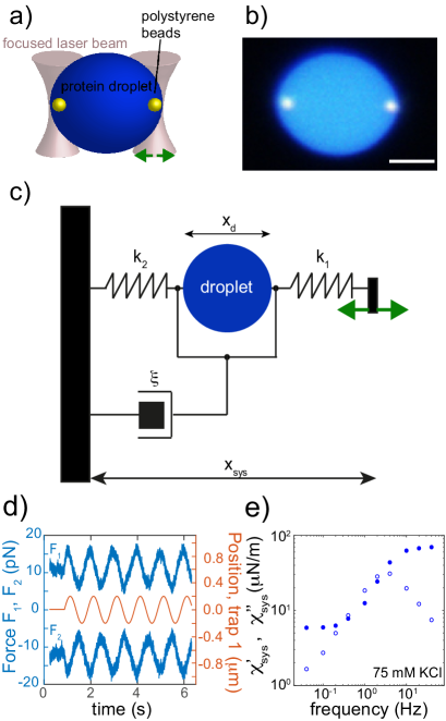
Force balance of droplet deformation. The microrheology setup based on a dual optical trap is shown in Fig. 1a,b. The droplet is deformed by moving one trap. The resulting force balance involves forces and exerted on the droplet by the traps with stiffnesses and , as well as viscous drag exerted on the droplet by the fluid with friction coefficient (Fig. 1c). In the over-damped regime, force balance requires
| (1) |
where is the droplet velocity. The difference of and is balanced by forces exerted by the deformed droplet. For time periodic forces and droplet diameter with angular frequency , we have
| (2) |
Here, the tilde denotes the complex amplitude of the Fourier mode at angular frequency and refers to the complex conjugate. In Eq. (2), characterizes the complex frequency-dependent spring constant of the droplet response, where and denote the real and imaginary part of the complex spring constant, respectively. For an oscillatory trap movement . The complex spring constant of the entire system (droplet in series with two optical traps) is
| (3) |
where is the distance between the trap centers (Fig. 1c). The distance can differ from the droplet diameter because the beads can move out of the trap centers. The complex spring constant of the droplet can be related to the complex spring constant of the system using
| (4) |
We find
| (5) |
Droplet mechanics in the optical trap. To determine the protein droplet’s surface tension as well as the complex frequency dependent shear modulus in the optical trap setup, we need to relate these material properties to the complex spring constant defined in Eq. (2). The mechanical droplet stress satisfies the force balance
| (6) |
For small deformations in the low Reynold’s number regime, the constitutive material equation of an incompressible droplet material can be written as
| (7) |
where denotes pressure and is the displacement vector with components . Stress boundary conditions on the surface of a spherical droplet of radius read in spherical coordinates , ,
| (8) | |||||
| (9) |
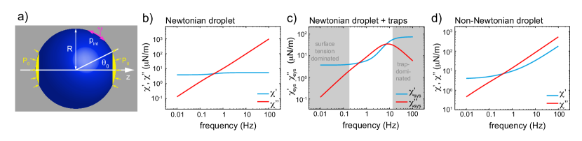
Here, is the local normal force per area exerted on the droplet surface and denotes the mean curvature of the weakly deformed droplet surface. Forces mediated by adherent beads of radius are captured by a time periodic pressure distribution with complex amplitude on the droplet surface (Fig. 2a), where denotes the Heaviside function and .
Using Eqn. (6-9), we calculate the time-dependent deformation field of droplet material using a stream function approach and an expansion in spherical harmonics, see Supplement. The time dependent droplet diameter can be expressed as and the difference of optical trap forces is given by . We then obtain the complex spring constant of the droplet response as a function of and using Eq. (2), see Supplement. This droplet spring constant is related to the spring constant of the entire optical trap system through Eq. (5).
Droplet spring constant in simple cases.
We first discuss the droplet and system spring constants for a droplet consisting of a Newtonian fluid. In this case, the complex shear modulus where is the viscosity.
Using the approach described above, we determine the droplet spring constant and the system spring constant . The droplet spring constant as a function of frequency is shown in Fig. 2b. The real part of the spring constant (blue) stems from surface tension. The imaginary part (red) is associated with viscosity.
The system spring constant for a Newtoninan droplet is shown in Fig. 2c. At low frequencies, the response of the system is dominated by the droplet surface tension, corresponding to a plateau in the real part (see gray low-frequency region Fig. 2c).
The intermediate frequency range is dominated by droplet viscosity. At high frequencies, the system response is dominated by the optical traps (see gray high-frequency region Fig. 2c).
We also consider a droplet composed of a non-Newtonian fluid with a power law rheology in both loss and storage moduli with exponent . The corresponding droplet spring constant reveals droplet surface tension at low frequencies and power-law rheology at high frequencies, Fig. 2d.
Salt dependent material properties of PGL-3 droplets.
We performed measurements on PGL-3 protein droplets formed in buffer (25mM HEPES, 1mM DTT, pH 7.5) at four different salt concentrations (75 mM, 115 mM, 150 mM and 180 mM KCl). Polystyrene beads of m diameter added to the buffer were brought into adhesive contact with protein droplets (see Supplement). Radii of droplets used were between m.
Droplets were deformed at frequencies between and Hz. Forces exerted by both optical traps and the position of the mobile trap center were recorded. From this data, we first determined the system spring constant using Eq. (3) (Fig. 1e). The frequency dependent spring constant exhibits the three regimes discussed above (compare Fig. 1e and Fig. 2c): a low frequency regime dominated by surface tension, an intermediate regime and a high frequency regime dominated by the traps (Fig. 1e).
We then determined the complex spring constant of uniaxial droplet elongation using Eq. (5) (Fig. 3a).
From this data, we first determined the surface tension in the low frequency regime using
| (10) |
valid for small (see Supplement). We then obtain the complex shear modulus by accounting for surface tension effects from the droplet spring constant (Fig. 3b and Supplement). For modulus and small we use
| (11) |
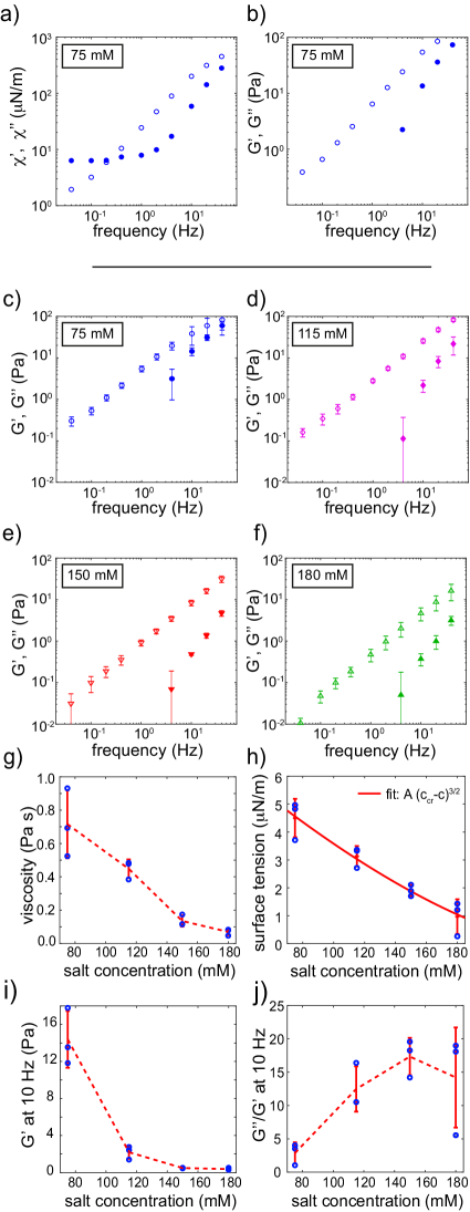
(see Supplement). Fig. 3c-f show storage and loss moduli as a function of frequency averaged over several experiments for four different salt concentrations. Loss moduli increased approximately linearly for increasing frequency (Fig. 3b-f). Corresponding viscosity values range from to s. Storage moduli at a reference frequency of Hz ranged between values of and Pa. Both, viscosities and storage moduli decreased with increasing salt concentration (Fig. 3g and i). The associated loss tangent exhibited a sharp drop when salt concentration was reduced to 75 mM, suggesting a more solid-like rheology at lower salt concentrations (Fig. 3j).
The estimated surface tension was about N/m for mM salt and decreased steadily for increasing salt concentrations with N/m at mM salt (Fig. 3h and Supplement).
To test our new method, we measured viscosity and surface tension using two established methods. Using passive bead-tracking micorheology, we estimated a viscosity Pa s at 75 mM salt which is in excellent agreement with our active microrheology results (Fig. 3g and Supplement). Furthermore, we analysed thermal fluctuations of droplet shape to measure surface tension N/m at 180 mM salt, consistent with our new method.
The surface tension and viscosity of polyelectrolyte condensates have been studied theoretically Spruijt et al. (2010a, b). In Fig. 3h, we compare the predicted salt-dependence of surface tension to our data and find good agreement. Furthermore, the salt dependence of a characteristic time is also in agreement with this theory (see supplemental Fig. S6).
Discussion. Here, we used a novel technique with optical traps to characterize the rheology of PGL-3 protein droplets. We find that newly formed PGL-3 droplets consist of a viscoelastic material with liquid-like material properties that depend on salt concentration. Viscosities range from to Pa s for decreasing salt concentrations from to mM. The storage modulus is smaller than the loss modulus, but approaches the loss modulus from below for frequencies larger than Hz.
Furthermore, we obtain more precise values of surface tension than previous methods. Values depend on salt concentration and range from 1 N/m to 5 N/m for decreasing salt concentrations from to mM. Such low values of surface tension are consistent with values estimated for colloidal systems with weak interactions but much smaller than typical air-water or oil-water systems Lekkerkerker et al. (2008a, b). Notably, our viscosity and surface tension estimates for PGL-3 droplets in vitro are consistent with estimates for P-granules in vivo suggesting that in vitro PGL-3 is a good model system for the physics and material properties of P-granules in vivo Brangwynne et al. (2009).
Interestingly, our viscosity and surface tension values for PGL-3 are between one to two orders of magnitude smaller than estimates for droplets of the P-granule protein LAF-1 formed in vitro Elbaum-Garfinkle et al. (2015).
Viscosities of PGL-3 droplets exhibit a strong salt dependence qualitatively similar to salt-dependent viscosity estimates reported for phase-separated LAF-1 by passive bead-tracking micro-rheology (Fig. 3g) Elbaum-Garfinkle et al. (2015).
The decrease in storage moduli, loss moduli and surface tensions for increasing salt concentrations suggests that screened electrostatic interactions play an important role in protein condensates and their material properties. As salt concentration is increased, interactions become weaker because of screening effects.
Interestingly, synthetic polyelectrolytes show a similar dependence of rheology and surface tension on salt concentration as the protein condensates studied here Spruijt et al. (2010a, b).
In conclusion, our measurements show that protein condensates are complex polymeric liquids with viscoelastic material properties that can be regulated by salt concentration. We obtain our results using a novel optical trap based rheometer suitable for micron-sized probes. This method does not rely on thermodynamic equilibrium. We expect that our work will trigger more studies that lead to a deep understanding of the physics of protein droplets and their phase behavior in biological cells.
I Acknowledgments
We would like to thank Marcus Jahnel and Stephan Grill for useful discussions and the staff of LUMICKS for their technical assistance. Furthermore, we would like to thank the following Services and Facilities of the MPI-CBG for their support: The Protein Expression Purification and Characterization (PEPC) facility.
References
- Hyman et al. (2014) A. A. Hyman, C. A. Weber, and F. Jülicher, Annual Review of Cell and Developmental Biology 30, 39 (2014).
- Brangwynne et al. (2009) C. P. Brangwynne, C. R. Eckmann, D. S. Courson, A. Rybarska, C. Hoege, J. Gharakhani, F. J licher, and A. A. Hyman, Science 324, 1729 (2009).
- Brangwynne et al. (2011) C. P. Brangwynne, T. J. Mitchison, and A. A. Hyman, Proceedings of the National Academy of Sciences 108, 4334 (2011).
- Gilks et al. (2004) N. Gilks, N. Kedersha, M. Ayodele, L. Shen, G. Stoecklin, L. M. Dember, and P. Anderson, Molecular Biology of the Cell 15, 5383 (2004).
- Patel et al. (2015) A. Patel, H. O. Lee, L. Jawerth, S. Maharana, M. Jahnel, M. Y. Hein, S. Stoynov, J. Mahamid, S. Saha, T. M. Franzmann, A. Pozniakovski, I. Poser, N. Maghelli, L. A. Royer, M. Weigert, E. W. Myers, S. Grill, D. Drechsel, A. A. Hyman, and S. Alberti, Cell 162, 1066 (2015).
- Saha et al. (2016) S. Saha, C. A. Weber, M. Nousch, O. Adame-Arana, C. Hoege, M. Y. Hein, E. Osborne-Nishimura, J. Mahamid, M. Jahnel, L. Jawerth, A. Pozniakovski, C. R. Eckmann, F. J licher, and A. A. Hyman, Cell 166, 1572 (2016).
- Lin et al. (2015) Y. Lin, D. S. W. Protter, M. K. Rosen, and R. Parker, Molecular Cell 60, 208 (2015).
- Banani et al. (2016) S. F. Banani, A. M. Rice, W. B. Peeples, Y. Lin, S. Jain, R. Parker, and M. K. Rosen, Cell 166, 651 (2016).
- kami et al. (2015) T. kami, S. Qamar, J. Q. Lin, G. S. K. Schierle, E. Rees, A. Miyashita, A. R. Costa, R. B. Dodd, F. T. S. Chan, C. H. Michel, D. Kronenberg-Versteeg, Y. Li, S.-P. Yang, Y. Wakutani, W. Meadows, R. R. Ferry, L. Dong, G. G. Tartaglia, G. Favrin, W.-L. Lin, D. W. Dickson, M. Zhen, D. Ron, G. Schmitt-Ulms, P. E. Fraser, N. A. Shneider, C. Holt, M. Vendruscolo, C. F. Kaminski, and P. St George-Hyslop, Neuron 88, 678 (2015).
- Nott et al. (2015) T. Nott, E. Petsalaki, P. Farber, D. Jervis, E. Fussner, A. Plochowietz, T. D. Craggs, D. Bazett-Jones, T. Pawson, J. Forman-Kay, and A. Baldwin, Molecular Cell 57, 936 (2015).
- Aoki et al. (2016) S. T. Aoki, A. M. Kershner, C. A. Bingman, M. Wickens, and J. Kimble, Proceedings of the National Academy of Sciences 113, 1279 (2016).
- Elbaum-Garfinkle et al. (2015) S. Elbaum-Garfinkle, Y. Kim, K. Szczepaniak, C. C.-H. Chen, C. R. Eckmann, S. Myong, and C. P. Brangwynne, Proceedings of the National Academy of Sciences 112, 7189 (2015).
- Molliex et al. (2015) A. Molliex, J. Temirov, J. Lee, M. Coughlin, A. P. Kanagaraj, H. J. Kim, T. Mittag, and J. P. Taylor, Cell 163, 123 (2015).
- Brangwynne et al. (2015) C. P. Brangwynne, P. Tompa, and R. V. Pappu, Nature Physics 11, 899 (2015).
- Lekkerkerker et al. (2008a) H. N. W. Lekkerkerker, V. W. A. De Villeneuve, J. W. J. De Folter, M. Schmidt, Y. Hennequin, D. Bonn, J. O. Indekeu, and D. Aarts, The European Physical Journal B 64, 341 (2008a).
- Lekkerkerker et al. (2008b) H. N. W. Lekkerkerker, V. W. A. d. Villeneuve, J. W. J. d. Folter, M. Schmidt, Y. Hennequin, D. Bonn, J. O. Indekeu, and D. G. a. L. Aarts, European Physical Journal B 64, 341 (2008b).
- Spruijt et al. (2010a) E. Spruijt, J. Sprakel, M. Lemmers, M. A. C. Stuart, and J. van der Gucht, Physical Review Letters 105, 208301 (2010a).
- Spruijt et al. (2010b) E. Spruijt, J. Sprakel, M. A. C. Stuart, and J. v. d. Gucht, Soft Matter 6, 172 (2010b).