Why large icosahedral viruses need scaffolding proteins: The interplay of Gaussian curvature and disclination interactions.
Abstract
While small single stranded viral shells encapsidate their genome spontaneously, many large viruses, such as the Herpes virus or Infectious Bursal Disease Virus (IBDV), typically require a template, consisting of either scaffolding proteins or inner core. Despite the proliferation of large viruses in nature, the mechanisms by which hundreds or thousands of proteins assemble to form structures with icosahedral order (IO) is completely unknown. Using continuum elasticity theory, we study the growth of large viral shells (capsids) and show that a non-specific template not only selects the radius of the capsid, but leads to the error-free assembly of protein subunits into capsids with universal IO. We prove that as a spherical cap grows, there is a deep potential well at the locations of disclinations that later in the assembly process will become the vertices of an icosahedron. Furthermore, we introduce a minimal model and simulate the assembly of viral shell around a template under non-equilibrium conditions and find a perfect match between the results of continuum elasticity theory and the numerical simulations. Besides explaining available experimental results, we provide a number of new predictions. Implications for other problems in spherical crystals are also discussed.
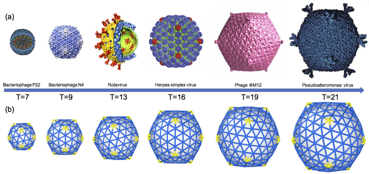
More than fifty years ago, Caspar and Klug Caspar and Klug (1962) made the striking observation that the capsids of most spherical viruses display icosahedral order(IO), defined by twelve five coordinated units (disclinations or pentamers) occupying the vertices of an icosahedron surrounded by hexameric units, see Fig. 1. While many studies have shown that this universal IO is favored under mechanical equilibrium Bruinsma (2006); Fejer et al. (2010); Rapaport (2004), the mechanism by which these shells grow, circumventing many possible activation barriers, and leading to the perfect IO remains mainly unknown.
Under many circumstances, small icosahedral capsids assemble spontaneously around their genetic material, often a single-stranded viral RNA Perlmutter et al. (2013); Comas-Garcia et al. (2012); Sikkema et al. (2007); Sun et al. (2007); Ning et al. (2016). Yet, larger double-stranded (ds) RNA or DNA viruses require what we generically denote as the template: scaffolding proteins (SPs) or an inner core Thuman-Commike et al. (1998); Earnshaw and King (1978); Aksyuk et al. (2015); Saad et al. (1999); Saugar et al. (2010); Coulibaly et al. (2005). The focus of this paper is on these large viruses that require a template for successful assembly.
The major difficulty in understanding the pathway towards IO is apparent from the results of the generalized Thomson problem, consisting of finding the minimum configuration for interacting -point particles constrained to be on the surface of a sphere. Simulation studies show that the number of metastable states increase exponentially with Erber and Hockney (1991), and only with the help of sophisticated optimization algorithms at relatively small values of Morris et al. (1996); Zandi et al. (2004); Perlmutter et al. (2013); Hagan and Zandi (2016), it is possible to obtain IO ground states. These situations, typical of spherical crystals, become even more difficult when considering the assembly of large capsids, in which once protein subunits are attached and a few bonds are made, it becomes energetically impossible for them to re-arrange: Should a single pentamer appear in an incorrect location, IO assembly would fail.
The combined effect of irreversibility and the inherent exponentially large number of metastable states typical of curved crystals puts many drastic constraints on IO growth. The complexity of the problem may be visualized by the various viral shells illustrated in Fig. 1, characterized by a structural index, the T number Caspar and Klug (1962); Wagner and Zandi (2015); Chen et al. (2007); Vernizzi and Olvera de la Cruz (2007) , with and arbitrary integers, such that the crystal includes monomers or hexamers and 12 pentamers (disclinations).
A possible mechanism to successfully self-assemble a desirable structure might consist of protein subunits with chemical specificity, very much like in DNA origami Rothemund (2006) where structures with complex symmetries are routinely assembled. In viruses, however, capsids are built either from one or a few different types of proteins, so specificity cannot be the driving mechanism leading to IO Yu et al. (2013); Ning et al. (2016); Nguyen et al. (2006); Chen et al. (2007); Zandi et al. (2004). In this paper, we show that a “generic” template provides a robust path to self- assembly of large shells with IO. This is consistent with many experimental data in that regardless of amino acid sequences and folding structures of virus coat and/or scaffolding proteins, due to the “universal” topological and geometrical constraints, large spherical viruses need scaffolding proteins to adopt IO, see Fig. 1. Although the focus of the paper is on virus assembly, the implication of our study goes far beyond and extend to many other problems where curved crystals are involved, a point that we we further elaborate in the conclusion Lidmar et al. (2003); Vernizzi et al. (2011).
The distinct feature of spherical crystals is that their global structure is constrained by topology. More concretely, if is the disclination density, then
| (1) |
where is the Euler characteristic ( for a sphere). However a capsid closes only at the end of the assembly, and thus, Eq. 1 does not really restrict the number of disclinations during the growth process, as pentamers or other disclinations may be created or destroyed at the boundaries. For a complete shell, the easiest way to fulfill Eq. 1 is with twelve disclinations, and this is the case we will follow hereon.
A minimal model for spherical crystals consists of a free energy
| (2) | |||||
where is the strain tensor. The coefficients are the Lame coefficients, which depend on the microscopic underlying interactions. Here is the extrinsic curvature of the template, the spontaneous curvature and the bending rigidity. By integrating the phonon degrees of freedom, we can recast the term in Eq. 2 as a non-local theory of interacting disclinations, with free energy Bowick et al. (2000)
| (3) | |||||
where is the Gaussian curvature and is the Young modulus. The disclination density has as variables the positions of 12 disclinations, each of charge . The function is the inverse of the Laplacian square Bowick et al. (2000). All previous studies for the model in Eq. 3 have been done for curved crystals without a boundary. In this paper, we provide, for the first time, the necessary formalism to include the presence of a boundary.
A discrete version of Eq. 2 is given by Seung and Nelson (1988); Vernizzi et al. (2011); Bowick et al. (2000); Lidmar et al. (2003)
| (4) |
with a preferred angle, related to the spontaneous curvature . The stretching energy sums over all bonds with the equilibrium bond length and the bending energy is between all neighboring trimers indexed with . We further assume that there is an attractive force between the trimers and the preformed scaffolding layer (inner core) (see Fig.4), which, consistent with our minimal model, involves a simple LJ-potential with the depth of the potential and the position of minimum energy corresponding to optimal distance between the center of the core and subunits. In the next section, we associate a dynamics to these models, which corresponds to following a local minimum energy pathway.
Methods
Discrete model
The growth of the shells is based on the following assumptions Comas-Garcia et al. (2012); Ning et al. (2016); Hagan and Zandi (2016); Yu et al. (2013): At each step of growth, a new trimer is added to the location in the boundary which makes the maximum number of bonds with the neighboring subunits. This is consistent with the fact that protein-protein attractive interaction is weak and a subunit can associate and dissociate till it sits in a position that forms a few bounds with neighboring proteins. These interactions eventually become strong for the subunits to dissociate and trimer attachment becomes irreversible Perlmutter et al. (2013). The attractive interactions between subunits, whose strength depends on electrostatic and hydrophobic forces, are implicit in the model. Note that pH and salt can modify the strength of protein-protein and protein-template interactions and thus the growth pathway. The impact of pH and salt on the shell assembly will be pursed elsewhere.
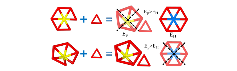
A crucial step in the assembly process is the formation of pentamers, which occurs only if the local energy is lowered, as illustrated in Fig. 2. After the addition of each subunit or the formation of a pentamer, using HOOMD package Anderson et al. (2008); Glaser et al. (2015), we allow the triangular lattice to relax and to find its minimum energy configuration Wagner and Zandi (2015).
The proposed mechanism follows a sequential pathway where trimers () attach to the growing capsid () according to the reaction
| (5) |
with characteristic rates and . The rate is diffusion limited, with the trimer radius and its diffusion coefficient, so that the reaction speed is linear in trimer concentration , is the detachment rate as the trimer searches for the local minimum, and is the irreversible rate of attachment of the trimer to the capsid. The combined reaction rate is therefore . Once the second reaction in Eq. Discrete model takes place, there is no possibility for correcting mistakes: if a pentamer forms in the incorrect location, IO is frustrated. With some additional assumptions about the dependence of on the coordination of the growing capsid, it is possible to derive overall rates for capsid formation, a problem that will be pursued elsewhere.
Two important parameters arises in discussing spherical crystals with the model Eq. 4. One is the Foppl von-Karman (FvK) number Lidmar et al. (2003)
| (6) |
which measures the ratio of stretching to bending moduli. When the FvK number is large, the protein subunits optimize stretching and bend away from their preferred radius of curvature showing some degree of faceting, which is the case of large viruses, see Figure 1. For the case of template driven self-assembly, we introduce a new parameter
| (7) |
which measures the relative strength of the bending rigidity to the attraction of the trimers to the template. For small , the proteins follow the core curvature during growth at all the time, regardless of proteins spontaneous curvature. For large , the shell detaches from the core and follow its preferred curvature. In this paper we will be mostly interested in the regime and , where the template, rather than the spontaneous curvature dictates the size of the capsid.
Continuum model
We now consider the model given in Eq. 3 on a spherical cap with an aperture angle , so that its geodesic radius is , see Fig. 4b. The Lame term () in Eq. 3 can then be written as
| (8) |
where is the metric defining the surface and the Laplacian is , with the Airy Stress function that satisfies
| (9) |
In SI Appendix, we provide the detailed calculations. We note that approximate solutions of Eq. 9 are available under the assumption that the Laplacian is computed with a flat metric, see Ref Grason (2012), which immediately leads to directly violating the topological constraint Eq. 1. Therefore previous results Castelnovo (2017) are limited to small curvatures or aperture angles (). The generalization of Eq. 3 to include boundaries proceeds by defining the stress tensor by the expression . We now include a stress free condition at the boundary, where is the normal to the boundary. For a spherical cap, see Fig. 4b, we use the metric . Note that following the simulation outcomes, we ignore boundary fluctuations. This is mainly because of the strength of protein-protein interactions and line tension implicit in the growth model and is consistent with the simulation results.
With the above definitions, the topological constraint Eq. 1 is satisfied exactly for a sphere. The free energy Eq. 3 then becomes
| (10) | |||||
with is the free energy of the hexamers, the interplay between Gaussian curvature and pentamers and describes disclination(pentamer) interactions. It is convenient to separate this last term as
| (11) | |||||
where is the disclination self-energy, which depends on the location of a pentamer relative to the boundary.
Results
Consistent with the assumptions describing the dynamics of growth noted in previous section, we consider the spherical cap in Fig. 4b with an aperture angle, which monotonically varies from to (sequential growth) as a function of time . Then, for each value of we calculate the free energy Eq. 10 and compare it to the one with an additional new defect (local condition). Once the latter one is favorable, the new defect is added.
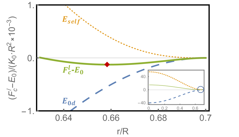
For small values of , the cap grows defect free. In Fig. 3 we plot the energy of a spherical cap for . The dotted line in the figure shows the disclination self-energy , the dashed line the Gaussian curvature-disclination interactions and the solid line is the sum of both energies as a function of the location of disclination in the cap. The diamond in the figure corresponds to the minimum of energy and indicates the location of the first (and only) disclination appearing in the cap, around . This value is very close to the geodesic distance following from the local “screening” of the Gaussian curvature such that . Somewhat counter intuitively, the first disclination does not appear at the center of the cap, which is the result of the competition between the disclination self-energy whose minimum is at the boundary and the Gaussian curvature-disclination interaction with its minimum energy occurring at the cap center, see Fig. S2 in SI Appendix where the contourplots of the different elastic free energies as a function of the location of the first disclination, are shown. As the shell grows, the appearance of a new disclination becomes energetically favorable, a new energy valley for the formation of a new disclination emerges, as illustrated in Fig. 4b, where we show the contour plots of total elastic energies for spherical caps with through . The bigger ball in each plot indicates the position of the latest energy well, which is where the addition of the next disclination takes place. Remarkably, both in the continuum model and simulations, during the growth process, the disclinations always appear in the positions that eventually become the vertices of an icosahedron.
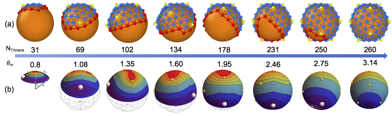
Results with the discrete model Eq. 4 are shown in Fig. 4a. Here again, the disclinations universally appear at the vertices of an icosahedron, in complete agreement with the analytical calculation. The simulations were performed for all values between and and in all cases the IO was achieved without a single error. The size of the core in Fig. 4a is commensurate with structures. We note that for these simulations the proteins spontaneous radius is much smaller than the core radius, (), a point that is discussed in more detail further below. In SI Appendix we provide a movie illustrating the growth of a structure, which includes 420 triangles.
The role of stretching and bending rigidity
Figure 5 shows the stretching energy vs (number of subunits assembled) as a shell grows for six different values of FvK . We note that for large spontaneous radius of curvature and small when bending rigidity is dominant, no large icosahedral shell assembles successfully. Rather interestingly, there are conspicuous differences in the dynamics as a function of the FvK parameter .
For small values of (thick black line in Fig. 5) the shell elastic energy grows almost linearly as a function of but does not show IO. This takes place for higher -values. The arrows in Fig. 5 indicate a drop in the elastic energy associated with the appearance of pentamers, see SI Appendix for more details. At the beginning of the growth, the shells with different values of might follow different pathways and thus, the number of hexamers vary before the first few pentamers form. However, as the shell grows, the pentamers appear precisely at the same place, independently of . Note that the bending energy of the shells always grows linearly as a function of number of subunits for any (see SI Appendix).
Discussion
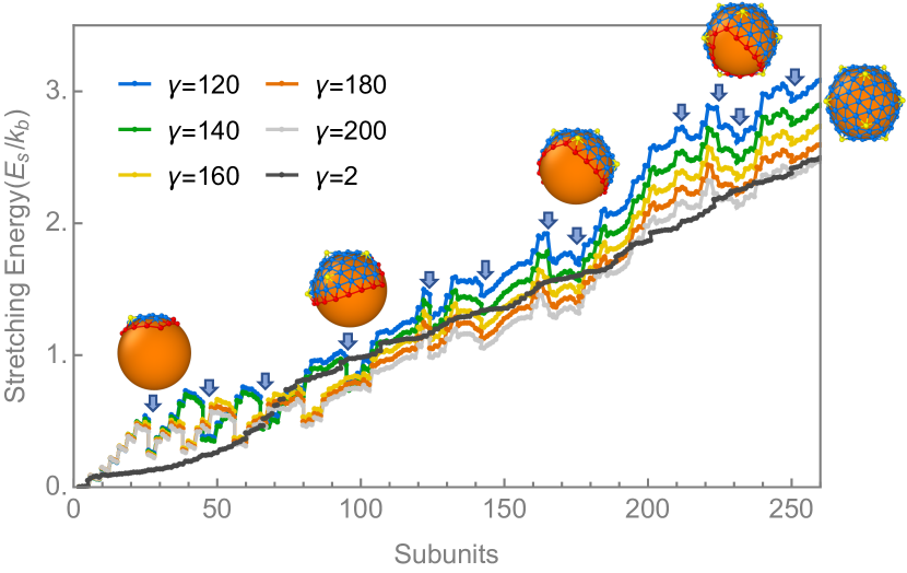
Our results show that for large shells () successful assembly into IO requires a non-specific attractive interaction between protein subunits and a template. This interaction is implicit in the continuum model and is included as a generic attractive Lennard-Jones potential in the simulations. Furthermore, we find that the location of pentamers are completely controlled by the stretching energy as it is the case in the continuum elasticity theory.
In the absence of the template, small spherical crystals ( and ) assemble spontaneously, for almost any FvK parameter . However, as we increase the spontaneous radius of curvature, the final structure depends on the value of . For small , large spherical shells without any specific symmetry form, and at large curved hexagonal sheets, which eventually assemble into tubular or conical structures are obtained. Thus our results predict that large shells with IO cannot grow without template.
A template can have a significant impact on the structure and symmetry of the shell. While a weak subunit-core attractive interaction has a minimal role in the shell shape, a very strong subunit-core interaction will override the mechanical properties of proteins. The subunits sit tightly on the template to form a sphere with no specific symmetry. We were able to observe large shells with IO only for but at high . In this regime, in order for pentamers to overcome the core attraction and form in the “correct” position, they must assume a symmetric shape and buckle up (see Fig. 1b). Indeed a strong bending energy is needed to overcome the shell adsorption. We find that without decreasing (increasing ) but with increasing spontaneous curvature, the bending energy associated with the deviation from the preferred curvature of subunits adsorbed to the core becomes strong enough to make the pentamers buckle and assume a smooth shape. Quite interestingly, we find that this is the strategy that the nature has taken to form large shells with IO.
The role of the inner core or the preformed scaffold layer presented above is very similar to the role of SPs, which assemble at the same time as the capsid proteins (CPs), i.e., the template grows simultaneously with the capsid (see Fig. 6). In fact one can think of the inner core as a permanent “inner scaffold” Coulibaly et al. (2005). For example, Bacteriophage P22 has a T=7 structure, but in the absence of scaffolding (Fig. 1, P22) often a smaller forms. Similarly, Herpesvirus makes a structure but without the SPs, a assembles. More relevant to the present study is the case of Infectious Bursal Disease Virus (IBDV) a dsRNA virus that in the presence of SPs forms a capsid but in the absence the subunits assemble to form a capsid. This is exactly the condition for formation of the structure in Fig. 4 where the preferred curvature between subunits is such that in the absence of scaffold they form a structure. Reoviridae virus family also form but they have multi-shell structures, which act as inner cores. For instance, in this family Bluetongue virus is a double capsid particle, outer (necessary for infection) and inner capsid (encloses RNA genome). The inner capsid, termed as “core” has two protein layers. The surface layer (or shell) is a capsid that assembles around the inner shell, a structure (an inner core). Interestingly, it has been suggested that there is an evolutionary connection between SPs of IBDV and inner capsid of Bluetongue virus Coulibaly et al. (2005).
Conclusions
Our model establishes that successful self-assembly of components into a spherical capsid with IO requires a template that determines the radius of the final structure. This template is very non-specific, and in its absence, protein subunits assemble into either smaller capsids or structures without IO.
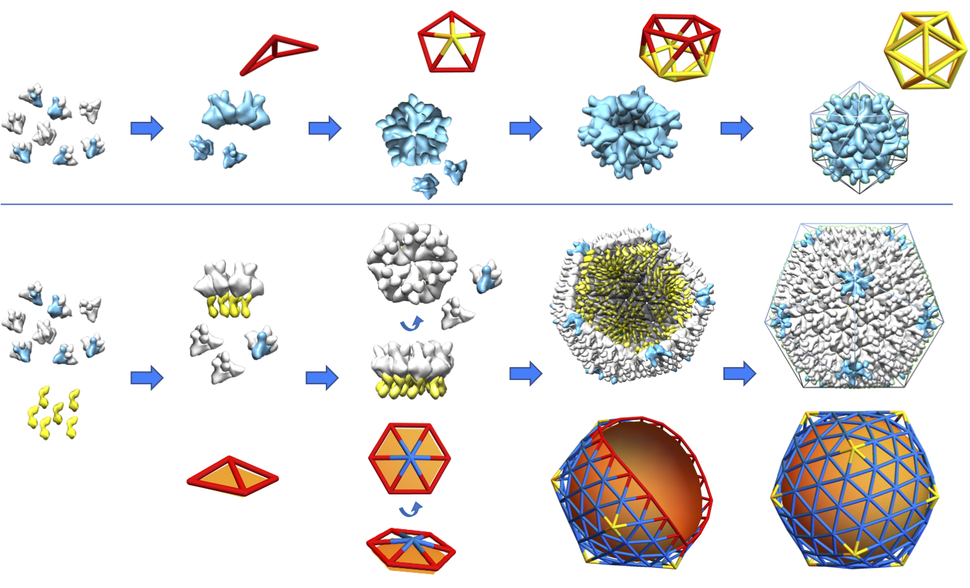
Even though the focus of the above study was on the impact of the preformed scaffolding layer, based on the experimental observations we conclude that the SPs, which assemble simultaneously with CPs (Fig. 6), play basically the same role as the inner core in the assembly of large icosahedral shells. Figure 6 shows that in the absence of SPs, CPs of IBDV form a structure but when the same IBDV proteins co-assemble with SPs (yellow units) a forms. The figure also shows the pathway of formation of and structures obtained in our simulations. We emphasize that the mechanical properties of subunits are the same for both shells, the difference in structures arises from the substrate or SPs.
The contribution of the SPs is twofold. The CPs of many viruses including bluetongue virus noted above do not assemble in the absence of SPs. On the one hand, it appears that SPs lower the energy barrier and help capsid subunits to aggregate. On the other hand, by forcing the CPs to assemble into a structure larger than their spontaneous radius of curvature, they contribute to preserving IO.
Examples of the role of templates on the formation of spherical crystals are not limited to viruses, but include crystallization of metals on nanoparticles Bao et al. (2011), solid domains on vesicles Korlach et al. (1999); Guttman et al. (2016), filament bundles Grason (2012) and colloidal assemblies at water-oil interfaces Bausch et al. (2003). Nevertheless, it has been shown Meng et al. (2014) that sufficiently rigid crystals grow as almost flat sheets free of defects, unable to assemble with IO. This regime, however, seems not to be accessible to viral capsids, as the hydrophobic interaction between monomers force close-packing structures that are incompatible with grain boundaries.
This study shed light at fundamental scale on the role of mechanical properties of building blocks and scaffolding proteins. The proposed mechanism is consistent with available experiments on viruses involving either scaffolding proteins or inner capsids. Further experiments will be necessary to validate many predictions of our described mechanism.
acknowledgements
The authors would like to thank Greg Grason for many helpful discussions. The S.L. and R.Z. work were supported by NSF Grant No. DMR-1719550 and A.T. by NSF Grant No. DMR-1606336. PR is funded through Senior Investigator Award, Welcome Trust under Grant No: 100218. A.T. and R.Z. thank the Aspen Center for Physics where part of this work was done with the support of the NSF Grant No.PHY-1607611.
References
- Caspar and Klug (1962) D. L. Caspar and A. Klug, Cold Spring Harbor Symp. Quant. Biol. 27, 1 (1962).
- Bruinsma (2006) R. F. Bruinsma, Euro. Phys. J. E 19, 303 (2006).
- Fejer et al. (2010) S. Fejer, D. Chakrabarti, and D. Wales, Nano Lett 4, 219 (2010).
- Rapaport (2004) D. C. Rapaport, Phys Rev E 70, 051905 (2004).
- Perlmutter et al. (2013) J. D. Perlmutter, C. Qiao, and M. F. Hagan, Elife 2 (2013), 10.7554/eLife.00632.
- Comas-Garcia et al. (2012) M. Comas-Garcia, R. D. Cadena-Nava, A. Rao, C. M. Knobler, and W. M. Gelbart, Journal of virology 86, 12271 (2012).
- Sikkema et al. (2007) F. D. Sikkema, M. Comellas-Aragones, R. G. Fokkink, B. J. M. Verduin, J. Cornelissen, and R. J. M. Nolte, Org. Biomol. Chem. 5, 54 (2007).
- Sun et al. (2007) J. Sun, C. DuFort, M.-C. Daniel, A. Murali, C. Chen, K. Gopinath, B. Stein, M. De, V. M. Rotello, A. Holzenburg, C. C. Kao, and B. Dragnea, Proceedings of the National Academy of Sciences 104, 1354 (2007).
- Ning et al. (2016) J. Ning, G. Erdemci-Tandogan, E. L. Yufenyuy, J. Wagner, B. A. Himes, G. Zhao, C. Aiken, R. Zandi, and P. Zhang, Nature Communications 7, 13689 (2016).
- Thuman-Commike et al. (1998) P. A. Thuman-Commike, B. Greene, J. A. Malinski, J. King, and W. Chiu, Biophysical journal 74, 559 (1998).
- Earnshaw and King (1978) W. Earnshaw and J. King, Journal of molecular biology 126, 721 (1978).
- Aksyuk et al. (2015) A. A. Aksyuk, W. W. Newcomb, N. Cheng, D. C. Winkler, J. Fontana, J. B. Heymann, and A. C. Steven, MBio 6, e01525 (2015).
- Saad et al. (1999) A. Saad, Z. H. Zhou, J. Jakana, W. Chiu, and F. J. Rixon, Journal of virology 73, 6821 (1999).
- Saugar et al. (2010) I. Saugar, N. Irigoyen, D. Luque, J. L. Carrascosa, J. F. Rodríguez, and J. R. Castón, Journal of Biological Chemistry 285, 3643 (2010).
- Coulibaly et al. (2005) F. Coulibaly, C. Chevalier, I. Gutsche, J. Pous, J. Navaza, S. Bressanelli, B. Delmas, and F. A. Rey, Cell 120, 761 (2005).
- Erber and Hockney (1991) T. Erber and G. M. Hockney, Journal of Physics A: Mathematical and General 24, L1369 (1991).
- Morris et al. (1996) J. R. Morris, D. M. Deaven, and K. M. Ho, Phys. Rev. B 53, R1740 (1996).
- Zandi et al. (2004) R. Zandi, D. Reguera, R. F. Bruinsma, W. M. Gelbart, and J. Rudnick, Proceedings of the National Academy of Sciences 101, 15556 (2004).
- Hagan and Zandi (2016) M. F. Hagan and R. Zandi, Curr. Opin. Virol. 18, 36 (2016).
- Wagner and Zandi (2015) J. Wagner and R. Zandi, Biophysical journal 109, 956 (2015).
- Chen et al. (2007) T. Chen, Z. Zhang, and S. C. Glotzer, Proceedings of the National Academy of Sciences 104, 717 (2007).
- Vernizzi and Olvera de la Cruz (2007) G. Vernizzi and M. Olvera de la Cruz, Proceedings of the National Academy of Sciences 104, 18382 (2007).
- Rothemund (2006) P. W. Rothemund, Nature 440, 297 (2006).
- Yu et al. (2013) Z. Yu, M. J. Dobro, C. L. Woodward, A. Levandovsky, C. M. Danielson, V. Sandrin, J. Shi, C. Aiken, R. Zandi, T. J. Hope, et al., Journal of molecular biology 425, 112 (2013).
- Nguyen et al. (2006) T. Nguyen, R. Bruinsma, and W. Gelbart, Physical review letters 96, 078102 (2006).
- Lidmar et al. (2003) J. Lidmar, L. Mirny, and D. R. Nelson, Physical Review E 68, 051910 (2003).
- Vernizzi et al. (2011) G. Vernizzi, R. Sknepnek, and M. O. De La Cruz, Proceedings of the National Academy of Sciences 108, 4292 (2011).
- Jordan et al. (2016) P. C. Jordan, D. P. Patterson, K. N. Saboda, E. J. Edwards, H. M. Miettinen, G. Basu, M. C. Thielges, and T. Douglas, Nature chemistry 8, 179 (2016).
- Choi et al. (2008) K. H. Choi, J. McPartland, I. Kaganman, V. D. Bowman, L. B. Rothman-Denes, and M. G. Rossmann, Journal of molecular biology 378, 726 (2008).
- Pesavento et al. (2003) J. B. Pesavento, M. K. Estes, and B. Prasad, in Viral Gastroenteritis, Perspectives in Medical Virology, Vol. 9 (Elsevier, 2003) pp. 115–127.
- Zhou et al. (2000) Z. H. Zhou, M. Dougherty, J. Jakana, J. He, F. J. Rixon, and W. Chiu, Science 288, 877 (2000).
- Stroupe et al. (2014) M. E. Stroupe, T. E. Brewer, D. R. Sousa, and K. M. Jones, Virology 450, 205 (2014).
- Oksanen et al. (2017) H. M. Oksanen et al., Journal of General Virology 98, 888 (2017).
- Bowick et al. (2000) M. J. Bowick, D. R. Nelson, and A. Travesset, Physical Review B 62, 8738 (2000).
- Seung and Nelson (1988) H. Seung and D. R. Nelson, Physical Review A 38, 1005 (1988).
- Anderson et al. (2008) J. A. Anderson, C. D. Lorenz, and A. Travesset, Journal of Computational Physics 227, 5342 (2008).
- Glaser et al. (2015) J. Glaser, T. D. Nguyen, J. A. Anderson, P. Lui, F. Spiga, J. A. Millan, D. C. Morse, and S. C. Glotzer, Computer Physics Communications 192, 97 (2015).
- Grason (2012) G. M. Grason, Physical Review E 85, 031603 (2012).
- Castelnovo (2017) M. Castelnovo, Phys. Rev. E 95, 052405 (2017).
- Bao et al. (2011) H. Bao, W. Peukert, and R. K. Taylor, Advanced Materials 23, 2644 (2011).
- Korlach et al. (1999) J. Korlach, P. Schwille, W. W. Webb, and G. W. Feigenson, Proc Natl Acad Sci USA 96, 8461 (1999).
- Guttman et al. (2016) S. Guttman, Z. Sapir, M. Schultz, A. V. Butenko, B. M. Ocko, M. Deutsch, and E. Sloutskin, Proceedings of the National Academy of Sciences 113, 493 (2016).
- Bausch et al. (2003) A. R. Bausch, M. J. Bowick, A. Cacciuto, A. D. Dinsmore, M. F. Hsu, D. R. Nelson, M. G. Nikolaides, A. Travesset, and D. A. Weitz, Science 299, 1716 (2003).
- Meng et al. (2014) G. Meng, J. Paulose, D. R. Nelson, and V. N. Manoharan, Science 343, 634 (2014).