Infrared electric-field sampled frequency comb spectroscopy
Abstract
Molecular spectroscopy in the mid-infrared portion of the electromagnetic spectrum (3–25 µm) has been a cornerstone interdisciplinary analytical technique widely adapted across the biological, chemical, and physical sciences. Applications range from understanding mesoscale trends in climate science via atmospheric monitoring to microscopic investigations of cellular biological systems via protein characterization. Here, we present a compact and comprehensive approach to infrared spectroscopy incorporating the development of broadband laser frequency combs across 3–27 µm, encompassing the entire mid-infrared, and direct electric-field measurement of the corresponding near single-cycle infrared pulses of light. Utilizing this unified apparatus for high-resolution and accurate frequency comb spectroscopy, we present the infrared spectra of important atmospheric compounds such as ammonia and carbon dioxide in the molecular fingerprint region. To further highlight the ability to study complex biological systems, we present a broadband spectrum of a monoclonal antibody reference material consisting of more than 20,000 atoms. The absorption signature resolves the amide I and II vibrations, providing a means to study secondary structures of proteins. The approach described here, operating at the boundary of ultrafast physics and precision spectroscopy, provides a table-top solution and a widely adaptable technique impacting both applied and fundamental scientific studies.
Over the past fifty years, advances in infrared (IR) technologies have played a vital role in shaping our understanding of the internal structure of molecular compounds via linear and nonlinear spectroscopic methods Stuart (2004). Specifically, the advent of the Fourier-transform infrared spectrometer (FTIR) has enabled studying vital chemical and biological processes as nearly all the relevant molecules exhibit unique absorption signatures corresponding to their ro-vibrational degrees of freedom in the molecular fingerprint region Griffiths and de Haseth (2007). Probing the properties of matter in this scientifically critical spectral region has been fruitful for a wide variety of scientific and industrial applications including atmospheric monitoring Nozière et al. (2015), food quality control Sun (2009), and conservation and composition studies of natural and synthetic materials Chukanov and Chervonnyi (2016); Derrick et al. (1999). Importantly, studying biological systems via IR spectroscopy has revealed structural and conformational information in complex organic compounds such as proteins, which is of fundamental significance for applications such as drug synthesis and delivery Barth (2007). While the broadband incoherent radiation in conventional FTIR has been sufficient for many of these studies, several fundamental scientific applications and next-generation analytical techniques require high-brightness, coherent sources of MIR light, along with commensurate rapid acquisition and high signal-to-noise ratio (SNR) measurement methodologies Pires et al. (2015).
Recently, coherent IR spectro-imaging of biological specimens has been proposed as a complementary technique in clinical pathology that could result in stain-free histology and label-free cellular analysis Katon (1996); Fernandez et al. (2005); Bellisola and Sorio (2011); Clemens et al. (2014). Moreover, near-field analytical techniques such as atomic force microscopy in the infrared (AFM-IR) enable nanoscale imaging of single proteins Keilmann and Hillenbrand (2004); Dazzi and Prater (2017). In addition to biologically relevant molecular compounds, near-field probing of synthetic polymers and exotic quantum matter using techniques such as scattering scanning near-field optical microscopy (sSNOM) also reveal fundamental structural information useful for material science Bechtel et al. (2014). These revolutionary applications place exacting criteria on the combination of the infrared sources and detection apparatus—requiring high-brightness coherent broadband light and high-SNR readout of the IR spectra. This has meant accessing IR synchrotron beamlines and using cryogenically-cooled detectors for low-noise operation, thereby placing significant constraints on accessibility, mobility, and cost Khatib et al. (2018).
Circumventing these limitations, we present an ultrabroadband source of laser frequency combs spanning 3–27 µm, which produces near-single-cycle pulses of light. Importantly, due to the enhanced peak power in such pulses, we use a nonlinear optical readout based on electro-optic sampling (EOS) to directly measure the IR electric fields with room-temperature telecom-grade InGaAs photodiodes. This high-SNR electric field measurement enables high-sensitivity and broadband IR spectroscopy with a large dynamic range. Moreover, we incorporate a dual frequency comb implementation of EOS (henceforth dual-comb EOS) Bartels et al. (2007), enabling electric-field measurement at a rate of 50 Hz, with high temporal (5 fs) and spectral (100 MHz) resolutions. This unique combination of video-rate acquisition and high resolution represents a significant improvement (by at least an order-of-magnitude) over prior MIR-EOS demonstrations Sell et al. (2008); Riek et al. (2015); Pupeza et al. (2017). This cohesive approach is a compact, table-top alternative to large IR synchrotron beamlines and broadband incoherent thermal sources in conventional FTIR systems.
Generation of the broadband infrared light relies on nonlinear frequency conversion processes driven by stabilized erbium fiber frequency combs. The frequency comb architecture enables transferring the exceptional frequency accuracy and stability of the 100 MHz spaced NIR comb teeth to all portions of the electromagnetic spectrum Schliesser et al. (2012). Harnessing these advantages, dual frequency comb spectroscopy (DCS) has been demonstrated as a high-resolution, fast-acquisition alternative to FTIR Coddington et al. (2016). Recently, high-resolution quantitative spectroscopy has been demonstrated in both the 3–5 µm and the 6–12 µm portions of the mid-infrared using DCS Kara et al. (2017); Maidment et al. (2018); Muraviev et al. (2018); Ycas et al. (2018); Timmers et al. (2018). However, for low-noise operation, DCS experiments have all required cryogenically cooled infrared photodetectors, which exhibit a limited range of linearity—typically limited to sub-milliwatt optical powers. This fundamentally limits the achievable signal-to-noise ratio Newbury et al. (2010).
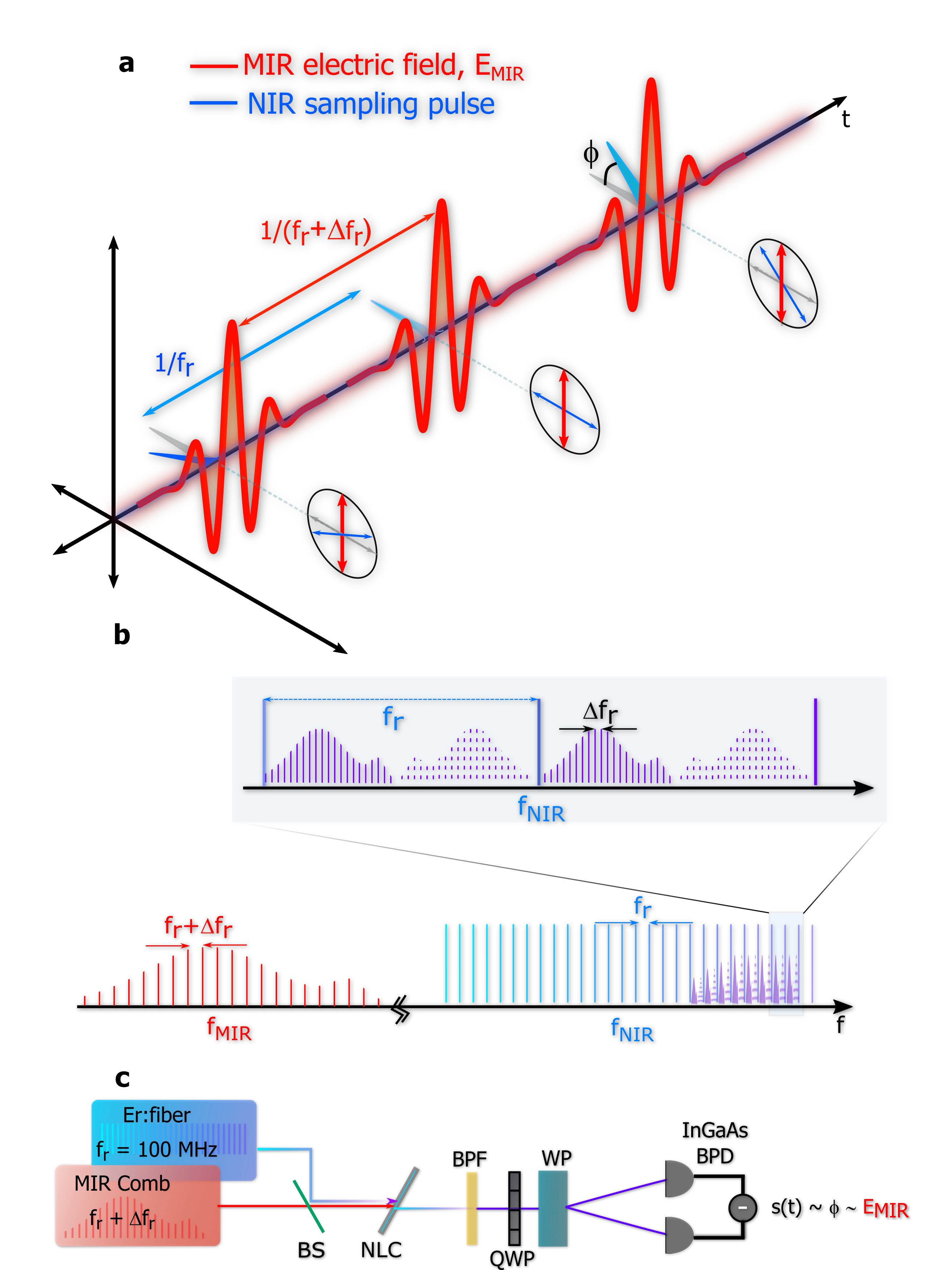
Here, we utilize the combination of difference-frequency generation (DFG) and sum-frequency generation (SFG) to generate and detect the infrared frequency combs. The generation of the infrared light is based on intra-pulse DFG in quadratic nonlinear media, which provides ultrashort infrared pulses with an inherently stabilized carrier-envelope phase (CEP) Baltuška et al. (2002). The CEP stability enables a direct nonlinear optical readout of the MIR electric field via electro-optic sampling Wu and Zhang (1995); Sell et al. (2008). In EOS, the MIR electric field, with a carrier frequency , modulates the polarization of an ultra-short NIR sampling pulse in an electro-optic crystal, as shown in Fig. 1(a). The polarization modulation is directly proportional to the MIR field amplitude and occurs due to SFG between the NIR and MIR pulses. The sum-frequency NIR components and the input sampling pulse are orthogonally polarized, which provides a polarization rotation Sell et al. (2008); Pupeza et al. (2015). As the time delay is scanned between the two pulses, a complete measurement of the electric field is made.
Previous EOS experiments have shown its advantages for spectroscopy Sell et al. (2008) and measurements with spectral dynamic range 110 dB have been reported Pupeza et al. (2017). Additionally, it has improved sensitivity relative to FTS Huber et al. (2017) with the ability to directly measure vacuum fluctuationsRiek et al. (2015). However, in all such cases the delay between the NIR and MIR pulses is varied using a mechanical translation stage, restricting the range of measurement and acquisition time to centimeter- and second-scales, respectively. On the other hand, in dual-comb EOS, fast acquisition of high-resolution infrared spectra is feasible as the sampling pulse and the MIR light are acquired from two different, mutually phase-locked Er:fiber laser frequency combs with a small offset in repetition rates ( = 50 Hz) as shown in Fig. 1(a). Unlike conventional frequency-comb-enabled linear optical sampling Coddington et al. (2009a, b, 2010), the down-sampled comb folds the entire MIR spectrum into each FSR of the NIR comb in dual frequency comb EOS (Fig. 1(b),Stead et al. (2012)). This is similar to the case of the stimulated Raman experiments performed with a dual comb configuration Ideguchi et al. (2013). Thus, the coherent multi-heterodyne beating occurs in the NIR, with the sampling pulse serving as a local-oscillator comb. For this reason, the sampling pulse must have a bandwidth greater than the highest MIR frequency component to be sampled Riek et al. (2015). In the time-domain, this means the sampling pulse duration is short compared to the optical cycle . The experimental implementation of EOS is shown in Fig. 1(c). We utilize a 10 fs, 1.55-µm sampling pulse Timmers et al. (2018), ensuring Nyquist-limited sampling of electric fields corresponding to 3 µm (3300 cm-1) light.
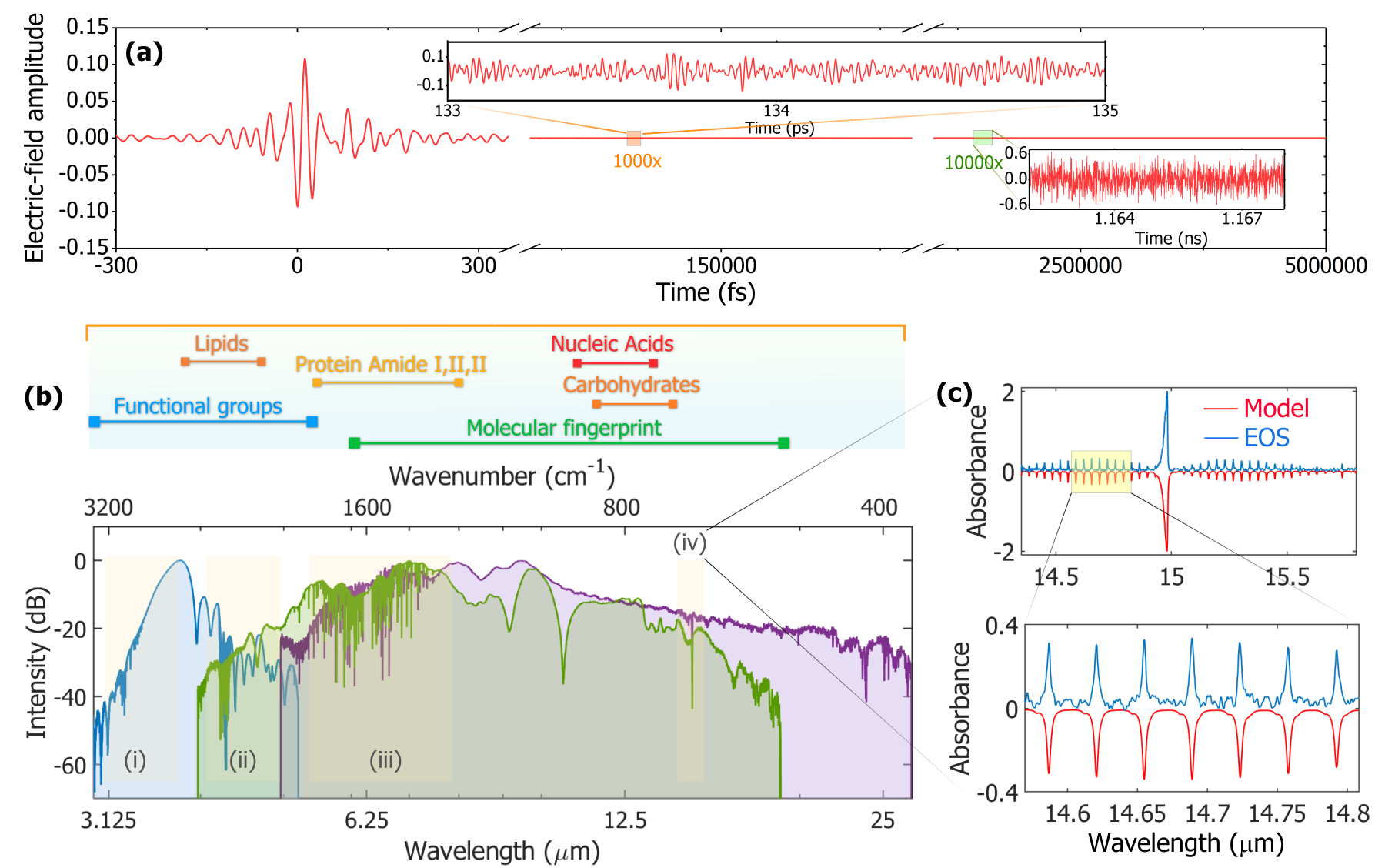
Coherent infrared radiation is generated via intra-pulse DFG in three different nonlinear crystals—periodically poled lithium niobate (PPLN), orientation-patterned gallium phosphide (OP-GaP), and gallium selenide (GaSe). We leverage quasi phase-matching in PPLN and OP-GaP and birefringence phase-matching in GaSe. Due to favorable dispersion and millimeter-scale crystal propagation lengths, the broadband phase-matching bandwidths are realized in all configurations. Correspondingly, in the time-domain, ultrashort femtosecond MIR pulses are generated. Owing to the enhanced peak power, the nonlinear optical readout of the electric field occurs with signal-to-noise ratio (SNR) exceeding 104 in 20 minutes of averaging, corresponding to a single-shot SNR . For the MIR comb generated in OP-GaP, the anomalous dispersion from crystal propagation is compensated by the normal dispersion from the germanium beam-splitter, and results in a near-single-cycle oscillation (Fig. 2a). The observed time-domain pulse is 1.2 cycles in duration, corresponding to 29 fs, with a carrier wavelength of 7.6 µm (1316 cm-1). Unique to dual-comb EOS, the electric field is measured with a 5-fs resolution over a 10-ns window, corresponding to a dynamic range in temporal duration of 60 dB (Fig. 2a).
The measurements described here are exceptionally broad in bandwidth, with the spectral coverage rivaling a thermal source in a commercial Fourier-transform infrared spectrometer. The frequency domain SNR scales with power, bandwidth, and spectral resolution and for 100 MHz resolution, we report SNR = 0.9 Hz1/2 over a recovered bandwidth of nearly 75 THz. The noise in the system stems from the shot noise of the sampling pulse and excess electrical noise from the balanced photo-detector. High-linearity InGaAs photodetectors would alleviate this issue. Due to the expansive bandwidth in the system, the SNR is smaller compared to dual-comb spectrometers Ycas et al. (2018); Timmers et al. (2018). However, the figure-of-merit (FOM, ), accounting for the number of comb-teeth (M) is comparable to other demonstrations. For the OP-GaP IR comb, corresponding approximately to 106 comb teeth, the FOM is 106, which is within a factor of 2 from the best MIR DCS performance.
Owing to the high SNR in the temporal measurement, the molecular free-induction decay of trace absorbents such as methane and ethane, along with ambient water vapor and carbon dioxide are also obtained via direct electric-field sampled spectroscopy (Fig. 2b). In particular, 1 GHz (0.033 cm-1) resolution vibrational spectra of the P, Q, and R branches of the O–C–O bending vibration around 15 µm (670 cm-1) are presented in Fig. 2c, along with a comparison to theory (HITRAN2012). Notably, this spectral region is beyond the detection range of current state-of-the-art high-speed mercury cadmium telluride (HgCdTe) photodetectors. While 1 GHz resolution is sufficient for pressure-broadened gases, higher resolution is desired for many precision measurements.
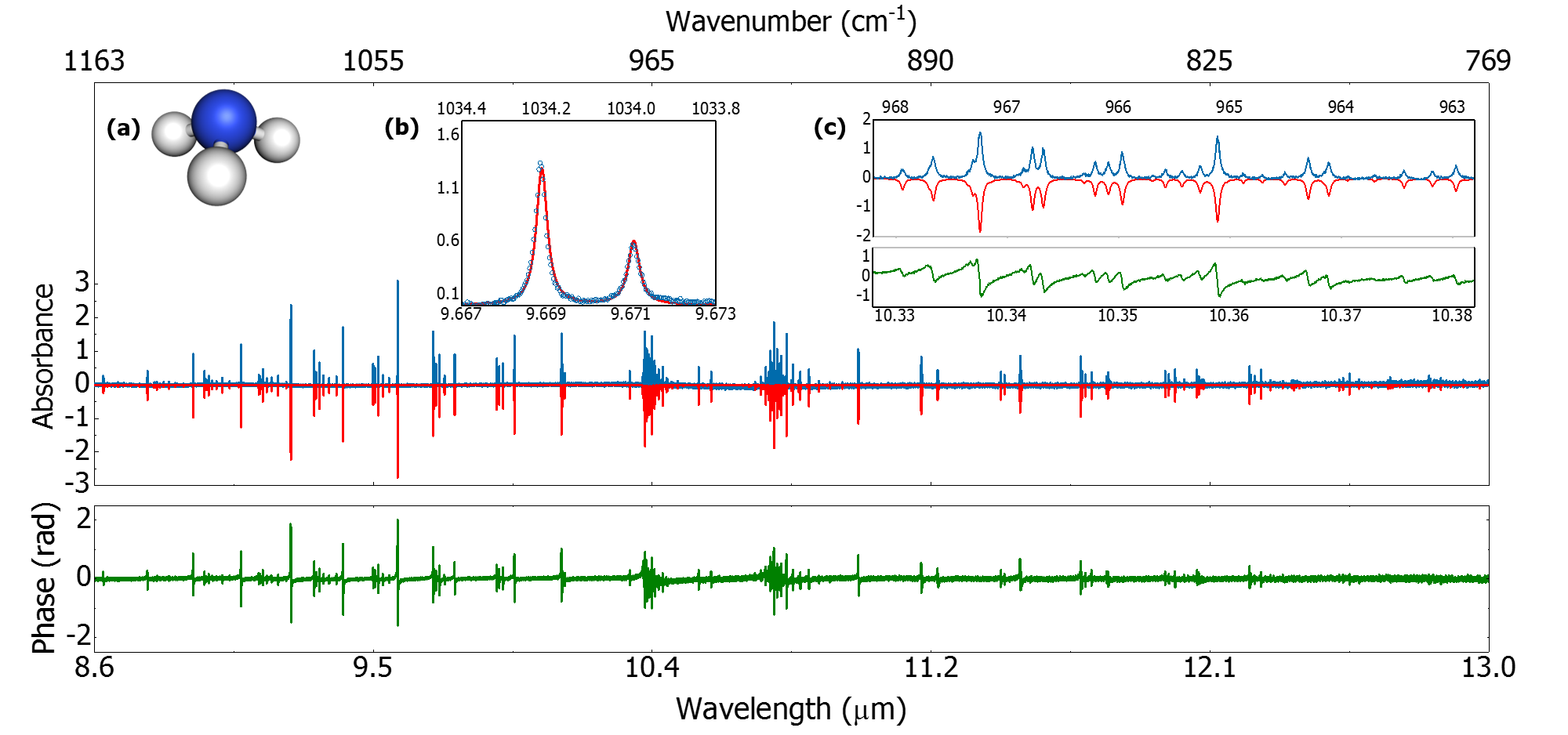
To demonstrate this, we present high-resolution absortion spectra of the vibration (A1 symmetric bend) in ammonia with 100 MHz resolution (Fig. 3). Ammonia is a harmful substance widely used in the agricultural and bio-pharmaceutical industries Timmer et al. (2005); Schliesser et al. (2005), and is an important contributor to atmospheric aerosol formation Kirkby et al. (2011). We observe the splitting in the Q-branch due to pyramidal inversion of the NH3 molecule, where the nitrogen atom undergoes room-temperature quantum tunneling through the potential barrier formed by the H3 plane. To acquire this broadband spectrum, the OP-GaP MIR comb is transmitted through a 15-cm-long gas cell, filled with NH3 (7.5 mbar partial pressure) and ambient air (150 mbar background pressure). The transmitted light is detected via EOS. Fourier transformation of the sampled electric field and subtraction of a reference spectrum collected without the gas cell yields the high-resolution absorption spectrum and phase response shown in Fig. 3a. In this spectrum spanning 8.6–13 µm, 118000 comb teeth are measured. In Fig. 3b, every absorption feature is resolved with individual comb teeth, which are stabilized to the 10 kHz level. The frequency accuracy of the stabilized comb teeth is , limited only by the microwave reference for the repetition rate, providing excellent agreement with the HITRAN2012 theoretical model (Fig. 3c).
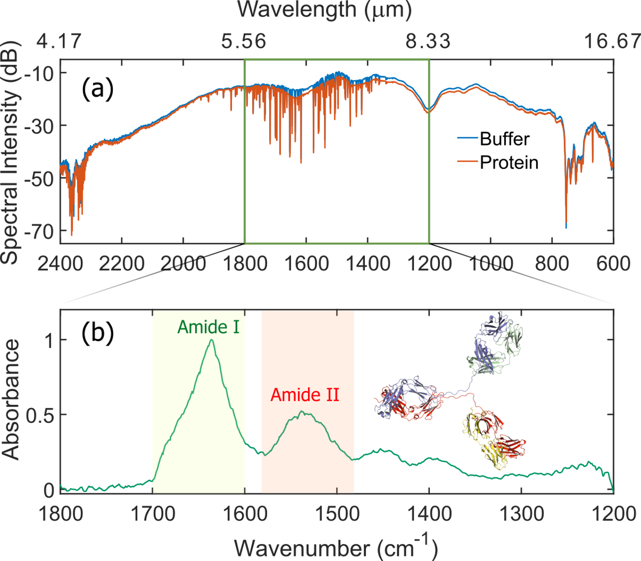
Gas-phase frequency comb spectroscopy of small molecules has been a useful diagnostic tool for atmospheric monitoring, probing complex molecules at cryogenic temperatures, and for studying temporal dynamics of chemical reactions Cossel et al. (2017); Spaun et al. (2016); Fleisher et al. (2014). In contrast, biochemically relevant compounds, such as proteins, often have tens of thousands of atoms, and exhibit interesting infrared spectroscopic signatures in liquid- and solid phases. Here, we use the high-brightness frequency comb to perform infrared spectroscopy of the NISTmAb (Fig. 4), an important reference antibody used in the biopharmaceutical industries Dong et al. (2018). Studying the amide vibrations can aid in determining the protein folding, unfolding, and aggregating mechanisms, important for example, in characterizing drug synthesis and delivery Barth (2007); Baker et al. (2008, 2014). We present the absorption spectra of the amide I and II bands in a NISTmAb sample (100 mg/mL), dissolved in L-Histidine buffer (pH: 6.0). The antibody was deposited as a thin film on a 3-mm-thick ZnSe window. A reference was acquired by depositing the buffer onto a separate ZnSe window. Due to the broad features, the electric field signal was temporally apodized to provide 1 cm-1 (30 GHz) resolution for the 600 cm-1-span spectrum. The experiment was conducted in room-temperature ambient conditions and resulting water vapor lines are removed via referencing (Fig. 4, top panel). The center frequency of the amide I band at 1636 cm-1, indicative of a -sheet structure, and the amide II band at 1549 cm-1 agree well with prior measurements and theoretical predictions Gokarn et al. (2015). The IR spectrum in Fig. 4 is the average of three measurements, corresponding to a total averaging time of 66 minutes. Notably, the NISTmAb consists of greater than 20,000 atoms, making it one of the largest complex molecules probed with frequency comb spectroscopy.
In conclusion, we presented a high-brightness ultrafast source of mid-infrared radiation and presented a versatile detection technique using dual frequency comb electro-optic sampling. Inheriting the robustness and simplicity from the near-infrared pulses derived from erbium fiber lasers, the compact source provides a table-top footprint for IR spectroscopy in the molecular fingerprint region and beyond. Furthermore, the use of room-temperature InGaAs photodetectors renders this system flexible and reliable for field-based IR spectroscopy and metrology. Along with precision, high-resolution spectroscopy of atmospheric carbon dioxide and ammonia gas in a spectral region beyond the reach of high-speed HgCdTe detectors, we presented the IR absorption spectrum of the NISTmAb with resolved amide vibrations, crucial for understanding and characterizing its secondary structure. As next steps, we anticipate that combined with novel imaging techniques such as AFM-IR and sSNOM, the apparatus described here can enable in-situ spectro-imaging of biological specimens. Moreover, the near single-cycle infrared pulses can serve as robust seeds for optical parametric chirped-pulse amplifiers in strong-field physics Wolter et al. (2015).
We thank John Schiel and Trina Mouchanoir for providing the NISTmAb samples and Daniel Lesko for providing the NH3 sample. We thank David Carlson, Nathan Newbury, Marissa Weichman, Curtis Meuse, and Kevin Cossel for providing useful comments on the manuscript, and Albrecht Bartels and Alfred Leitenstorfer for advice on the experiments. The funding for this work was provided by Defense Advanced Research Projects Agency (DARPA), National Institute of Standards and Technology (NIST), National Science Foundation (NSF) (1708743), National Research Council (NRC), and the Air Force Office of Scientific Research (AFOSR) (FA9550-16-1-0016). U.E. and J.B. acknowledge funding through the “Severo Ochoa” Programme for Centres of Excellence in R&D (SEV-2015-0522), MINECO “FIS2017-89536-P”, AGAUR “2017 SGR 1639” and ERC Advanced Grant “TRANSFORMER”, (788218).This work is a contribution of the United States government and is not subject to copyright in the United States of America.
References
- Stuart (2004) B. Stuart, Infrared Spectroscopy: Fundamentals and Applications, 1st ed., Analytical Techniques in the Sciences (Wiley Interscience, 2004).
- Griffiths and de Haseth (2007) P. G. Griffiths and J. A. de Haseth, Fourier transform infrared spectrometry, 2nd ed. (Wiley Interscience, 2007).
- Nozière et al. (2015) B. Nozière, M. Kalberer, M. Claeys, J. Allan, B. D’Anna, S. Decesari, E. Finessi, M. Glasius, I. Grgić, J. F. Hamilton, T. Hoffmann, Y. Iinuma, M. Jaoui, A. Kahnt, C. J. Kampf, I. Kourtchev, W. Maenhaut, N. Marsden, S. Saarikoski, J. Schnelle-Kreis, J. D. Surratt, S. Szidat, R. Szmigielski, and A. Wisthaler, Chemical Reviews 115, 3919 (2015).
- Sun (2009) D.-W. Sun, Infrared Spectroscopy for Food Quality Analysis and Control, 1st ed. (Elsevier, 2009).
- Chukanov and Chervonnyi (2016) N. V. Chukanov and A. D. Chervonnyi, Infrared Spectroscopy of Minerals and Related Compounds, 1st ed., Springer Minerology (Springer International Publishing, 2016).
- Derrick et al. (1999) M. Derrick, D. Stulik, and J. M. Landry, Infrared Spectroscopy in Conservation Science, Scientific Tools for Conservation (The Getty Conservation Institute, 1999).
- Barth (2007) A. Barth, Biochimica et Biophysica Acta (BBA) - Bioenergetics 1767, 1073 (2007).
- Pires et al. (2015) H. Pires, M. Baudisch, D. Sanchez, M. Hemmer, and J. Biegert, Progress in Quantum Electronics 43, 1 (2015).
- Katon (1996) J. E. Katon, Micron 27, 303 (1996).
- Fernandez et al. (2005) D. C. Fernandez, R. Bhargava, S. M. Hewitt, and I. W. Levin, Nature Biotechnology 23, 469 (2005).
- Bellisola and Sorio (2011) G. Bellisola and C. Sorio, American Journal of Cancer Research 2, 1 (2011).
- Clemens et al. (2014) G. Clemens, J. R. Hands, K. M. Dorling, and M. J. Baker, Analyst 139, 4411 (2014).
- Keilmann and Hillenbrand (2004) F. Keilmann and R. Hillenbrand, Philosophical Transactions of the Royal Society of London A: Mathematical, Physical and Engineering Sciences 362, 787 (2004).
- Dazzi and Prater (2017) A. Dazzi and C. B. Prater, Chemical Reviews 117, 5146 (2017).
- Bechtel et al. (2014) H. A. Bechtel, E. A. Muller, R. L. Olmon, M. C. Martin, and M. B. Raschke, Proceedings of the National Academy of Sciences 111, 7191 (2014).
- Khatib et al. (2018) O. Khatib, H. A. Bechtel, M. C. Martin, M. B. Raschke, and G. L. Carr, ACS Photonics 5, 2773 (2018).
- Bartels et al. (2007) A. Bartels, R. Cerna, C. Kistner, A. Thoma, F. Hudert, C. Janke, and T. Dekorsy, Review of Scientific Instruments 78, 035107 (2007).
- Sell et al. (2008) A. Sell, R. Scheu, A. Leitenstorfer, and R. Huber, Applied Physics Letters 93, 251107 (2008).
- Riek et al. (2015) C. Riek, D. V. Seletskiy, A. S. Moskalenko, J. F. Schmidt, P. Krauspe, S. Eckart, S. Eggert, G. Burkard, and A. Leitenstorfer, Science 350, 420 (2015).
- Pupeza et al. (2017) I. Pupeza, M. Hubert, W. Schweinberger, M. Trubetskov, S. A. Hussain, L. Vamos, O. Pronin, F. Habel, V. Pervak, N. Karpowicz, E. Fill, A. Apolonski, M. Zigman, A. M. Azzeer, and F. Krausz, in 2017 Conference on Lasers and Electro-Optics Europe European Quantum Electronics Conference (CLEO/Europe-EQEC) (2017) pp. 1–1.
- Schliesser et al. (2012) A. Schliesser, N. Picque, and T. W. Hansch, Nat Photon 6, 440 (2012).
- Coddington et al. (2016) I. Coddington, N. Newbury, and W. Swann, Optica 3, 414 (2016).
- Kara et al. (2017) O. Kara, L. Maidment, T. Gardiner, P. G. Schunemann, and D. T. Reid, Optics Express 25, 32713 (2017).
- Maidment et al. (2018) L. Maidment, O. Kara, P. G. Schunemann, J. Piper, K. McEwan, and D. T. Reid, Applied Physics B 124, 143 (2018).
- Muraviev et al. (2018) A. V. Muraviev, V. O. Smolski, Z. E. Loparo, and K. L. Vodopyanov, Nature Photonics 12, 209 (2018).
- Ycas et al. (2018) G. Ycas, F. R. Giorgetta, E. Baumann, I. Coddington, D. Herman, S. A. Diddams, and N. R. Newbury, Nature Photonics 12, 202 (2018).
- Timmers et al. (2018) H. Timmers, A. Kowligy, A. Lind, F. C. Cruz, N. Nader, M. Silfies, G. Ycas, T. K. Allison, P. G. Schunemann, S. B. Papp, and S. A. Diddams, Optica 5, 727 (2018).
- Newbury et al. (2010) N. R. Newbury, I. Coddington, and W. Swann, Optics Express 18, 7929 (2010).
- Baltuška et al. (2002) A. Baltuška, T. Fuji, and T. Kobayashi, Physical Review Letters 88, 133901 (2002).
- Wu and Zhang (1995) Q. Wu and X.-C. Zhang, Applied Physics Letters 67, 3523 (1995).
- Pupeza et al. (2015) I. Pupeza, D. Sánchez, J. Zhang, N. Lilienfein, M. Seidel, N. Karpowicz, T. Paasch-Colberg, I. Znakovskaya, M. Pescher, W. Schweinberger, V. Pervak, E. Fill, O. Pronin, Z. Wei, F. Krausz, A. Apolonski, and J. Biegert, Nature Photonics 9, 721 (2015).
- Huber et al. (2017) M. Huber, W. Schweinberger, M. Trubetskov, S. A. Hussain, O. Pronin, L. Vamos, E. Fill, A. Apolonski, M. Zigman, F. Krausz, and I. Pupeza, in 2017 Conference on Lasers and Electro-Optics Europe European Quantum Electronics Conference (CLEO/Europe-EQEC) (2017) pp. 1–1.
- Coddington et al. (2009a) I. Coddington, W. C. Swann, L. Nenadovic, and N. R. Newbury, Nature Photonics 3, 351 (2009a).
- Coddington et al. (2009b) I. Coddington, W. C. Swann, and N. R. Newbury, Optics Letters 34, 2153 (2009b).
- Coddington et al. (2010) I. Coddington, W. C. Swann, and N. R. Newbury, Optics Letters 35, 1395 (2010).
- Stead et al. (2012) R. A. Stead, A. K. Mills, and D. J. Jones, JOSA B 29, 2861 (2012).
- Ideguchi et al. (2013) T. Ideguchi, S. Holzner, B. Bernhardt, G. Guelachvili, N. Picqué, and T. W. Hänsch, Nature 502, 355 (2013).
- Timmer et al. (2005) B. Timmer, W. Olthuis, and A. van den Berg, Sensors and Actuators B: Chemical 107, 666 (2005).
- Schliesser et al. (2005) A. Schliesser, M. Brehm, F. Keilmann, and D. W. van der Weide, Opt. Express 13, 9029 (2005).
- Kirkby et al. (2011) J. Kirkby, J. Curtius, J. Almeida, E. Dunne, J. Duplissy, S. Ehrhart, A. Franchin, S. Gagné, L. Ickes, A. Kürten, A. Kupc, A. Metzger, F. Riccobono, L. Rondo, S. Schobesberger, G. Tsagkogeorgas, D. Wimmer, A. Amorim, F. Bianchi, M. Breitenlechner, A. David, J. Dommen, A. Downard, M. Ehn, R. C. Flagan, S. Haider, A. Hansel, D. Hauser, W. Jud, H. Junninen, F. Kreissl, A. Kvashin, A. Laaksonen, K. Lehtipalo, J. Lima, E. R. Lovejoy, V. Makhmutov, S. Mathot, J. Mikkilä, P. Minginette, S. Mogo, T. Nieminen, A. Onnela, P. Pereira, T. Petäjä, R. Schnitzhofer, J. H. Seinfeld, M. Sipilä, Y. Stozhkov, F. Stratmann, A. Tomé, J. Vanhanen, Y. Viisanen, A. Vrtala, P. E. Wagner, H. Walther, E. Weingartner, H. Wex, P. M. Winkler, K. S. Carslaw, D. R. Worsnop, U. Baltensperger, and M. Kulmala, Nature 476, 429 (2011).
- Cossel et al. (2017) K. C. Cossel, E. M. Waxman, I. A. Finneran, G. A. Blake, J. Ye, and N. R. Newbury, JOSA B 34, 104 (2017).
- Spaun et al. (2016) B. Spaun, P. B. Changala, D. Patterson, B. J. Bjork, O. H. Heckl, J. M. Doyle, and J. Ye, Nature 533, 517 (2016).
- Fleisher et al. (2014) A. J. Fleisher, B. J. Bjork, T. Q. Bui, K. C. Cossel, M. Okumura, and J. Ye, The Journal of Physical Chemistry Letters 5, 2241 (2014).
- Dong et al. (2018) Q. Dong, Y. Liang, X. Yan, S. P. Markey, Y. A. Mirokhin, D. V. Tchekhovskoi, T. H. Bukhari, and S. E. Stein, mAbs 10, 354 (2018).
- Baker et al. (2008) M. J. Baker, E. Gazi, M. D. Brown, J. H. Shanks, P. Gardner, and N. W. Clarke, British Journal of Cancer 99, 1859 (2008).
- Baker et al. (2014) M. J. Baker, J. Trevisan, P. Bassan, R. Bhargava, H. J. Butler, K. M. Dorling, P. R. Fielden, S. W. Fogarty, N. J. Fullwood, K. A. Heys, C. Hughes, P. Lasch, P. L. Martin-Hirsch, B. Obinaju, G. D. Sockalingum, J. Sulé-Suso, R. J. Strong, M. J. Walsh, B. R. Wood, P. Gardner, and F. L. Martin, Nature Protocols 9, 1771 (2014).
- Gokarn et al. (2015) Y. Gokarn, S. Agarwal, K. Arthur, A. Bepperling, E. S. Day, D. Filoti, D. G. Greene, D. Hayes, R. Kroe-Barrett, T. Laue, J. Lin, B. McGarry, V. Razinkov, S. Singh, R. Taing, S. Venkataramani, W. Weiss, D. Yang, and I. E. Zarraga, in State-of-the-Art and Emerging Technologies for Therapeutic Monoclonal Antibody Characterization Volume 2. Biopharmaceutical Characterization: The NISTmAb Case Study, ACS Symposium Series, Vol. 1201 (American Chemical Society, 2015) pp. 285–327.
- Wolter et al. (2015) B. Wolter, M. G. Pullen, M. Baudisch, M. Sclafani, M. Hemmer, A. Senftleben, C. D. Schröter, J. Ullrich, R. Moshammer, and J. Biegert, Physical Review X 5, 021034 (2015).