Solvation Effects Alter the Photochemistry of 2-Thiocytosine
Abstract
Radiationless deactivation channels of 2-thiocytosine in aqueous environment are revisited by means of quantum-chemical simulations of excited-state absorption spectra, and investigations of potential energy surfaces of the chromophore clustered with two water molecules using the algebraic diagrammatic construction method to the second-order (ADC(2)), and multireference configuration interaction with single and double excitations (MR-CISD) methods. We argue that interactions of explicit water molecules with thiocarbonyl group might enable water-chromophore electron transfer (WCET) which leads to formation of intersystem crossing that was not considered previously. This is the first example of a WCET process occurring in the triplet manifold of electronic states. This phenomenon might explain nonradiative decay of the triplet state population observed in thiopyrimidines in the absence of molecular oxygen. According to our calculations this WCET process might also entail a subsequent, virtually barrierless, electron-driven proton transfer (EDPT) resulting in the formation of hydroxyl radical which could further participate in photohydration or deamination reactions.
1 Introduction
Photochemistry and photophysics of thiated nucleobases has recently attracted increasing attention, owing to to their potential use as photosensitizers in pharmacological applications [1], nanotechnology [2], and their intriguing properties in promoting prebiotically credible chemical reactions and processes [3, 4, 5, 6, 7]. For instance, -2-thioribocytidine was recently demonstrated to be a crucial intermediate in the photochemical synthesis of -ribonucleosides under the conditions of the early Earth [7]. The prebiotic background of thiated nucleobases is also supported by presence of naturally occurring thiobases in mitochondrial tRNA of some bacteria and yeasts [8, 9, 10]. However, the main focus on thiated nucleobases stems from their potential applications in photochemotherapy [11, 12, 13], photodynamic therapies in treating skin disorders [14], superficial tumors [15, 16] and bladder cancer [12]. It is worth noting that thioguanine is already used as a therapeutic agent for lymphoblastic leukemia [17] and breast cancer [18], while 2-thiocytosine influences the mitosis of human lymphocytes [19] and shows cytotoxic [20], anticancer [21] and antibacterial activity [22].
Characterization of photochemical and photophysical properties of thiated nucleobases is crucial for understanding the mechanisms governing their phototherapeutic activity and photoinduced chemical reactions. In the past decade, various spectroscopic and theoretical studies aimed to scrutinize the photophysics and photochemistry of thiocytosine, thioguanine, thiouracil, and thiothymine [23, 24, 25, 26, 27, 28, 29, 30, 31, 13, 32, 33, 34]. It is now well established that the most attractive features of thionucleobases are their structural similarity to canonical nucleobases combined with considerable singlet-triplet spin-orbit couplings (SOC) that enables efficient population of long-lived triplet states and efficient generation of singlet oxygen [35, 1]. Recently joint transient absorption spectroscopy (TAS) and computational studies of Mai et al. [33] revealed that near unity triplet yields often observed in thionucleobases may also be explained by relatively high energies and consequently lower accessibility of S1/S0 conical intersections when compared to the lowest-energy intersystem crossings.
Although many aspects of the photophysics of thionucleobases are now quite well understood, previous computational studies focused predominantly on isolated chromophores. This may be a serious limitation obscuring interpretation of the experimental results, since recently there is a growing amount of data suggesting that water molecules may actively participate in photochemistry of hydrated heterocycles by modifying state crossings and, more importantly, opening new photorelaxation channels which are not accessible in the gas phase. The examples include microhydrated adenine [36, 37, 38, 39], cytosine and cytidine [40], eumelanine [41], and azole chromophores [42, 43]. For instance, Barbatti demonstrated that water molecules may stabilize the excited state in 7H-adenine through the interaction of the orbital of adenine with the pz orbital of the neighbouring water molecule [37]. This permits radiationless deactivation through the / state crossing, which is induced by water-to-chromophore electron transfer (WCET). More recently, some of us suggested that the long-lived dark state observed experimentally in aqueous cytidines might correspond to the excited state also involved in WCET [40]. Consequently, understanding how the surrounding water molecules could affect the photochemistry of thionucleobases (e.g. thiocytosine) is the obvious next step in providing their complete photochemical characteristics in native environments.
In this work, we show that microhydration of 2-thiocytosine by two water molecules [2tCyt-] may enable a similar WCET process, which could lead to the formation of yet another intersystem crossing in microsolvated 2tCyt that was not considered previously. This phenomenon might also explain nonradiative decay of the triplet state population observed in thiopyrimidines in the absence of molecular oxygen [35].
2 Computational methods
The ground-state minimum energy geometries and harmonic vibrational frequencies of microhydrated 2tCyt were computed at the MP2/cc-pVTZ level [44, 45, 46, 47]. The stationary points on excited state potential energy surfaces were located using the algebraic diagrammatic construction to the second order method [ADC(2)] method [48, 49, 50] and the cc-pVTZ basis set as implemented in the Turbomole 7.1 package [51].
Vertical excitation energies were obtained at the ADC(2)/cc-pVTZ level, assuming the S0 geometry optimized using the MP2/cc-pVTZ method. Solvation effects on the vertical excitation energies exerted by bulk water were estimated using the non-equilibrium polarizable continuum solvation model (PCM) combined with ADC(2)/cc-pVTZ method which are implemented in the Q-Chem 5.0 package [52]. The perturbed state specific (ptSS) approach was used either in a fully consistent perturbation-energy-and-density (PTED) or perturbation-density (PTD) variant [53, 54]. The orbital character of the considered excited states was assigned based on natural transition orbitals [55] (NTOs) obtained using TheoDore package [56]. The potential energy (PE) profiles for WCET mechanism were calculated using the ADC(2) and MP2 methods for the excited and ground electronic states, respectively, using the Turbomole 7.1 package. The PE scans were performed based on linear interpolation in internal coordinates (LIIC) between the appropriate stationary geometries.
The minimum-energy crossing points (MECPs) were located using the sequential penalty constrained optimization method proposed by Levine et al. [57] employing energies and analytical gradients computed at the ADC(2) and MP2 levels, for the excited and ground state, respectively, and assuming the cc-pVTZ basis set. The MECPs were optimized using CIOpt package [57] which was interfaced with the Turbomole 7.1 program. This approach was also employed to locate T2()/T1() MECP, which in terms of non-adiabatic transition state theory [58] represents the saddle point (transition state) separating the ring-puckered and WCET minima on the T1 hypersurface. The transition state structure was verified by the assignment of the orbital character of the degenerated T2 and T1 states, and the dominant contributions were associated with the (ring-puckered) and (WCET) characters respectively.
To validate whether the ADC(2) method is appropriate for the studied case the T1 () minimum-energy and T1/S0 MECI structures of microhydrated 2tCyt obtained using ADC(2) method were reoptimized at MR-CISD(2,3)/6-31++G(d,p) level using the Columbus 7.0 package [59]. The reference configurations were obtained by distributing two electrons among occupied and two virtual orbitals. The Davidson size-extensivity correction was applied to all MR-CISD energies (MR-CISD+Q). The spin-orbit coupling (SOC) was computed for the S1/T2 MECP ISC at the MS-CASPT2/cc-pVTZ-DK level using MOLCAS 8.0 package [60]. The 2nd order Douglas–Kroll–Hess Hamiltonian was adopted in these computations to include scalar relativistic effects.
The excited-state absorption (ESA) spectra of isolated and microhydrated 2tCyt were simulated employing the nuclear ensemble method [61], with 500 initial conditions generated from a Wigner distribution for all vibrational normal modes calculated for the appropriate minimum-energy excited state structures using the ADC(2) method and the cc-pVTZ basis set. The required excitation energies and oscillator strengths for the 11 lowest excited states were obtained using ADC(2) method and the same basis set. The ESA spectra were simulated using the modified Newton-X 2.0 program [62] which was interfaced with the Turbomole 7.1 package.
3 Results and Discussion
3.1 Vertical excitations in the light of UV-absorption spectrum of 2-thiocytosine
The stationary absorption spectrum of 2tCyt in water solution shows a broad band with maxima at 270 nm (4.59 eV), 240 nm (5.17 eV) and a shoulder at about 220 nm (5.64 eV) [63]. Based on the results of ab initio calculations, the first peek was assigned by Mai et al. to S4 and S2 states having similar character (centered around the thiocarbonyl group but associated with different orbitals), and the remaining features to the S6 and S8 states, respectively, arising from transitions within the aromatic ring [63]. The 2tCyt water cluster used in these calculations was constructed by saturating all the possible water-chromophore hydrogen bonds, and the vertical excitation energies were computed using the multi-state complete active space perturbation theory method (MS-CASPT2) including the non-equilibrium polarizable continuum (PCM) implicit solvation model.
| State / Transition | ADC(2) | MS-CASPT2 | |||
|---|---|---|---|---|---|
| Eexc/[eV] | fosc | Eexc/[eV] | fosc | ||
| S1 | 4.00 | 2.1 | 4.50 | 0.02 | |
| S2 | 4.30 | 0.10 | 4.23 | 0.01 | |
| S3 | 4.40 | 1.7 | 5.12 | 0.02 | |
| S4 | 4.52 | 0.30 | 4.60 | 0.40 | |
| T1 | 3.73 | - | - | - | |
| T2 | 3.93 | - | - | - | |
| T3 | 4.00 | - | - | - | |
The vertical excitations of the 2tCyt- cluster considered in present contribution (cf. Fig. 1), computed using the ADC(2)/cc-pVTZ method with non-equilibrium ptSS-PCM implicit solvation model of bulk water in a fully self-consistent PTED variant, are shown in Tab. 1. The corresponding gas-phase data are shown in Tab. S1 in the Supplementary Information (SI). Generally, our results are in agreement with earlier MS-CASPT2 predictions with one notable exception. In both ADC(2) and MS-CASPT2 calculations the low-lying and states are destabilized in polarizable medium but to different extent. That is why we do not observe the change of ordering of S1 and S2 states in our ADC(2) results that is predicted by the MS-CASPT2/PCM approach [63]. It is worth noting that the reaction field in MS-CASPT2 calculations was computed at the CASSCF level, thus lacking the dynamic correlation effects, while the ADC(2)/ptSS-PCM(PTED) calculations were self-consistent with the correlated electron density. Our assumption is further confirmed by the fact that the ordering of the and states is exchanged only in the MS-CASPT2 calculations including the PCM solvation model, while inclusion of five explicit water molecules without polarizable dielectric environment in the MS-CASPT2 calculation yielded a result which is in qualitative agreement with the ADC(2)/ptSS-PCM(PTED) calculations [63].
Since the ordering of states is important for further discussion, we decided to reoptimize the structure with five explicit water molecules from Ref. 63 at the MP2/PCM/cc-pVTZ level. This geometry was further used to compute vertical excitation energies using the ADC(2)/ptSS-PCM(PTED)/cc-pVTZ approach. The results indicate that the transition is still the lowest-lying state (4.40 eV) and the second one is the state (4.54 eV), albeit the energy difference between these excitations has decreased. We also performed calculations with the same cluster surrounded by more than 200 water molecules represented by the effective fragment potentials (EFP) and implicit solvent model at the ADC(2)/EFP/ptSS-PCM(PTD)/cc-pVDZ level which also show the same ordering of the low-lying states (cf. SI for details).
3.2 Nonradiative deactivation channels of microsolvated 2-thiocytosine
The experimental transient absorption spectra (TAS) recorded for 2tCyt show broad and featureless absorption during the early 100 fs which was tentatively assigned to population of singlet and states [33]. After the initial 120 fs, the spectrum exhibits two distinct absorption maxima at about 350 and 550 nm. The former maximum is decaying after 320 fs while the latter is continually rising during the first 3.0 ps. These bands were assigned to the singlet and triplet states, respectively (possibly with some contribution from transition), and the latter peak does not decay within 20 ps which confirms its assignment to the long-lived triplet state [1]. Two time constants of 210 and 480 fs were fitted for the initial dynamics, and tentatively assigned to the S1/T2 intersystem crossing and the T2/T1 internal conversion, and near unity triplet yield was reported [33]. Consequently, a substantial population of the T1 () state was reported after the initial 400 fs. Much less is known about deactivation of this reactive triplet state. Generally, for all thiobases solvent quenching dominates the deactivation of T1 () state in polar solvents while triplet self-quenching is otherwise important [35].
In order to further interpret the experimental TAS results [33] and the possible effects of explicit solvation we optimized stationary points and identified relevant MECPs of microhydrated 2tCyt at the ADC(2)/cc-pVTZ level. Here, we discuss the plausible radiationless relaxation channels (Fig. 2) upon photoexcitation at 274 nm (4.52 eV). According to Kasha’s rule, the initial population of optically bright S4 state (4.52 eV) should undergo ultrafast internal conversion to the S2 () state. The population of the latter state may further lead to the efficient population of the S1 state, which might be attained through the S2/S1 MECP at 3.04 eV. A competitive relaxation channel might drive the system towards the T2 triplet state which could be reached by intersystem crossing in the vicinity of the S2()/T2() MECP, which is isoenergetic with the aforementioned S2/S1 conical intersection (3.04 eV). The lowest-lying T1 triplet state can be also accessed through ISC at S1()/T2() MECP at 3.04 eV with SOC of about 163 cm-1 and the subsequent T2/T1 conical intersection at 2.98 eV. In fact, the S1T2 ISC should be very efficient due to the molecular orbital character change, what is confirmed by sizable SOC matrix elements and nearly degenerate energy levels at the stationary points lying close to the S1/T2 MECP. We located ring-puckered and S-out-of-plane minimum-energy structures having the energies of 2.91 and 2.95 eV (with respect to the ground state geometry), on the T1 hypersurface of the 2tCyt- cluster. These results show that the ADC(2) picture of radiationless deactivation of microsolvated 2tCyt system is compatible with the previous interpretation of TAS results and MS-CASPT2 calculations [33].
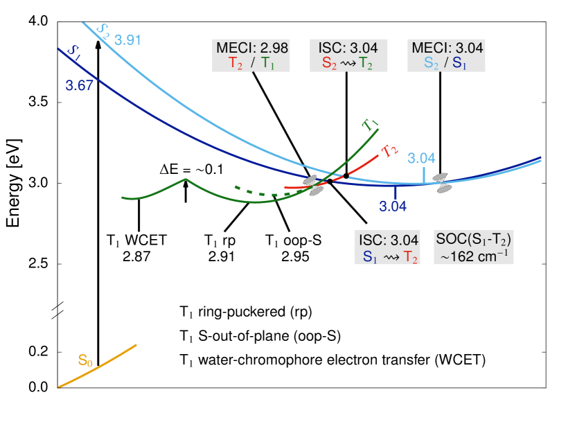
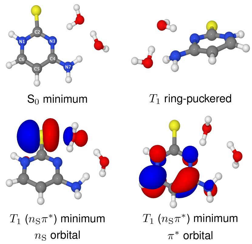
Interestingly, the ring-puckered T1 minimum energy structure at 2.91 eV shown in Fig. 1, displaying character, was also observed by Mai et al. [33]. However, according to our calculations, water molecules may significantly stabilize the component of the T1 state through the water-chromophore electron transfer (WCET) phenomenon which leads to formation of another minimum energy structure at 2.87 eV that is accessible from the ring puckered T1 minimum after passing through a modest energy barrier of 0.1 eV. This WCET process might, in fact, facilitate a radiationless deactivation channel which ensues quenching of triplet of states in aqueous 2tCyt. Below, we show a possible mechanism of such deactivation via a consecutive electron-driven proton transfer mechanism.
3.3 Water-chromophore electron transfer in microhydrated 2-thiocytosine
Fig. 1 shows minimum-energy structures of the ground and lowest-lying triplet electronic states of 2tCyt- cluster. The WCET minimum is characterized by substantial rearrangement of water molecules with respect to the ground-state geometry. More specifically, the water molecule which in the ground state was hydrogen-bonded to the N3 atom of 2tCyt is partially rotated in a way allowing attractive interaction between its lone-pair orbital and the orbital of thiocarbonyl group (see also Tab. S2 in the SI). This dispersion-like interaction is associated with charge transfer from the water molecule to the chromophore which amounts to approximately 0.15 electron (according to particle-hole analysis of the electronic wave-function at ADC(2)/cc-pVTZ level) [56]. The corresponding distance between the sulphur and the interacting oxygen atoms amounts to 2.40 Å and is noticeably shorter than \ceH2OS distance in the electronic ground state. In contrast, the O–H bond in this water molecule, interacting through a hydrogen bond with the N3 atom of 2tCyt is extended to 1.07 Å. This structure is similar to the WCET minimum-energy crossing points reported previously for 7H-adenine and cytosine [37, 40], however, it is the first time that this phenomenon is found also in the triplet manifold and with the involvement of the thiocarbonyl group.
The minimum-energy structure lies only 0.36 eV above the closed-shell ground state at the ADC(2) level, and the corresponding SOC between the and S0 amounts to 46 cm-1. This indicates that 2tCyt might in fact undergo relatively efficient ISC to the electronic ground state from this T1 minimum. The –S0 energy gap is further reduced to 0.22 eV when more explicit water molecules are considered (i.e. in [2tCyt(H2O)4] cluster, cf. the SI for details). Thus the observed charge transfer and elongation of the O–H bond might initiate a proton transfer from water molecule to the N3 atom of 2tCyt. According to our calculations, such electron-driven proton transfer results in the formation of the T1/S0 state crossing. The corresponding MECP geometry located at the ADC(2)/MP2/cc-pVTZ level is shown as inset in Fig. 3. This photochemical reaction leads to the formation of radical form of 2tCyt and hydroxyl radical which is known to be highly reactive and mobile. This implies that such mechanism might initiate photoinduced conversion of 2-thiocytosine to thiouracil after the subsequent migration of the hydroxyl radical to the C4 atom of the pyrimidine ring and extrusion of ammonia. The UV-induced deamination reaction was in fact observed experimentally in canonical cytosine and cytidine [64].
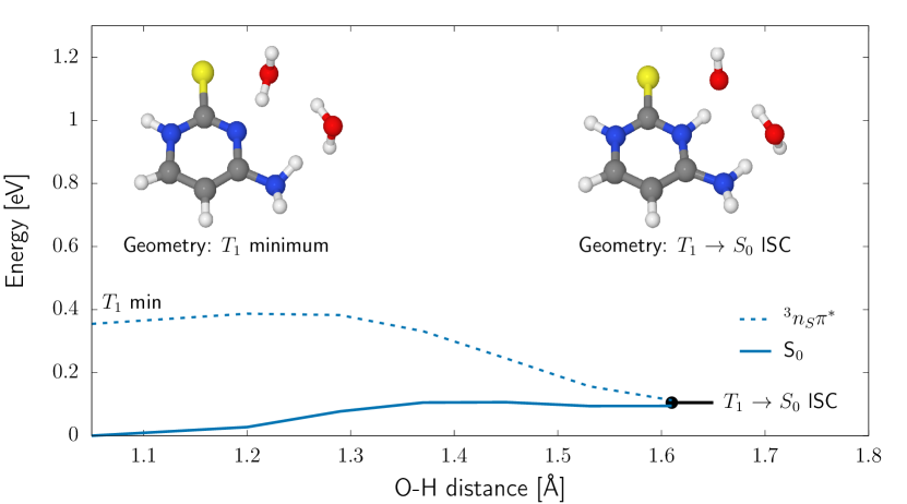
Fig. 3 shows potential energy profile corresponding to the photorelaxation pathway connecting the minimum-energy structure of state stabilized by WCET and the /S0 MECP ISC, all located at the ADC(2)/MP2/cc-pVTZ level. The shallow minimum visible on the left-hand side of the PE profile actually vanishes when the water cluster is extended to several explicit water molecules (cf. SI for details), indicating that in this case the electron-driven proton transfer may be a virtually barrierless process. The spin-orbit coupling (SOC) between the T1 and S0 states calculated for this MECP geometry amounts to 63 , what suggests that the closed-shell S0 state might be efficiently repopulated at this state crossing. It should be noted though that the foregoing photodeactivation channel may also lead to the formation a triplet biradical system.
To assess applicability of ADC(2) method to describe the partially biradical state of microhydrated system, we employed the multireference configuration interaction with single and double excitations MR-CISD(2,3)/6-31++G(d,p) method to reoptimize its minimum-energy structure and /S0 state crossing. Both of these MR-CISD optimizations resulted in geometries characterized by degenerate and S0 states. Even though the ADC(2) results suggested a T1-S0 energy gap of 0.36 eV in the minimum, such E is in fact in the infrared spectral region and consequently the corresponding state crossing might be accessible under ambient conditions. Therefore, despite small quantitative differences between the MR-CISD and ADC(2) methods, the qualitative features of the WCET minimum are correctly reproduced at the ADC(2) level. In contrast, the /S0 MECP associated with the proton transfer process and optimized at the MR-CISD level is higher in energy by 0.43 eV than the corresponding T1 WCET minimum. This suggests lower availability of the EDPT process than that implied by the ADC(2) method.
3.4 Simulations of excited-state spectra of 2-thiocytosine
In order to verify our findings and compare our results to previous experimental and theoretical results of Mai at al. [33], we performed simulations of excited-state absorption (ESA) spectra for isolated 2tCyt (Fig. 4a) and microhydrated 2tCyt (Fig. 4b) molecules employing the nuclear ensemble method. The simulated ESA spectra can be further used in assignment of transient absorption (TAS) UV measurements. The simulated ESA spectra for the isolated molecule were obtained using the minimum-energy structures corresponding to the S1, S2, T1 ring-puckered and T1 S-out-of-plane minima optimized at the ADC(2)/cc-pVTZ level, whereas in the case of microhydrated molecules the ESA spectra were simulated assuming S1, T1 ring-puckered, T1 S-out-of-plane and T1 WCET excited state potential energy surfaces.
Comparison of the ESA spectra (Fig. 4) obtained for both isolated and microhydrated 2tCyt shows similar features with somewhat redishifted absorption bands for the latter. According to the experimental TAS spectrum of 2tCyt in water solution [33], there are two distinct absorption maxima at about 350 and 525 nm which were assigned based on MS-CASPT2 calculations to the and states, respectively. Our simulated ESA spectra (Fig. 4a) of 2tCyt are generally consistent with these findings and show that these bands may refer to the S2 (or S1) and T1 states in ADC(2) calculations, respectively, for which the computed absorption maxima are located at about 310 and 400 nm, respectively. It is worth noting that the latter state (T1 ring-puckered minimum) has two components, mixing the and transitions, in line with what was suggested in the previous work conducted at the MS-CASPT2 level [33]. The disparity between experimental and simulated maxima (310 nm vs 350 nm) assigned to the singlet state (S2 or S1) may be due to lack or inappropriate description of solvent effects in the simulated ESA spectrum. It should be noted though that our ESA spectra are simulated from the corresponding minimum energy geometries distorted along vibrational normal modes, and thus do not correspond to fully relaxed PE surfaces. Even more pronounced differences in the experimental and theoretical absorption bands (400 nm compared to 525 nm) are apparent for the T1 ring-puckered state. However, vibrational cooling effects result in systematic blue-shift of this band during the excited state dynamics of the system and the position of the experimental maximum (525 nm) was reported at 3.7 ps. Consequently, we expect this band to undergo further hypsochromic shift at longer time delays.
The ESA spectrum simulated for the T1 ring-puckered state () of microhydrated 2tCyt-\ce(H2O)2 reveals slight redshift of the predicted absorption maximum to 450 nm. In addition, the simulated ESA spectrum (Fig. 4b) of the T1 S-out-of-plane (/) minimum-energy structure exhibits an overlapping band at 450 nm, similar to the ESA spectrum of the T1 ring-puckered state. This indicates that both the T1 ring-puckered and T1 S-out-of-plane minima could be populated during the excited-state dynamics of aquated 2tCyt. As shown in Fig. 4b the simulated ESA spectrum for the T1() WCET minimum is characterized by high absorption in the same spectral range as the S1 and S2 states of both microhydrated and isolated 2tCyt. Consequently, the absorption band visible at 350 nm between 2 and 4 ps in the experimental TAS spectrum might be also the result of partial population of the T1() WCET state [33]. Unfortunately, the corresponding excited-state absorption band overlaps with that of the T1 S-out-of-plane with dominant contribution from the configuration. Therefore, analysis of TAS measurements is probably not sufficient to provide unambiguous identification of the different states contributing to the excited-state absorption in the triplet manifold.
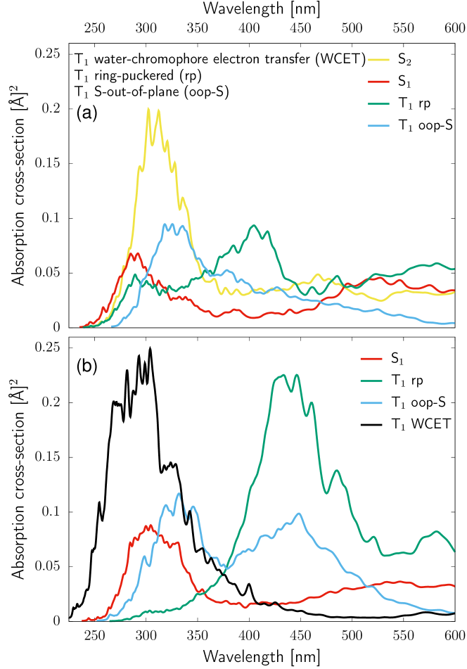
It is worth noting, that the WCET process is characterized by very small T1/S0 energy gaps and relatively high SOC matrix elements (46 cm-1) what suggests that it might not be observable in TAS measurements due to rather short lifetimes. However, population of the WCET state might lead to efficient proton transfer and formation of the hydroxyl radical or even unreactive repopulation of the electronic ground state. Another scenario induced by the characteristic WCET interaction may be related to the population of the S-out-of-plane T1 minimum, which according to our preliminary XMS-CASPT2 optimizations has sufficient contribution from the state to induce the characteristic \ceH2OS=C interaction (cf. SI for details). In each of these cases, we expect that the WCET photodeactivation channel described in detail in the previous paragraphs could efficiently quench triplet states of 2tCyt with the direct involvement of solvent molecules. Consequently the WCET process could explain the weak photosensitizing properties of aqueous 2tCyt solutions.
4 Conclusions
In order to elucidate radiationless deactivation of 2tCyt in aqueous environment, we performed computational explorations of excited-state potential energy surfaces of microhydrated 2tCyt model system. We discovered that water-chromophore electron transfer (WCET) might be the potential driving force responsible for quenching excited triplet states of the studied molecule. For the first time, we show that this characteristic WCET interaction may involve a thiocarbonyl group leading to an interaction between the orbital of water and the orbital of the chromophore. Furthermore, this is the first example of a WCET process occurring in the triplet manifold of electronic states [40, 37]. In particular, our results demonstrate that the T1 topography of aqueous thionucleobases cannot be limited to the sole consideration of S-out-of-plane and ring-puckered minima (as suggested by Bai et al.) [65], and in specific cases electronic states might come into play. As shown above, very low T1-S0 energy gap and considerable SOC matrix elements could provide an efficient channel for the repopulation of the closed-shell electronic ground state.
Since photoinduced electron transfer processes often entail subsequent proton transfer, we considered similar possibility from the WCET minimum of microhydrated 2tCyt. According to our ADC(2) simulations this virtually barrierless EDPT process might be resulting in the formation of hydroxyl radical which could further participate in photohydration or deamination reactions. It should be noted though that the additional MR-CISD results suggest existence of a modest energy barrier (below 0.5 eV) for this process. In fact, this phenomenon is closely related to UV-induced water splitting reactions observed for pyridine [66, 67], acridine [68] and heptazine [69]. Therefore, we suggest that further studies of thionated compounds with similar photochemical properties might be an interesting further direction for photochemical water splitting, particularly since, the aforementioned nitrogenous heterocycles exhibit predominantly singlet and not triplet reactive photochemistry.
Resuming, our study demonstrates that water molecules may modify the shapes of excited-state PE surfaces in thionucleobases, stabilize exciplex interactions and open photorelaxation channels which are not available in isolated chromophores. Since these effects cannot be reproduced theoretically by sole use of implicit solvation models or hybrid quantum mechanics/molecular mechanics (QM/MM) approach, we suggest that the inclusion of explicit QM water molecules might be the necessary next step in determining the important details of the photochemistry of aqueous thionucleobases.
Supplementary data
Supplementary data associated with this article can be found, in the online version including: vertical excitation energies of microhydrated 2-thiocytosine (2tCyt) in gas phase; detailed discussion of the T1 minimum energy structures of microhydrated 2-thiocytosine (2tCyt) located using various methods; properties of larger microhydrated clusters and the results of QM/EFP calculations.
Acknowledgments
This work was supported by a fellowship from the Simons Foundation (494188 to R.S.), financial support from the National Science Centre Poland (2016/23/B/ST4/01048 to R.W.G.) and a statutory activity subsidy from the Polish Ministry of Science and Higher Education for the Faculty of Chemistry of Wroclaw University of Science and Technology. M.J.J. acknowledges support within the “Diamond Grant” (0144/DIA/2017/46) from the Polish Ministry of Science and Higher Education. Computational grants from Interdisciplinary Centre for Mathematical and Computational Modelling (ICM, Grant No. G53-28) and Wrocław Centre of Networking and Supercomputing (WCSS) are also gratefully acknowledged.
References
References
- Arslancan et al. [2017] S. Arslancan, L. Martínez-Fernández, I. Corral, Photophysics and Photochemistry of Canonical Nucleobases’ Thioanalogs: From Quantum Mechanical Studies to Time Resolved Experiments, Molecules 22 (2017) 998.
- Wojciechowski and Leumann [2011] F. Wojciechowski, C. J. Leumann, Alternative DNA base-pairs: from efforts to expand the genetic code to potential material applications, Chem. Soc. Rev. 40 (2011) 5669–5679.
- Szostak [2012] J. W. Szostak, The eightfold path to non-enzymatic RNA replication, J. Syst. Chem. 3 (2012) 2.
- Zhang et al. [2013] S. Zhang, J. C. Blain, D. Zielinska, S. M. Gryaznov, J. W. Szostak, Fast and accurate nonenzymatic copying of an RNA-like synthetic genetic polymer, Proc. Natl. Acad. Sci. U.S.A. 110 (2013) 17732–17737.
- Heuberger et al. [2015] B. D. Heuberger, A. Pal, F. Del Frate, V. V. Topkar, J. W. Szostak, Replacing Uridine with 2-Thiouridine Enhances the Rate and Fidelity of Nonenzymatic RNA Primer Extension, J. Am. Chem. Soc. 137 (2015) 2769–2775.
- Weiss et al. [2016] M. C. Weiss, F. L. Sousa, N. Mrnjavac, S. Neukirchen, M. Roettger, S. Nelson-Sathi, W. F. Martin, The physiology and habitat of the last universal common ancestor, Nat. Microbiol. 1 (2016) 16116.
- Xu et al. [2017] J. Xu, M. Tsanakopoulou, C. J. Magnani, R. Szabla, J. E. Šponer, J. Šponer, R. W. Góra, J. D. Sutherland, A prebiotically plausible synthesis of pyrimidine -ribonucleosides and their phosphate derivatives involving photoanomerization, Nat. Chem. 9 (2017) 303–309.
- Carbon et al. [1968] J. Carbon, H. David, M. H. Studier, Thiobases in Escherchia coli Transfer RNA: 2-Thiocytosine and 5-Methylaminomethyl-2-thiouracil, Sci. 161 (1968) 1146–1147.
- Ajitkumar and Cherayil [1988] P. Ajitkumar, J. D. Cherayil, Thionucleosides in transfer ribonucleic acid: diversity, structure, biosynthesis, and function., Microbiol. Rev. 52 (1988) 103–113.
- Cherayil [1990] J. D. Cherayil, Transfer RNAs and Other Soluble RNAs, CRC Press, 1990.
- Attard and Karran [2011] N. R. Attard, P. Karran, UVA photosensitization of thiopurines and skin cancer in organ transplant recipients, Photochem. Photobiol. Sci. 11 (2011) 62–68.
- Pridgeon et al. [2011] S. W. Pridgeon, R. Heer, G. A. Taylor, D. R. Newell, K. O’Toole, M. Robinson, Y.-Z. Xu, P. Karran, A. V. Boddy, Thiothymidine combined with UVA as a potential novel therapy for bladder cancer, Br. J. Cancer 104 (2011) 1869.
- Pollum et al. [2015] M. Pollum, S. Jockusch, C. E. Crespo-Hernández, Increase in the photoreactivity of uracil derivatives by doubling thionation, Phys. Chem. Chem. Phys. 17 (2015) 27851–27861.
- Reelfs et al. [2011] O. Reelfs, P. Karran, A. R. Young, 4-thiothymidine sensitization of DNA to UVA offers potential for a novel photochemotherapy, Photochem. Photobiol. Sci. 11 (2011) 148–154.
- Massey et al. [2001] A. Massey, Y.-Z. Xu, P. Karran, Photoactivation of DNA thiobases as a potential novel therapeutic option, Curr. Biol. 11 (2001) 1142–1146.
- Zhang et al. [2007] X. Zhang, G. Jeffs, X. Ren, P. O’Donovan, B. Montaner, C. M. Perrett, P. Karran, Y.-Z. Xu, Novel DNA lesions generated by the interaction between therapeutic thiopurines and UVA light, DNA Repair 6 (2007) 344–354.
- Vora et al. [2006] A. Vora, C. D. Mitchell, L. Lennard, T. O. B. Eden, S. E. Kinsey, J. Lilleyman, S. M. Richards, Toxicity and efficacy of 6-thioguanine versus 6-mercaptopurine in childhood lymphoblastic leukaemia: a randomised trial, Lancet 368 (2006) 1339–1348.
- Johnson et al. [2011] R. W. Johnson, A. R. Merkel, S. Danilin, M. P. Nguyen, G. R. Mundy, J. A. Sterling, 6-Thioguanine Inhibition of Parathyroid Hormone-related Protein Expression Is Mediated by GLI2, Anticancer Res. 31 (2011) 2705–2712.
- Robin T. B. Rye et al. [1984] Robin T. B. Rye, Oswald S. Tee, Eva M. Kazdan, The mass spectra of pyrimidines. II. 2(1h)-Pyrimidinethiones and some N(1)-substituted derivatives, Can. J. Chem. 62 (1984).
- Lozzio [1971] C. B. Lozzio, Effect of thiopyrimidine ribonucleosides on DNA and RNA synthesis in synchronized mammalian cell cultures, Exp. Cell. Res. 69 (1971) 377–383.
- Vetter et al. [2009] C. Vetter, C. Wagner, G. N. Kaluderović, R. Paschke, D. Steinborn, Synthesis, characterization, and cytotoxicity of trimethylplatinum(IV) complexes with 2-thiocytosine and 1-methyl-2-thiocytosine ligands, Inorg. Chim. Acta. 362 (2009) 189–195.
- Fillat et al. [2011] M. F. Fillat, M. C. Gimeno, A. Laguna, E. Latorre, L. Ortego, M. D. Villacampa, Synthesis, Structure and Bactericide Activity of (Aminophosphane)gold(I) Thiolate Complexes, Eur. J. Inorg. Chem. 2011 (2011) 1487–1495.
- Harada et al. [2007] Y. Harada, T. Suzuki, T. Ichimura, Y.-Z. Xu, Triplet Formation of 4-Thiothymidine and Its Photosensitization to Oxygen Studied by Time-Resolved Thermal Lensing Technique, J. Phys. Chem. B 111 (2007) 5518–5524.
- Reichardt and Crespo-Hernández [2010a] C. Reichardt, C. E. Crespo-Hernández, Room-Temperature Phosphorescence of the DNA Monomer Analogue 4-Thiothymidine in Aqueous Solutions after UVA Excitation, J. Phys. Chem. Lett. 1 (2010a) 2239–2243.
- Reichardt and Crespo-Hernández [2010b] C. Reichardt, C. E. Crespo-Hernández, Ultrafast spin crossover in 4-thiothymidine in an ionic liquid, Chem. Commun. 46 (2010b) 5963–5965.
- Reichardt et al. [2011] C. Reichardt, C. Guo, C. E. Crespo-Hernández, Excited-State Dynamics in 6-Thioguanosine from the Femtosecond to Microsecond Time Scale, J. Phys. Chem. B 115 (2011) 3263–3270.
- Martínez-Fernández et al. [2012] L. Martínez-Fernández, L. González, I. Corral, An ab initio mechanism for efficient population of triplet states in cytotoxic sulfur substituted DNA bases: the case of 6-thioguanine, Chem. Commun. 48 (2012) 2134–2136.
- Cui and Fang [2013] G. Cui, W.-h. Fang, State-specific heavy-atom effect on intersystem crossing processes in 2-thiothymine: A potential photodynamic therapy photosensitizer, J. Chem. Phys. 138 (2013) 044315.
- Martínez-Fernández et al. [2014] L. Martínez-Fernández, I. Corral, G. Granucci, M. Persico, Competing ultrafast intersystem crossing and internal conversion: a time resolved picture for the deactivation of 6-thioguanine, Chem. Sci. 5 (2014) 1336–1347.
- Taras-Goślińska et al. [2014] K. Taras-Goślińska, G. Burdziński, G. Wenska, Relaxation of the T1 excited state of 2-thiothymine, its riboside and deoxyriboside-enhanced nonradiative decay rate induced by sugar substituent, J. Photochem. Photobiol. A 275 (2014) 89–95.
- Gobbo and Borin [2014] J. P. Gobbo, A. C. Borin, 2-Thiouracil deactivation pathways and triplet states population, Comp. Theor. Chem. 1040–1041 (2014) 195–201.
- Mai et al. [2015] S. Mai, P. Marquetand, L. González, A Static Picture of the Relaxation and Intersystem Crossing Mechanisms of Photoexcited 2-Thiouracil, J. Phys. Chem. A 119 (2015) 9524–9533.
- Mai et al. [2016a] S. Mai, M. Pollum, L. Martínez-Fernández, N. Dunn, P. Marquetand, I. Corral, C. E. Crespo-Hernández, L. González, The origin of efficient triplet state population in sulfur-substituted nucleobases, Nat. Commun. 7 (2016a) 13077.
- Mai et al. [2016b] S. Mai, P. Marquetand, L. González, Intersystem Crossing Pathways in the Noncanonical Nucleobase 2-Thiouracil: A Time-Dependent Picture, J. Phys. Chem. Lett. 7 (2016b) 1978–1983.
- Pollum et al. [2014] M. Pollum, L. Martínez-Fernández, C. E. Crespo-Hernández, Photochemistry of Nucleic Acid Bases and Their Thio- and Aza-Analogues in Solution, in: M. Barbatti, A. C. Borin, S. Ullrich (Eds.), Photoinduced Phenomena in Nucleic Acids I, volume 355 of Topics in Current Chemistry, Springer International Publishing, Berlin, Heidelberg, 2014, pp. 245–327.
- Szabla et al. [2017] R. Szabla, R. W. Góra, M. Janicki, J. Šponer, Photorelaxation of imidazole and adenine via electron-driven proton transfer along H2o wires, Faraday Discuss. 195 (2017) 237–251.
- Barbatti [2014] M. Barbatti, Photorelaxation Induced by Water–Chromophore Electron Transfer, J. Am. Chem. Soc. 136 (2014) 10246–10249.
- Chaiwongwattana et al. [2015] S. Chaiwongwattana, M. Sapunar, A. Ponzi, P. Decleva, N. Došlić, Exploration of Excited State Deactivation Pathways of Adenine Monohydrates, J. Phys. Chem. A 119 (2015) 10637–10644.
- Wu et al. [2016] X. Wu, T. N. V. Karsili, W. Domcke, Excited-State Deactivation of Adenine by Electron-Driven Proton-Transfer Reactions in Adenine–Water Clusters: A Computational Study, ChemPhysChem 17 (2016) 1298–1304.
- Szabla et al. [2017] R. Szabla, H. Kruse, J. Šponer, R. W. Góra, Water–chromophore electron transfer determines the photochemistry of cytosine and cytidine, Phys. Chem. Chem. Phys. 19 (2017) 17531–17537.
- Nogueira et al. [2017] J. J. Nogueira, A. Corani, A. El Nahhas, A. Pezzella, M. d’Ischia, L. González, V. Sundström, Sequential proton-coupled electron transfer mediates excited-state deactivation of a eumelanin building block, J. Phys. Chem. Lett. 8 (2017) 1004–1008.
- Szabla et al. [2014] R. Szabla, J. E. Šponer, J. Šponer, A. L. Sobolewski, R. W. Góra, Solvent effects on the photochemistry of 4-aminoimidazole-5-carbonitrile, a prebiotically plausible precursor of purines, Phys. Chem. Chem. Phys. 16 (2014) 17617–17626.
- Szabla et al. [2013] R. Szabla, D. Tuna, R. W. Góra, J. Šponer, A. L. Sobolewski, W. Domcke, Photochemistry of 2-Aminooxazole, a Hypothetical Prebiotic Precursor of RNA Nucleotides, J. Phys. Chem. Lett. 4 (2013) 2785–2788.
- Weigend and Häser [1997] F. Weigend, M. Häser, RI-MP2: first derivatives and global consistency, Theor. Chem. Acta. 97 (1997) 331–340.
- Weigend et al. [1998] F. Weigend, M. Häser, H. Patzelt, R. Ahlrichs, RI-MP2: optimized auxiliary basis sets and demonstration of efficiency, Chem. Phys. Lett. 294 (1998) 143–152.
- Hättig et al. [2006] C. Hättig, A. Hellweg, A. Köhn, Distributed memory parallel implementation of energies and gradients for second-order Møller–Plesset perturbation theory with the resolution-of-the-identity approximation, Phys. Chem. Chem. Phys. 8 (2006) 1159–1169.
- Kendall et al. [1992] R. A. Kendall, T. H. Dunning, R. J. Harrison, Electron affinities of the first-row atoms revisited. Systematic basis sets and wave functions, J. Chem. Phys. 96 (1992) 6796–6806.
- Schirmer [1982] J. Schirmer, Beyond the random-phase approximation: A new approximation scheme for the polarization propagator, Phys. Rev. A 26 (1982) 2395–2416.
- Hättig [2005] C. Hättig, Structure Optimizations for Excited States with Correlated Second-Order Methods: CC2 and ADC(2), Adv. Quantum Chem. 50 (2005) 37–60.
- Dreuw and Wormit [2015] A. Dreuw, M. Wormit, The algebraic diagrammatic construction scheme for the polarization propagator for the calculation of excited states, WIREs Comput Mol Sci 5 (2015) 82–95.
-
TUR [2016]
TURBOMOLE V7.1, a development of University of Karlsruhe
and Forschungszentrum Karlsruhe GmbH, 1989-2007, TURBOMOLE GmbH, since
2007; available from
http://www.turbomole.com., 2016. - Shao et al. [2015] Y. Shao, Z. Gan, E. Epifanovsky, A. T. B. Gilbert, M. Wormit, J. Kussmann, A. W. Lange, A. Behn, J. Deng, X. Feng, D. Ghosh, M. Goldey, P. R. Horn, L. D. Jacobson, I. Kaliman, R. Z. Khaliullin, T. Kús, A. Landau, J. Liu, E. I. Proynov, Y. M. Rhee, R. M. Richard, M. A. Rohrdanz, R. P. Steele, E. J. Sundstrom, H. L. Woodcock III, P. M. Zimmerman, D. Zuev, B. Albrecht, E. Alguire, B. Austin, G. J. O. Beran, Y. A. Bernard, E. Berquist, K. Brandhorst, K. B. Bravaya, S. T. Brown, D. Casanova, C.-M. Chang, Y. Chen, S. H. Chien, K. D. Closser, D. L. Crittenden, M. Diedenhofen, R. A. DiStasio Jr., H. Dop, A. D. Dutoi, R. G. Edgar, S. Fatehi, L. Fusti-Molnar, A. Ghysels, A. Golubeva-Zadorozhnaya, J. Gomes, M. W. D. Hanson-Heine, P. H. P. Harbach, A. W. Hauser, E. G. Hohenstein, Z. C. Holden, T.-C. Jagau, H. Ji, B. Kaduk, K. Khistyaev, J. Kim, J. Kim, R. A. King, P. Klunzinger, D. Kosenkov, T. Kowalczyk, C. M. Krauter, K. U. Lao, A. Laurent, K. V. Lawler, S. V. Levchenko, C. Y. Lin, F. Liu, E. Livshits, R. C. Lochan, A. Luenser, P. Manohar, S. F. Manzer, S.-P. Mao, N. Mardirossian, A. V. Marenich, S. A. Maurer, N. J. Mayhall, C. M. Oana, R. Olivares-Amaya, D. P. O’Neill, J. A. Parkhill, T. M. Perrine, R. Peverati, P. A. Pieniazek, A. Prociuk, D. R. Rehn, E. Rosta, N. J. Russ, N. Sergueev, S. M. Sharada, S. Sharmaa, D. W. Small, A. Sodt, T. Stein, D. Stück, Y.-C. Su, A. J. W. Thom, T. Tsuchimochi, L. Vogt, O. Vydrov, T. Wang, M. A. Watson, J. Wenzel, A. White, C. F. Williams, V. Vanovschi, S. Yeganeh, S. R. Yost, Z.-Q. You, I. Y. Zhang, X. Zhang, Y. Zhou, B. R. Brooks, G. K. L. Chan, D. M. Chipman, C. J. Cramer, W. A. Goddard III, M. S. Gordon, W. J. Hehre, A. Klamt, H. F. Schaefer III, M. W. Schmidt, C. D. Sherrill, D. G. Truhlar, A. Warshel, X. Xua, A. Aspuru-Guzik, R. Baer, A. T. Bell, N. A. Besley, J.-D. Chai, A. Dreuw, B. D. Dunietz, T. R. Furlani, S. R. Gwaltney, C.-P. Hsu, Y. Jung, J. Kong, D. S. Lambrecht, W. Liang, C. Ochsenfeld, V. A. Rassolov, L. V. Slipchenko, J. E. Subotnik, T. Van Voorhis, J. M. Herbert, A. I. Krylov, P. M. W. Gill, M. Head-Gordon, Advances in molecular quantum chemistry contained in the q-chem 4 program package, Mol. Phys. 113 (2015) 184–215.
- Mewes et al. [2015] J.-M. Mewes, Z.-Q. You, M. Wormit, T. Kriesche, J. M. Herbert, A. Dreuw, Experimental Benchmark Data and Systematic Evaluation of Two a Posteriori, Polarizable-Continuum Corrections for Vertical Excitation Energies in Solution, J. Phys. Chem. A 119 (2015) 5446–5464.
- Mewes et al. [2017] J.-M. Mewes, J. M. Herbert, A. Dreuw, On the accuracy of the general, state-specific polarizable-continuum model for the description of correlated ground- and excited states in solution, Phys. Chem. Chem. Phys. 19 (2017) 1644–1654.
- Martin [2003] R. L. Martin, Natural transition orbitals, J. Chem. Phys. 118 (2003) 4775–4777.
- the [2017] F. Plasser, “TheoDORE 1.4: A package for theoretical density, orbital relaxation, and exciton analysis", 2017.
- Levine et al. [2008] B. G. Levine, J. D. Coe, T. J. Martínez, Optimizing Conical Intersections without Derivative Coupling Vectors: Application to Multistate Multireference Second-Order Perturbation Theory (MS-CASPT2), J. Phys. Chem. B 112 (2008) 405–413.
- Lorquet and Leyh-Nihant [1988] J. C. Lorquet, B. Leyh-Nihant, Nonadiabatic unimolecular reactions. 1. A statistical formulation for the rate constants, J. Phys. Chem. 92 (1988) 4778–4783.
- Lischka et al. [2012] H. Lischka, R. Shepard, I. Shavitt, R. M. Pitzer, M. Dallos, T. Müller, P. G. Szalay, F. B. Brown, R. Ahlrichs, H. J. Böhm, A. Chang, D. C. Comeau, R. Gdanitz, H. Dachsel, C. Ehrhardt, M. Ernzerhof, P. Höchtl, S. Irle, G. Kedziora, T. Kovar, V. Parasuk, M. J. M. Pepper, P. Scharf, H. Schiffer, M. Schindler, M. Schüler, M. Seth, E. A. Stahlberg, J.-G. Zhao, S. Yabushita, Z. Zhang, M. Barbatti, S. Matsika, M. Schuurmann, D. R. Yarkony, S. R. Brozell, E. V. Beck, J.-P. Blaudeau, M. Ruckenbauer, B. Sellner, F. Plasser, J. J. Szymczak, Columbus, Relase 7.0 2012, an Ab Initio Electronic Structure Program, 2012. URL: http://www.univie.ac.at/columbus.
- Aquilante et al. [2016] F. Aquilante, J. Autschbach, R. K. Carlson, L. F. Chibotaru, M. G. Delcey, L. De Vico, I. Fdez. Galván, N. Ferré, L. M. Frutos, L. Gagliardi, M. Garavelli, A. Giussani, C. E. Hoyer, G. Li Manni, H. Lischka, D. Ma, P. Malmqvist, T. Müller, A. Nenov, M. Olivucci, T. B. Pedersen, D. Peng, F. Plasser, B. Pritchard, M. Reiher, I. Rivalta, I. Schapiro, J. Segarra-Martí, M. Stenrup, D. G. Truhlar, L. Ungur, A. Valentini, S. Vancoillie, V. Veryazov, V. P. Vysotskiy, O. Weingart, F. Zapata, R. Lindh, Molcas 8: New capabilities for multiconfigurational quantum chemical calculations across the periodic table, J. Comput. Chem. 37 (2016) 506–541.
- Crespo-Otero and Barbatti [2012] R. Crespo-Otero, M. Barbatti, Spectrum Simulation and Decomposition with Nuclear Ensemble: Formal Derivation and Application to Benzene, Furan and 2-Phenylfuran, Theor. Chem. Acc. 131 (2012) 1–14.
- Barbatti et al. [2014] M. Barbatti, M. Ruckenbauer, F. Plasser, J. Pittner, G. Granucci, M. Persico, H. Lischka, Newton-X: A Surface-Hopping Program for Nonadiabatic Molecular Dynamics, Wiley Interdiscip. Rev.: Comput. Mol. Sci. 4 (2014) 26–33.
- Mai et al. [2017] S. Mai, B. Ashwood, P. Marquetand, C. E. Crespo-Hernández, L. González, Solvatochromic Effects on the Absorption Spectrum of 2-Thiocytosine, J. Phys. Chem. B 121 (2017) 5187–5196.
- Powner et al. [2009] M. W. Powner, B. Gerland, J. D. Sutherland, Synthesis of Activated Pyrimidine Ribonucleotides in Prebiotically Plausible Conditions, Nature 459 (2009) 239–242.
- Bai and Barbatti [2017] S. Bai, M. Barbatti, On the decay of the triplet state of thionucleobases, Phys. Chem. Chem. Phys. 19 (2017) 12674–12682.
- Liu et al. [2013] X. Liu, A. L. Sobolewski, R. Borrelli, W. Domcke, Computational investigation of the photoinduced homolytic dissociation of water in the pyridine–water complex, Phys. Chem. Chem. Phys. 15 (2013) 5957–5966.
- Liu et al. [2014] X. Liu, A. L. Sobolewski, W. Domcke, Photoinduced Oxidation of Water in the Pyridine–Water Complex: Comparison of the Singlet and Triplet Photochemistries, J. Phys. Chem. A 118 (2014) 7788–7795.
- Liu et al. [2015] X. Liu, T. N. V. Karsili, A. L. Sobolewski, W. Domcke, Photocatalytic Water Splitting with the Acridine Chromophore: A Computational Study, J. Phys. Chem. B 119 (2015) 10664–10672.
- Ehrmaier et al. [2017] J. Ehrmaier, T. N. V. Karsili, A. L. Sobolewski, W. Domcke, Mechanism of Photocatalytic Water Splitting with Graphitic Carbon Nitride: Photochemistry of the Heptazine–Water Complex, J. Phys. Chem. A 121 (2017) 4754–4764.