Fast water flow through graphene nanocapillaries: a continuum model approach involving the microscopic structure of confined water
Abstract
Water inside a nanocapillary becomes ordered, resulting in unconventional behavior. A profound enhancement of water flow inside nanometer thin capillaries made of graphene has been observed [B. Radha et.al., Nature (London) 538, 222 (2016)]. Here we explain this enhancement as due to the large density and the extraordinary viscosity of water inside the graphene nanocapillaries. Using the Hagen-Poiseuille theory with slippage-boundary condition and incorporating disjoining pressure term in combination with results from molecular dynamics (MD) simulations, we present an analytical theory that elucidates the origin of the enhancement of water flow inside hydrophobic nanocapillaries. Our work reveals a distinctive dependence of water flow in a nanocapillary on the structural properties of nanoconfined water in agreement with experiment, which opens a new avenue in nanofluidics.
Water flow through nanoscale channels, and the determination of the slip length, have been the subject of intensive studies1; 2; 3; 4; 5; 6; 7; 8; 9; 10; 11; 5; 6; 7; 8; 12; 13; 14; 15; 16; 17; 18; 19. In a recent study Radha et al.20 fabricated atomically flat 2D-capillaries and was able to control the water flux through the channel size. In Ref. [17], an unexpectedly fast flow (up to 1 m/s) of water through flat nanochannels was reported20. In addition to the large slip length, this unexpected phenomena might be due to the high disjoining pressures21 inside the nanochannel. The disjoining pressure is added to the well-known capillary pressure that causes oscillation in the meniscus pressure which for channels thinner than H=8Å, was found to be in the order of 1 kbar22; 23.
In the continuum limit, transport of water through a capillary is described by the Hagen-Poiseuille equation (HPE), however, for nanofluidics several modifications (beyond the no slip-boundary conditions) should be made20; 21; 22; 23; 24; 25; 26; 27; 28. There have been several studies on the ordering of water inside a hydrophobic nanocapillary29; 30; 31; 33; 34. Particularly monolayer/bilayer ice confined within a hydrophobic nanochannel has been studied using MD simulations30; 31; 33; 35; 36 and using density functional theory calculations37. Such an ordering of water molecules can change significantly the density8; 38; 39 and its viscosity31 inside the channel. Using molecular dynamics simulations the pressure-driven water flow through carbon nanotubes (CNTs) with diameters ranging from 0.83 to 1.66 nm were studied by Thomas et. al.40, where a transition from continuum to subcontinuum flow with decreasing CNT diameter was found. While the standard linear relationship in Darcy law is violated, they modified the Darcy (continuum) equation in order to explain their molecular dynamics simulations results.40
Here we will demonstrate the profound influence of the density and viscosity of water inside graphene nanocapillaries on the water flow rate. Our calculations are based on the well-known continuum model formalism but taking into account the ordering of the water molecules. We propose an analytical model to describe experimental results of Ref. [17], i.e. fast water flow in graphene nanocapillaries, employing aforementioned microscopic structure of confined water. It is entropically unfavorable for a hydrophobic surface to bind water molecules via ionic or hydrogen bonds resulting in low friction of water inside graphene capillaries. The water-solid wall slip length (at molecular scale) is much larger than the capillary size which results in different boundary conditions as compared to bulk water in macroscopic channels. It is well-known that the slip dynamics appears through three different length scales: i) individual molecular, ii) beyond-few molecules, i.e. actual slip at a liquid-solid boundary, and iii) apparent slip due to the motion over complex boundaries. Using a slip length of about 600Å, and a contact angle close to 90o, our analytical results agree very well with recent experiments on water flow through graphene nanochannels20.
The model. The Poiseuille flow solution using no-slip boundary condition for a channel with height , which is subjected to a pressure difference (P) along the length of the channel, is quadratic in velocity. For water flow through equal nanochannels when the effect due to the slip velocity () is taken into account, the volumetric flow rate is given by7
| (1) |
where is the slip length, “ is the density, is the viscosity, and is the length of each channel having a width . Notice that in Eq. (1) the slip term is dominant for . For water inside a nanochannel the density and viscosity vary with the capillary size41; 42; 43. The density increases due to the fact that the accessible volume for water molecules in a nanochannel is smaller than the geometrical volume – because of excluded volume effect near the confining walls. Furthermore, the interaction within the layers and with the hydrophobic confining walls induces structuring in water which significantly enhances the viscosity31; 44; 39. Such changes in the density and viscosity are expected to be pronounced for nanochannel with height H12 Å. We take and where gcm-3 and mPa.s are the bulk values for density and viscosity, respectively. Here and are exponential decay lengths for density and viscosity inside the channel where the two parameters and are determined from MD simulations. The two functions should approach when . We propose the following functions fulfilling these boundary conditions
| (2) |
where a, b, , and will be obtained by fitting to results from our MD simulations.


By assuming that the hydrostatic pressure is many orders of magnitude smaller than: i) the Laplace pressure23; 28 which corresponds to the traditional capillary pressure (here is the interfacial tension45 and is the contact angle between water and graphite/graphene. The latter has been measured, however the results vary widely which can be traced back to functional groups on the surface, i.e. 2; 46; 47; 48; 49). ii) The disjoining pressure (DP) 50 which is due to the ordered structure of water inside the nanochannel33; 31; 29 and the interaction of water with the channel wall. The DP can be one to three orders of magnitude larger than the capillary pressure and is a consequence of van der Waals (vdW) and entropic components22. Substituting aforementioned pressures in Eq. (1), and by including both the density and viscosity functions, we find
| (3) |
The parameter A= is taken as a scaling factor in our model (using experimental numbers 1300Å and Å for one channel, is about 1.2172 sÅ-2 which is very close to our obtained number from a fitting on the experimental data, i.e. 1.2174 sÅ-2). By measuring the chemical potential difference inside the capillary and the reservoirs, the entropic pressure is given by8 where RT=2494.2 J/mol, and =18 cm3/mol.
Computer simulations and experiments confirmed the existence of an ordered structure for confined water inside capillaries with H Å.29; 35; 31; 33; 37. The distance 2.8 Å in semi-squared lattice structure of confined water results in a water density of about 1.36 gcm-3.
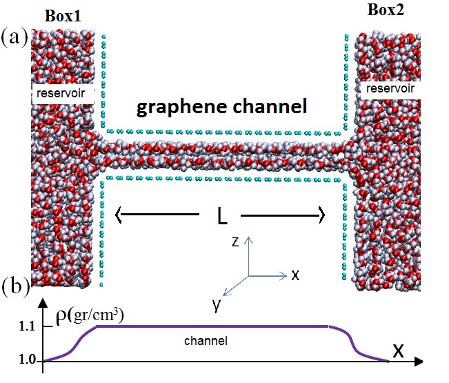
We performed extensive MD simulations using TIP3P force field51 to find the density and the structure of water inside nanochannels. Our simulation setup consists of three elements, i.e. a graphene nanochannel elongated in the x-direction having height H[6.5 Å to 16 Å], length Å, and width Å which connects two water reservoirs on both sides of the channel, see Fig. 1. The simulations are performed using LAMMPS package and we employed an NVT ensemble. The long-range electrostatic interactions were computed with the particle-particle particle-mesh (PPPM) method with a cutoff distance of 12 Å. The non-bonding interactions were modeled by using the Lennard-Jones(LJ) potential. In Fig. 1(a) we show a side view of confined water and the two reservoirs for a channel with height H=10 Å.
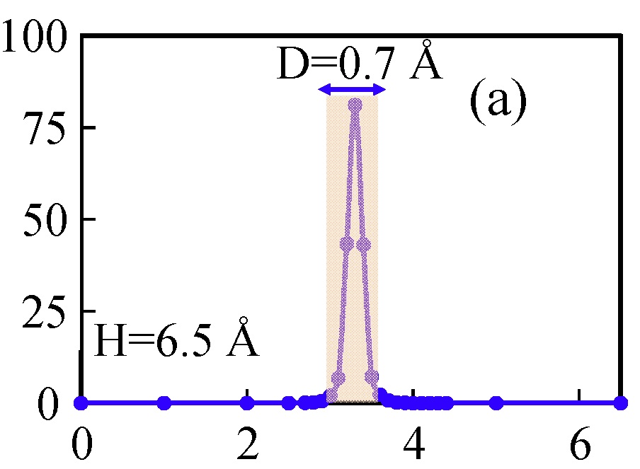
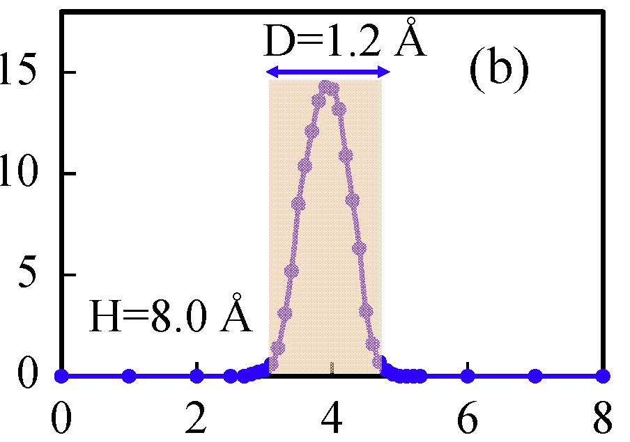
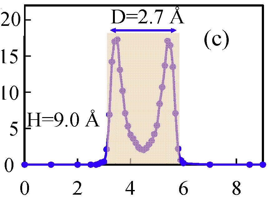
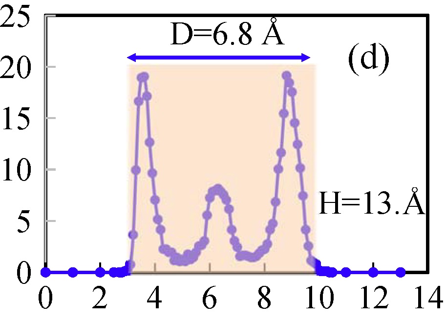
First we calculate the density profile across the channels (i.e. perpendicular to the graphene layers, i.e. along the z-axis). These profiles were calculated by counting the number of water molecules in the yz-plane (across the channel) and averaging along the channel, i.e. where is the number of molecules at point inside the channel. Four typical density profiles are shown in Fig. 2. We found that e.g. for H=6.5 Å, and 8 Å are Gaussian functions with decreasing width for decreasing H. For other H’s, is divided into two or more distinctive peaks. The middle peak (see Fig. 2(d)) disappears by increasing H and only two peaks remained close to the edges38. Note that beyond H=11.0 Å the number of layers and D (where distance ‘’ is effective height of the channel with geometrical height H) becomes larger. For H=13 Å the middle layer is smaller than those on both sides which plays an important role in the fast water flow. These results indicate that the accessible volume for water inside the nanochannels (grayed rectangles in Fig. 2) is smaller than the geometrical volume. Therefore, the accessible volume for water inside the channels is proportional to instead of . Here is the total number of water molecules per surface area inside the channel with size H. In most of the cases, regions of about 3.0 Å from both sides of the graphene walls are inaccessible for the water molecules38 (independent of H). They form an excluded volume due to the graphene wall-water interaction. Obviously, for larger H, the accessible volume approaches the geometrical volume, i.e. the relative difference .
Henceforth, the problem of determining the density is reduced to determining ‘D’ and the corresponding volume. D is taken such that 98 of water molecules are confined in the middle of the channels (i.e. within the colored rectangles shown in Fig. 2). We found H-D is almost the same for H Å and the corresponding density is close to bulk water. For the smallest H i.e. H=6.5 Å, we obtain almost planar and square-rhombic lattice structure at room temperature and lateral pressure of about 0.9GPa with Å, from which we determine a maximum density around 1.4 gcm-3. This can be found only if we use D0.3Å. Alternatively one may use the vdW radius of O and C atoms to define the effective height52; 39. These led us to conclude that the density of confined water is larger than the bulk density which is in agreement with previous reports39; 38. The circle symbols in Fig. 3 are the densities found from our MD simulations using the aforementioned D values. The corresponding function using the best fit of the MD data is shown by the solid line in Fig. 3 with =10.9 and =2.2 Å. The profile shown in Fig. 1(b) schematically shows the variation of the density inside and outside the channel with H=10 Å.
Our approach is general and can also be used to describe fast water flow in CNTs. Using an array of field effect transistors defined on individual CNTs, Qin et al.8 measured the water flow rate through individual CNTs and found a rate enhancement of 882 for CNTs with diameter of 8.1 Å. The water density in the CNT can be described by the same function where and is the diameter in units of Angstrom.
The dependence of the viscosity on H was reported in our previous work31. In general viscosity is direction dependent in confined systems where the major contribution of the viscosity is due to its xy-component, see Ref. [20]. The large viscosity (in ) is due to the layered/ordered structure of water The MD simulation results are shown in Fig. 3(b). A typical fit (which satisfies the above mentioned boundary requirements) on our MD data is shown by the solid curve in Fig. 3(b) with parameters b=6.23 and Å.
Using the obtained and , we first use (neglecting the vdW pressure). The scaling parameter A is the only fitting parameter and our theoretical results for Q are shown in Fig. 4 (red dashed curve) where the slip length was set to be 600 Å and mN/m which results in a contact angle of 89.96o by using . The symbols in Fig. 4 are the experimental data20 (the green error bars indicate the experimental detection limit) and dash-red curve is the calculated water flow rate. Our results are in very good agreement with the experimental results. In the inset of Fig. 3(a), we depict the ratio between and the capillary pressure which is larger than 40 for H13 Å. The capillary pressure is larger than the entropic pressure only for H22 Å.
Finally, it is worthwhile to investigate the effect of vdW pressure.22 To that end, we add the vdW pressure to the DP as
| (4) |
where 53 is the Hamaker constant for intercalated graphite with water. Using the previous and and Å we obtain the blue-dotted curve in Fig. 4. It is seen that, including vdW pressure significantly improves the results for large H (H80Å) while the results for H= 23 and 30Å are slightly overestimated. Therefore disjoining pressure causes a substantial enhancement of the water flux for channel heights around 13Å. Notice that previous MD simulations (see Fig. 3 in Ref. [20] found peak in the water-flow rate that are located in the [17-23]Å range which deviates from the experiment where the peak is around 13Å.
Notice that, we found that for large channel heights (H), the well-known contribution of capillary pressure dominates in Eq. (3) where 1 and 1, which yields a linear dependence H. Moreover in order to provide an independent check on the chosen slip length in our study, we can roughly estimate the slip length as follows. The Navier slip length is defined as where is the water-solid interfacial friction coefficient. We calculated using the method introduced in Ref. [54], and for H10 Å found it almost independent of H, i.e. . Using the obtained number for and those we already found for , the slip length is in the range [500-700]Å. All numbers in this range give a reasonable peak for Q around H=13 Å and give different Q with only 1 difference. Therefore the value Å as a mean value used in our analysis is reasonable.
Our modelling highlights the unique role of confinement on water flow for channel size H13Å and we found our results in very good agreement with the experiment of Ref. [20]. Below H=13 Å, the molecular regime dominates i.e. two layers of water are weakly bound to the walls of the channel and the available cross sectional flow area that remains between these two layers is much smaller than the molecular size of water leading to negligible flow. Around 13 Å, as shown in Fig. 2(d), a third layer of water appears between the two water layers which assemble into a two-dimensional structure for which neither the slip length nor the effective viscosity is well defined. Water molecules in the middle layer are interacting weakly with the other layers and the walls, and their density is slightly smaller than the one of bulk water. However, the total density of water inside the channel is larger than the bulk density. The water molecules in the middle layer diffuse freely. For larger H, although the two side layers of water are still present, the water molecules in the middle layer (having bulk density) randomly diffuse in the remaining space which results in a resistance against water flow and consequently a decrease in the flow rate. Note that the large viscosity and density we introduced in our analytical model is for water inside the whole of the channel. If we subtract the contribution of the two water layers adjacent to the walls, both density and viscosity are only slightly smaller than the one of bulk for channels of size around 13 Å and approach the bulk values for larger H. Notice that as shown in Fig. 3, both density and viscosity for the sub-continuum regime (H13 Å) and the continuum regime (H13 Å) are about the bulk values. This clearly indicates that for H 13Å the effect of the two layers adjacent to the walls are important and they are effectively included by having a large density and viscosity for H13 Å. Therefore, the peak at H =12-14Å arises from the rapidly rising disjoining pressure which is due to the crystal structure of the formed two water layers close to the walls and their induced larger density .
We explained the observed fast water flow through graphene nanochannels, which has been reported in a recent experiment20, which finds its origin in the large density and the large viscosity of water inside nanochannels. Our MD simulations confirm the ordered structure and the large density and viscosity of confined water between graphene channels.
Acknowledgments. We acknowledge fruitful discussion with Andre K. Geim and I. V. Grigorieva. This work was supported by the Flemish Science Foundation (FWO-Vl) and the Methusalem program. B.R. acknowledges the Royal Society Fellowship, the LOreal fellowship for women in science, and EPSRC Grant No. EP/R013063/1.
References
- (1) S. K. Kannam, B. D. Todd, J. S. Hansen, and P. J. Daivis, J. Chem. Phys. 138, 094701 (2013).
- (2) K. Wu, Z. Chen, J. Li, X. Li, J. Xu, and X. Dong, PNAS 114, 3358 (2017).
- (3) D. Mattiaand Y. Gogotsi. Microfluid. Nanofluid. 5, 289 (2008).
- (4) K. Falk, F. Sedlmeier, K. Joly, R. R. Netz, L. Bocquet. Nano Lett. 10, 4067 (2010).
- (5) M. Majumder, N. Chopra, R. Andrews, and B. J. Hinds, Nature 438, 44 (2005).
- (6) J. K. Holt, H. G. Park, Y. Wang, M. Stadermann, A. B. Artyukhin, C. P. Grigoropoulos, A. Noy, and O. Bakajin, Science 19, 5776 (2006).
- (7) J. K. Holt, Adv. Mater. 21, 3542 (2009).
- (8) X. Qin, Q. Yuan Q, Y. Zhao, S. Xie, and Z. Liu, Nano Lett. 11, 2173 (2011).
- (9) J. Köfinger and G. Hummer, and C. Dellago, PNAS 105, 13218 (2008).
- (10) J. A. Thomas and A. J. H. McGaughery, Nano Lett. 8, 2788 (2008).
- (11) J. C. Rasaiah, S. Garde, and D. Hummer, Ann. Rev. Phys. Chem. 59, 713 (2008).
- (12) L. Lacerda, S. Raffa, M. Prato, A. Bianco, and K. Kostarelos. Nano Today 2, 38 (2007).
- (13) A. Noy. Nano Today 2, 22 (2007).
- (14) A. Berezhkovskii and G. Hummer. Phys. Rev. Lett. 89, 064503 (2002).
- (15) S. Joseph and N. R. Aluru. Phys. Rev. Lett. 101, 064502 (2008).
- (16) J. A. Thomas and A. J. H. McGaughey. Phys. Rev. Lett. 102, 184502 (2009).
- (17) S. Joseph and N. R. Aluru, Phys. Rev. Lett. 101, 064502 (2008).
- (18) V. P. Sokhan and N. Quirke, Phys. Rev. E 78, 015301R (2008).
- (19) M. Whitby and N. Quirke, Nature Nanotechnol. 2, 87 (2007).
- (20) B. Radha, A. Esfandiar, F. C. Wang, A. P. Rooney, K. Gopinadhan, A. Keerthi, A. Mishchenko, A. Janardanan, P. Blake, L. Fumagalli, M. Lozada-Hidalgo, S. Garaj, S. J. Haigh, I. V. Grigorieva, H. A. Wu, and A. K. Geim, Nature (London) 538, 222 (2016).
- (21) J. N. Israelachvili, Intermolecular and Surface Forces, 3rd ed., (Elsevier Academic, New York, 2011).
- (22) C. Mathew Mate, IEEE Transactions on Magnetics 47, 124 (2011).
- (23) S. Gravelle, C. Ybert, L. Bocquet, and L. Joly, Phys. Rev. E 93, 033123 (2016).
- (24) C. H. Ahn, Y. Baek, C. Lee, S. O. Kim, S. Kim, S. Lee, S.-H. Kim, S. S. Bae, J. Park, and J. J. Yoon, Ind. Eng. Chem. 18, 1551 (2012).
- (25) M. Khademi and M. Sahimi. J. Chem. Phys. 135, 204509 (2011).
- (26) M. Khademi, R. K. Kalia, and M. Sahimi. Phys. Rev. E 92, 030301 (2015).
- (27) M. Khademi and M. Sahimi. J. Chem. Phys. 145, 024502 (2016).
- (28) F. Ebrahimi, F. Ramazani, and M. Sahimi. Scientific Reports 8, 7752 (2018).
- (29) G. Algara-Siller, O. Lehtinen, F. C. Wang, R. R. Nair, U. Kaiser, H. A. Wu, A. K. Geim, and I. V. Grigorieva, Nature (London) 519, 443 (2015).
- (30) H. Qiu, X. C. Zeng, and W. Guo, ACS Nano 9, 9877 (2015).
- (31) M. Neek-Amal, F. M. Peeters, I. V. Grigorieva, and A. K. Geim, ACS Nano 9, 3685 (2016).
- (32) Jesper S. Hansen, Jeppe C. Dyre, Peter Daivis, Billy D. Todd, and Henrik Bruus, Langmuir 31 (49), 13275 (2015).
- (33) M. Sobrino Fernandez, M. Neek-Amal, and F. M. Peeters, Phys. Rev. B 92, 245428 (2015).
- (34) M. Sobrino Fernández, F.M. Peeters, and M. Neek-Amal, Phys. Rev. B 94, 04546 (2016).
- (35) R. Zangi and A. E. Mark, J. Chem. Phys. 120, 7123 (2004).
- (36) Shun Chen, Adam Paul Draude, Xuechuan Nie, Haiping Fang, Niels R Walet, Shiwu Gao, and Jichen Li, to appear in J. Phys. Communications (2018).
- (37) F. Corsetti, J. Zubeltzu, and E. Artacho, Phys. Rev. Lett. 116, 085901 (2016).
- (38) H. Mosadeghi, S. Alavi, M. H. Kowsari, and Bijan Najafi, J. Chem. Phys. 137, 184703 (2012).
- (39) P. Kumar, S. V. Buldyrev, F. W. Starr, N. Giovambattista, and H. Eugene Stanley, Phys. Rev. E. 72, 051503 (2005).
- (40) John A. Thomas and Alan J. H. McGaughey, Phys. Rev. Lett 102, 184502, (2009).
- (41) H. Ye, H. Zhang, Z. Zhang, and Y. Zheng. Nanoscale Res. Lett. 6, 87 (2011).
- (42) J. A. Thomas, International Journal of Thermal Sciences 49, 281 (2010).
- (43) F. Calabrò, K. P. Lee, and D. Mattia. Applied Mathematics Letters 26, 991 (2013).
- (44) H. Itoh and H. Sakuma, J. Chem. Phys. 142, 184703 (2015).
- (45) C. T Nguyen and B. Kim, International J. of Precision Engineering and Manufacturing 17, 503 (2016).
- (46) F. Taherian, V. Marcon, and N. F. A. van der Vegt, Langmuir 29, 1457 (2013).
- (47) C. Mücksch, C. Rösch, C. Müller−Renno, C. Ziegler, and H. M. Urbassek, J. Phys. Chem. C 119, 12496 (2015).
- (48) W. A. Zisman, Advances in Chemistry, 43, 1-51 (1964).
- (49) T. Werder, J. H. Walther, R. L. Jaffe, T. Halicioglu, F. Noca, and P. Koumoutsakos, Nano Letters 1 697, (2001).
- (50) C. M. Mate, IEEE Trans. Magn. 47, 124, (2011); B. V. Derjaguin and N. V. Churaev, Journal of Colloid and Interface Science 49, 249 (1974).
- (51) W. L. Jorgensen, J. Chandrasekhar, J. D. Madura, R. W. Impey, and M. L. Klein, J. Chem. Phys. 79, 926 (1983).
- (52) J. Zubeltzu and E. Artacho, arXiv:1705.05270 (2017).
- (53) J.-L Li, J. Chun, N. S. Wingreen, R. Car, I. A. Aksay, and D. A. Saville, Phys. Rev. B. 71, 235412 (2004).
- (54) Bladimir Ramos-Alvarado, Satish Kumar, and G. P. Peterson, Phys. Rev. E 93 023101 (2016).