Local Magnetic States of the Weakly Ferromagnetic Iron-Based Superconductor Sr2VFeAsO3-δ Studied by X-ray Magnetic Circular Dichroism
Abstract
We have performed x-ray magnetic circular dichroism (XMCD) measurements on the iron-based superconductor Sr2VFeAsO3-δ to study the origin of weak ferromagnetism (WFM) reported for this compound. While Fe 3 electrons show a magnetic response similar to the other iron pnictides, signals from V 3 electrons remain finite at zero magnetic field and may be responsible for the WFM.
Iron-based superconductors have attracted extensive interest since their discoveries. Despite the lack of thorough understanding on the microscopic mechanism of Cooper pairing, there is a consensus that the proximity to antiferromagnetism plays a key role as in the cuprates [1]. Among all the iron-based superconductors, Sr2VFeAsO3-δ is a rare case where ferromagnetism, which is usually destructive to superconductivity, emerges at low temperatures and co-exists with superconductivity [2, 3]. A small spontaneous ferromagnetic moment of for the compound [4] is enhanced to – by the introduction of oxygen vacancies [2, 3]. The Sr2VFeAsO3-δ lattice consists of an alternate stack of FeAs and perovskite-like Sr2VO3 layers [5]. While the FeAs layer serves as a stage for high-temperature superconductivity below 37 K for [5], Mott insulating state has been proposed for V 3 electrons in the Sr2VO3 layer [6].
The central issue for Sr2VFeAsO3-δ is therefore the nature of the magnetic states. Cao et al. [4] concluded from the Fe57 Mössbauer spectra that no magnetic transitions occur at Fe in the range of –300 K, and hence proposed that V gives rise to the weak ferromagnetism (WFM) through canting of the antiferromagnetically ordered moments [7]. In contrast, an antiferromagnetic transition at 165 K was suggested at Fe sites from a nuclear magnetic resonance (NMR) study by Ueshima et al. [8] Ok et al. [9] instead ascribed the transition at 155 K to non-magnetic charge/orbital ordering of Fe and regarded the ferromagnetic signal as a side effect. Thus not only the origin of the WFM but also the nature of the magnetic state below the transition temperature remains unclear. In this short note, we apply x-ray magnetic circular dichroism (XMCD), which provides element-specific information about the spin and orbital magnetic moments. samples with larger ferromagnetic moments than were measured. The results suggest that the magnetic moments of V 3 rather than Fe 3 contribute to the WFM in Sr2VFeAsO3-δ.
Bulk single crystals of Sr2VFeAsO3-δ () were grown by the self-flux method. Superconducting and ferromagnetic transition temperatures were determined from magnetic susceptibility measurements using a SQUID magnetometer to be 20 K and 210 K, respectively. At 50 K, a remanence magnetization of /f.u. was detected. X-ray absorption spectroscopy (XAS) and XMCD measurements were conducted at beamline BL23SU of SPring-8 in the total electron-yield (TEY) mode at K. Magnetic fields of 0.2–10 T were applied parallel to the -axis of the crystal. The samples were cleaved in vacuum ( Torr) to obtain clean surfaces.
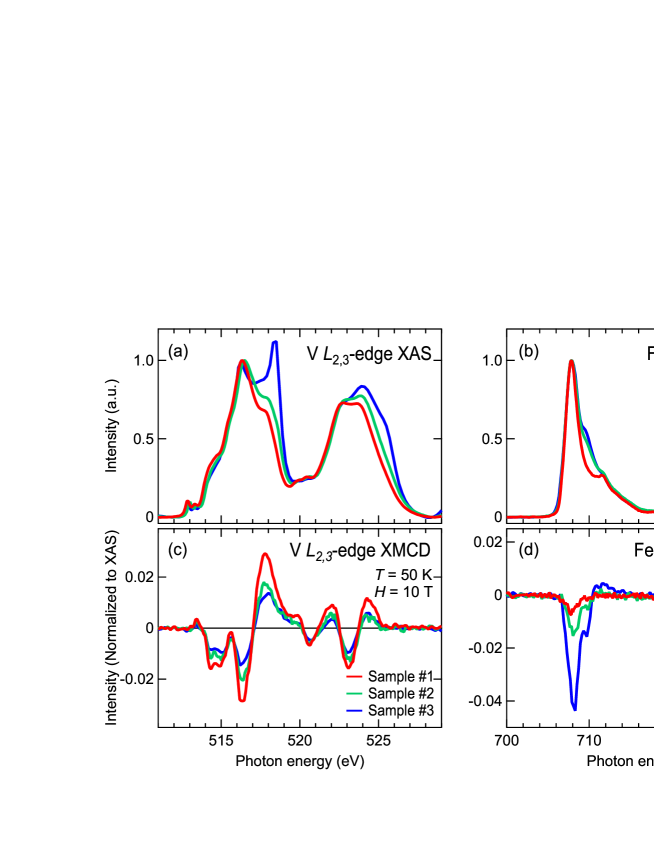
Figures 1(a) and (b) show V and Fe -edge XAS spectra for different samples, i.e., different cleaved surfaces. The spectral line shape apparently depends on the sample. While the V XAS spectrum of sample #1 resembles that of V2O3 [10], consistent with dominant 3+ valence state reported in previous studies [4], additional spectral weight emerges at higher photon energies for samples #2 and #3, suggesting the existence of higher valence state such as V5+. The spectrum gains weight at higher energies also for Fe XAS of the same samples. These results suggest that the cleaved surfaces on samples #2 and #3 are somewhat oxidized probably because the crystals originally had cracks and were cleaved along them. The oxidation also affects the magnetic states as evident from XMCD spectra [Figs. 1(c) and (d)]. For V, the oxidation decreases the magnitude of the XMCD signal. This is consistent with the fact that V5+ has no 3 electrons and hence no magnetic moments. In contrast, the XMCD signal is enhanced for Fe in samples #2 and #3 probably due to the increase of ferromagnetic iron oxides. In order to discuss the intrinsic magnetic properties of Sr2VFeAsO3-δ, we will focus on the data for sample #1 in the following.
XMCD spectra obtained from sample #1 with varying magnetic field are presented in Figs. 2(a) and (b). While finite XMCD is found at T for Fe, the signal goes below the detection limit when the magnetic field is decreased down to 1 T. Applying the sum rule to the T spectrum assuming the electron configuration, the spin, orbital, and total magnetic moments are deduced to be , , and / Fe atom, respectively, and the magnetic susceptibility of emu/Oe mol is obtained. The susceptibility value is of similar magnitude to BaFe2As2 or its Co-doped compounds ( emu/Oe mol) [11], and no signature of WFM is observed for Fe. This result is compatible with the previous Mössbauer study [4], which reported no ordered moments for Fe, and also with the NMR study [8], where the antiferromagnetic transition similar to BaFe2As2 was observed.
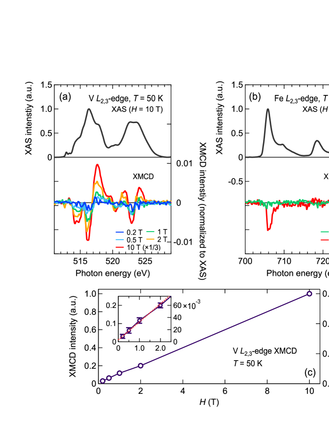
As for V, the XMCD signal remains finite down to –0.5 T. Although the spin sum rule cannot be applied to light transition-metal elements such as V due to the overlap between the and edges, one can still discuss the magnetic moment from the magnitude of the XMCD signal. We first assumed that the XMCD line shape does not depend on magnetic field. This is reasonable since relevant parameters such as Coulomb interaction and transfer integrals should not change significantly by the application of magnetic field. Then, all the V XMCD spectra were fitted to the T line shape with varying its magnitude. Thus extracted XMCD magnitude is plotted as a function of the magnetic field in Fig. 2(c) with an error bar defined as 2 of the fitting. The plot is almost linear, but extrapolation to T (red dashed line) leaves finite XMCD signal. One method to obtain quantitative information about the V magnetic moment is comparison with configuration-interaction cluster calculation that yields the spin and orbital magnetic moments. In Sr2VFeAsO3-δ, oxygen atoms sit at octahedral sites around the V atoms with one apical oxygen atom missing. Still, the XAS and XMCD spectra are similar to what obtained for compounds where V atoms are octahedrally coordinated by O atoms [12]. By comparing the present XMCD spectra with cluster-model calculation [12], we obtain an approximate value for the V magnetic moment of 0.3 at T. Then from Fig. 2(c), one finds the remnant magnetic moment of , which is consistent with the present SQUID measurement. This implies that the V magnetic moments make a significant contribution to the WFM in Sr2VFeAsO3-δ.
Very recently, Tojo et al. [3] reported oxygen-vacancy dependent phase diagram of Sr2VFeAsO3-δ. They concluded from first-principles calculations that oxygen vacancies induce ferrimagnetic order of V and hence give rise to WFM, whereas their Mössbauer measurements indicated that Fe is paramagnetic for superconducting compositions [3]. Our results are consistent with their study and further provide experimental and quantitative information about V magnetic moment. Canting of the antiferromagnetically ordered V moments [7, 8] also remains another possible explanation for the WFM.
In conclusion, we have conducted XMCD measurements on Sr2VFeAsO3-δ () and studied its magnetic states element-specifically. Magnetic susceptibility of Fe was found to be comparable to that of the other iron pnictides such as Ba(Fe,Co)2As2, and no signature of ferromagnetism was observed. One order of magnitude larger magnetic moment was detected for V, which appears to remain finite () even under zero magnetic field. Our results are thus compatible with scenarios that V is the origin of the WFM.
This work was performed under the Shared Use Program of Japan Atomic Energy Agency (JAEA) Facilities (Proposal No. 2015A-E28) supported by JAEA Advanced Characterization Nanotechnology Platform as a program of gNanotechnology Platform h of the Ministry of Education, Culture, Science and Technology (MEXT) (Proposal No. A-15-AE-0026). The synchrotron radiation experiments were performed at the JAEA beamline BL23SU in SPring-8 (Proposal No. 2015A3881). The measurements using a SQUID magnetometer were made at the Cryogenic Research Center, the University of Tokyo. This work was supported by JSPS KAKENHI Grant Nos. 15H02109 and 14J09200. M.H., Y.K.W., and S.S. acknowledge support from the Program for Leading Graduate Schools. M.H. acknowledges support from the JSPS Fellowship for Young Scientists.
References
- [1] A. Chubukov: Annu. Rev. Condens. Matter Phys. 3 (2012) 57.
- [2] A. S. Sefat, D. J. Singh, V. O. Garlea, Y. L. Zuev, M. A. McGuire, and B. C. Sales: Physica C 471 (2011) 143 .
- [3] Y. Tojo, T. Shibuya, T. Nakamura, K. Shoji, H. Fujioka, M. Matoba, S. Yasui, M. Itoh, S. Iimura, H. Hiramatsu, H. Hosono, S. Hirai, W. Mao, S. Kitao, M. Seto, and Y. Kamihara: arXiv:1802.03907 .
- [4] G.-H. Cao, Z. Ma, C. Wang, Y. Sun, J. Bao, S. Jiang, Y. Luo, C. Feng, Y. Zhou, Z. Xie, F. Hu, S. Wei, I. Nowik, I. Felner, L. Zhang, Z. Xu, and F.-C. Zhang: Phys. Rev. B 82 (2010) 104518.
- [5] X. Zhu, F. Han, G. Mu, P. Cheng, B. Shen, B. Zeng, and H.-H. Wen: Phys. Rev. B 79 (2009) 220512.
- [6] T. Qian, N. Xu, Y.-B. Shi, K. Nakayama, P. Richard, T. Kawahara, T. Sato, T. Takahashi, M. Neupane, Y.-M. Xu, X.-P. Wang, G. Xu, X. Dai, Z. Fang, P. Cheng, H.-H. Wen, and H. Ding: Phys. Rev. B 83 (2011) 140513.
- [7] H. Nakamura and M. Machida: Phys. Rev. B 82 (2010) 094503.
- [8] K. Ueshima, F. Han, X. Zhu, H.-H. Wen, S. Kawasaki, and G.-q. Zheng: Phys. Rev. B 89 (2014) 184506.
- [9] J. M. Ok, S.-H. Baek, C. Hoch, R. Kremer, S. Park, S. Ji, B. Buchner, J.-H. Park, S. Hyun, J. Shim, Y. Bang, E. Moon, I. Mazin, and J. S. Kim: Nat. Commun. 8 (2017) 2167.
- [10] M. Abbate, H. Pen, M. Czyżyk, F. de Groot, J. Fuggle, Y. Ma, C. Chen, F. Sette, A. Fujimori, Y. Ueda, and K. Kosuge: J. Electron Spectrosc. Relat. Phenom. 62 (1993) 185 .
- [11] A. S. Sefat, R. Jin, M. A. McGuire, B. C. Sales, D. J. Singh, and D. Mandrus: Phys. Rev. Lett. 101 (2008) 117004.
- [12] Y. Nonaka, G. Shibata, R. Koborinai, K. Ishigami, S. Sakamoto, K. Ikeda, Z. Chi, T. Koide, A. Tanaka, T. Katsufuji, and A. Fujimori: Phys. Rev. B 97 (2018) 205126.