22institutetext: Department of Neuro-oncology, Massachusetts General Hosptial, Harvard Medical School, Boston MA, USA.
DeepNeuro: an open-source deep learning toolbox for neuroimaging
Abstract
Translating neural networks from theory to clinical practice has unique challenges, specifically in the field of neuroimaging. In this paper, we present DeepNeuro, a deep learning framework that is best-suited to putting deep learning algorithms for neuroimaging in practical usage with a minimum of friction. We show how this framework can be used to both design and train neural network architectures, as well as modify state-of-the-art architectures in a flexible and intuitive way. We display the pre- and postprocessing functions common in the medical imaging community that DeepNeuro offers to ensure consistent performance of networks across variable users, institutions, and scanners. And we show how pipelines created in DeepNeuro can be concisely packaged into shareable Docker containers and command-line interfaces using DeepNeuro’s pipeline resources.
Keywords:
deep learning, neuroimaging, software, open-source, preprocessing, reproducibility1 Introduction
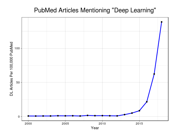
Deep learning is a generic term that defines an increasingly popular approach to machine learning that commonly involves learning abstract representations from datasets using learning architectures titled neural networks. With the advent of powerful graphical processing units and flexible coding frameworks, deep learning approaches have become the standard approach for computer vision tasks, and increasingly used in speech and text analysis tasks[1, 2, 3, 4]. Naturally, deep learning is also gaining popularity in medical applications, in an era where medical imaging and written patient records are a key component of many medical workflows (Figure 1). Deep learning has been successfully applied to the automated diagnosis of skin cancer, macular degeneration, diabetic retinopathy, and retinopathy of prematurity [5, 6, 7, 8]. Specifically within the the field of neuroimaging, deep learning has been shown to have high utility for addressing pathologies as varied as Alzheimer’s disease, stroke, glioma, schizophrenia, and others [9, 10, 11, 12, 13, 14]. In each of these problem spaces, deep learning has shown the potential for algorithms to reach accuracy and efficiency previously thought to be limited to human operators.
Despite progress at the intersection of deep learning and medical imaging, there remain practical challenges that prevent the widespread translation of algorithms into research and clinical practice. Deep learning algorithms and neural networks are often fully-described in academic research, but the source code for implementations of these algorithms are seldom made available to other clinicians and researchers. When source code is made available, documentation is either absent, or presumes a level of familiarity with deep learning software beyond the expertise of the typical neuroimaging researcher. Even if source code for deep learning algorithms is both public and well-documented, operating system requirements and dependencies on other software packages may make their usage impractical without extensive technical support. Each of these failure points adds friction to the process of translating academic deep learning discoveries in into research practice, and further delays the point at which deep learning can be evaluated within a clinical setting.
.
Provided that these software challenges are overcome, the unique nature of medical imaging data produces additional barriers to the neuroscience researcher. Medical images often require numerous and highly-specialized pre- and post-processing techniques, each of which can unpredictably affect the performance of deep learning algorithms. Imaging data is often in higher resolution, as with digital pathology, or in higher dimensions, as with magnetic resonance (MR) imaging data, than traditional imaging datasets. As a result, images may need to be divided into patches, slices, or other representations before being input to a deep learning algorithm, and the specific implementation of these methods can have a significant impact on that algorithms’ performance. Post-processing techniques, particularly for segmentation algorithms, can have a significant effect on an algorithm’s practical utility in the clinic. All of these problems are even more highly elaborated in the field neuroimaging, which often has imaging sequence-specific or disease-specific processing steps for medical data. Even when such processing steps are described, a subtle change in their implementation can have significant effects on the accuracy and consistency of a deep learning algorithm.
Other software packages have attempted to address certain aspects of these issues. NiftyNet is a software package under active development that serves as a framework and templating tool for medical imaging datasets, as well as a model repository for individual use cases [15]. DLTK also serves as a framework for deep learning with medical imaging, and also has a repository for trained neural networks [16]. ModelHub.ai is an open-source, contributor-driven framework for sharing deep learning models created with any framework via Docker containers, and has an online interface for model testing. DeepInfer is a module that allows deep learning algorithms to be used within the context of 3DSlicer, a popular graphical platform for medical imaging used by researchers and clinicians alike [17, 18].
While these packages all taken together provide a strong foundation for both sharing and designing deep learning algorithms, few have the ability to do both simultaneously, and few natively provide utilities for working with data retrieved from the clinic. In this paper, we present DeepNeuro, a deep learning framework that is best-suited to putting deep learning algorithms for neuroimaging in practical usage with a minimum of friction. We will show how this framework can be used to both design and train neural network architectures, as well as modify state-of-the-art architectures in a flexible and intuitive way. We will display the pre- and postprocessing functions common in the medical imaging community that DeepNeuro offers to ensure consistent performance of networks across variable users, institutions, and scanners. And we will show how pipelines created in DeepNeuro can be concisely packaged into shareable Docker containers and command-line interfaces using DeepNeuro’s pipeline resources.
2 Data Processing
.
2.1 Data loading
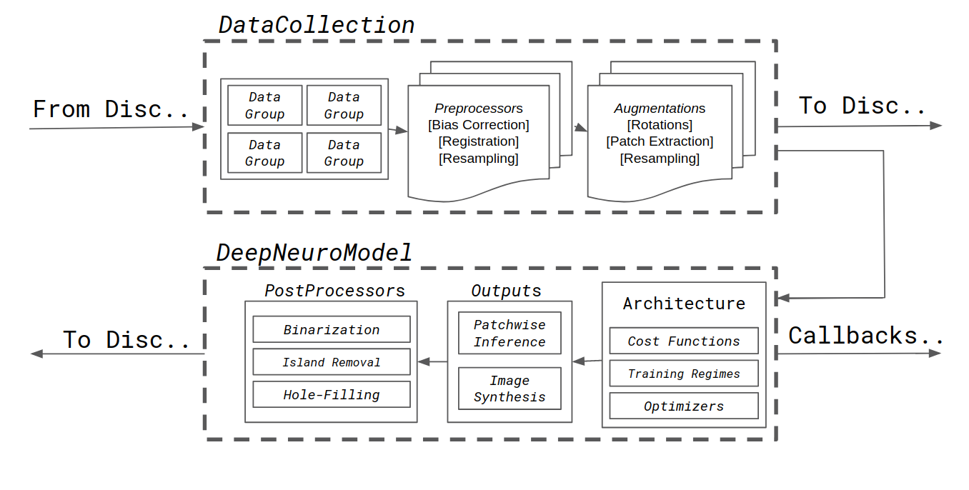
The DeepNeuro scripting language is centered on the DataCollection class. DataCollections are Python objects that stores listed information about any data to be input into later processing steps (Figure 2). Each dataset is conceptualized as a series of “cases”, or individual groups of images and metadata upon which other DeepNeuro methods act. DataCollections have flexible data loaders to associate and stack different medical imaging inputs (e.g. sequences, modalities) into stacked NumPy arrays from regular folder structures or .csv lists to file paths [19]. DataCollections store both information about the location of data on disk, derived attributes of the data itself – e.g. data shape, dimension, and intensity range, and metadata if provided in the original disk data format.
DataCollections list separate Python objects called DataGroups. DataGroups are subsets of a given patient case that may be input in different sections of a model. The most typical DataGroups designations are input_data, i.e. the input node of a neural network, and ground_truth data, against which the cost function of neural network can be evaluated. However, other groups can be specified for more complex architectures, such as networks that contain inputs at multiple nodes. DataCollections can sample data such that the same data from each DataGroup is sampled in each batch, or such that DataGroups are sampled randomly, as in the case of unpaired generative adversarial networks.
DataCollections have the ability to save their inputs into HDF5 file format, and load from HDF5 file format without changes in data organization. DataCollections utilize lazy loading, only loading data to generate attributes upon request.
.
2.2 Preprocessors and Postprocessors
2.2.1 Preprocessing
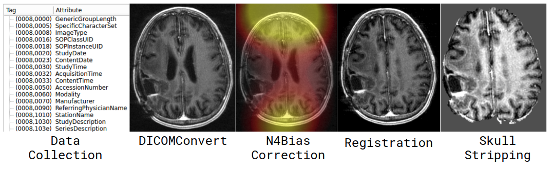
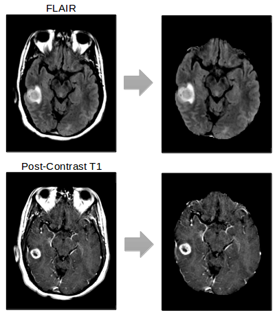
Standardized preprocessing methods are essential to deep learning pipelines that operate on medical images, as slight differences in preprocessing methods can lead to catastrophic prediction failures. DeepNeuro allows the user to preprocess data before inference using the Python object Preprocessor. Transformations applied in Preprocessor objects can be pure Python implementations, inference via neural networks, or links to outside programs such as 3DSlicer or ANTs [20]. These transformations can be applied to data held either in memory or loaded from a provided filepath, and can be returned as either Numpy arrays or stored back to disc. Preprocessors can be concatenated sequentially into preprocessing pipelines, or applied selectively to certain data objects and not others. Current Preprocessor objects available include 3D image registration (3DSlicer), 3D image resampling (3DSlicer), N4 Bias Correction (3DSlicer, ANTs), and skull stripping using a model trained with DeepNeuro (Figure 3).
2.2.2 Postprocessing
Postprocessor objects share the same structure and capabilities as Preprocessor objects, but are applied to DeepNeuroModel objects (described below) instead of DataCollections. Postprocessors are used to apply transformations to data that has been generated by a model. Postprocessor transformations include island-removal and hole-filling for binary outputs, and binarization for scalar outputs.
2.3 Data Augmentation
Data augmentation is used to functionally increase the size of datasets fed into machine learning models via spatial or contrast-based data transformations. Data augmentation is especially important in medical imaging, as medical image datasets tend to be far smaller than datasets of natural images. DeepNeuro addresses this need with Augmentation objects, which can be assigned to DataCollection objects. Each Augmentation object specifies a data augmentation to be applied to input data either before writing to HDF5, or lazily during training. Augmentations can be applied for all DataGroups, or selectively for specific DataGroups. Augmentations are specified in a sequential fashion, and a recursive method is used to concatenate transformations on a single data input. Data can be augmented before saving to HDF5 format, and augmented data is randomly shuffled before sampling in batches.
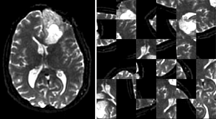
Current Augmentation objects available include 2D and 3D flips and rotations, intensity scaling and shifting, 3D patch extraction, channel-wise dropout, and nearest-neighbor downsampling. Patch extraction can be performed to preferentially select patches that match certain criteria, such as being near a tissue of interest (Figure 4).
2.4 Additional Utilities
2.4.1 Medical Image Data Formats
Data loading features for NiFTI, NRRD, and DICOM files are also included as standalone functions, as well as data saving functions for both NiFTI and DICOM Segmentation Objects (DSO) via the external package dcmqi [21].
3 Model Design
3.1 DeepNeuroModels
Language abstraction is common in programming frameworks for neural networks. Keras, one of the most popular frameworks, is an abstraction that can be run with several different backends, including TensorFlow, Theano, and MXNet [22, 23, 24]. These, in turn, are Python abstractions over lower-level languages. Still other families exist outside of the Keras framework, such as the popular academic deep learning framework PyTorch [25]. Inadvertently, this wide variety of languages has created some difficulty in making models open-source, as researchers split between TensorFlow, PyTorch, or another framework may have difficulty readily implementing each others’ code.
The DeepNeuroModel is another level of abstraction designed to address this problem within the practical context of DeepNeuro pipelines. DeepNeuroModels are objects that take DataCollections as inputs, and can process the data stored in that object via a set of standardized functions shared across deep learning frameworks. These include model training, model saving, generating model callbacks for training loss and other features, and performing inference. This minimum set of functions is presently implemented via subclasses for Keras and Tensorflow, and can be called agnostically of the original framework from DeepNeuro’s interface.
3.1.1 Model Customization
When possible, all models are constructed to be implemented in 2D or 3D. The models available in DeepNeuro have several options for parameter customization, and thus hyperparameter optimization. These parameters include model depth, filter number, dropout ratios, batch normalization, activation type, cost functions, kernel size, and stride size. Models can take advantage of custom cost functions more common in medical imaging, such as the soft-dice cost function and the Wasserstein gradient penalty. Parameter specific to training regimes can also be specified, including batch sizes, learning rates, and optimizers.
3.1.2 Model Implementations
For segmentation applications, we have implemented the U-Net architecture in both 2-D and 3-D [26, 27]. For image synthesis applications, we have implemented both a traditional generative adversarial network architecture (GAN) and the progressively growing GAN architecture proposed by Karras et. al [28].
To improve performance of the U-Net, we incorporate state-of-the-art components that have improved neural network architectures for classification tasks, namely inception modules, residual connections, dense connections, and squeeze-and-excitation modules. [29, 30, 31, 32] Inception modules have multiple pathways with different convolution filter sizes. Residual connections are “shortcut” connections that allow for bypass of convolution layers. Dense connections allow feature maps from every convolutional layer to be carried forward. Squeeze-and-excitation modules allow learning of relationships between different feature maps. We also allow for easy modification of the neural network architectures for changing the ordering of batch normalization, convolutional, and activation layers. [33]
3.1.3 Inference and Outputs
DeepNeuro includes Output objects, which are processes that can be generated from DeepNeuroModel objects. The primary subclass is Inference, which computes the output of a Model after being fed input data. Inference itself is subclassed, for example with ModelPatchesInference for patch-based models. ModelPatchesInference provides options such as the degree of patch overlap (i.e., how many patches to extract and average) and whether to pad the input data to ensure full coverage.
4 Pipeline Distribution
DeepNeuro is designed to be packaged into human-readable modules, and then distributed through Docker containers. The pipelines module of DeepNeuro contains several example pipelines (detailed in “Sample Applications”) that give a simple framework for building DeepNeuro training and inference modules.
DeepNeuro also contains utilites to package Docker containers into command line utilities, with examples in the pipelines module. These command-line modules process datasets individually, are data format agnostic, and let the user specify the number of preprocessing steps required for their particular dataset. DeepNeuro Docker containers are based on the nvidia-docker runtime in order to take advantage of NVIDIA GPUs for neural network inference. Docker containers exist for each module, containing the base DeepNeuro container and the models necessary to run that particular module.
Models are stored outside of DeepNeuro in a cloud-based data management system, and either come pre-downloaded via Docker containers, or can be downloaded via the load modules in DeepNeuro. Models are stored within the DeepNeuro library, and can be deleted via the load module in the scenario where the user wishes to train a newly initialized model.
5 Sample Modules
5.1 Brain Extraction
Brain extraction or “skull-stripping” is a common image preprocessing step that is essential for many neuroimaging applications, including cortical parcellation and surface reconstruction. [34] Many effective, computationally-efficient tools exist for performing brain extraction. [35, 36, 37, 38, 39] However, their generalizability is limited in the presence of varying acquisition parameters or abnormal pathology, such as tumors. Without manual correction, poor brain extraction can introduce errors in downstream analysis. Using DeepNeuro’s U-Net architecture with the following parameters, we trained on a dataset of 30 glioma patients from a multi-institutional cohort, with manually-segmented brain masks (Figure 5). This patient cohort contained both pre-operative and post-operative patients.
5.2 Glioblastoma Segmentation
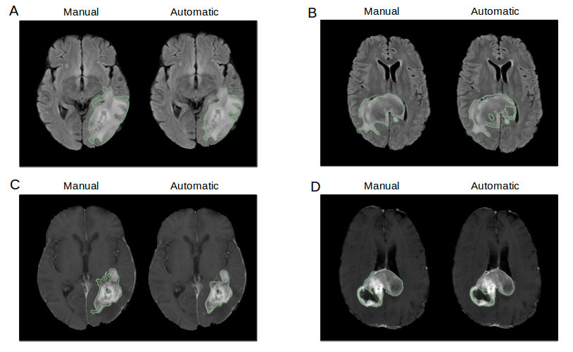
Pathological volume monitoring, and thus pathological tissue segmentation, is essential for assessment of treatment response and prognosis in glioblastoma treatment. [40, 41] Furthermore, volumes and imaging features derived from delineated tumor regions can be used for downstream prediction of molecular biomarkers, treatment response, progression, and survival.[42, 43, 44, 45]. Unfortunately, manual delineation of tumor boundaries can be challenging and subject to inter- and intra-rater variability, resulting in low reproducibility even among expert radiologists and oncologists. Additionally, it is a laborious task especially for high-resolution scans which can have numerous image slices. This diverts clinicians’ time away from other clinical and research tasks, as well as other patients. There are two tumor regions that are of key interest to the clinician. The first is the whole tumor, which consists of edematous tissue, non-enhancing, enhancing tumor, and necrosis. This is best seen on the T2 FLAIR sequence and represents the total tumor burden. The second is contrast-enhancing tumor, which represents regions of breakdown of the blood brain barrier. [46]
The Glioblastoma Segmentation module uses two 3D U-Net architectures. The first creates a binary labelmap of a region-of-interest defined as whole tumor. The output of this network is fed as an additional channel into a second network, which predicts a binary labelmap of enhancing tumor alone (Figure 6). Both networks take in 32x32x32mm patches extracted from FLAIR, pre-contrast T1, and post-contrast T1 patient MR sequences, stacked channel-wise.
Both U-Nets have a depth of four max-pooling layers, with two convolutional layers between each pooling layer, leading to a U-Net architecture with 18 convolutional layers. The network is trained on the BRATS 2017 dataset as well as a clinical trial patient cohort from the Massachusetts General Hospital [11, 12, 47, 48].
6 Future Directions
We present a Python package and model distribution system entitled DeepNeuro. It is a framework for generating and training neural network architectures across multiple programming backends, an all-in-one data preprocessing tool for neuroimaging, and a templating and distribution system for end-to-end deep learning algorithms in neuroimaging.
We will continue to add features to DeepNeuro, and encourage contributions from the users of DeepNeuro in the form of both features and additional modules. We particularly anticipate expanding DeepNeuro’s support for PyTorch, expanding the breadth of model templates available for users to train on, and creating a GUI interface for creating DeepNeuro pipelines for those without scripting proficiency. We also plan to expand the subclasses of DeepNeuroModel to include models created with MXNet and PyTorch, to facilitate the rapid development of pipelines in these languages.
7 Acknowledgements
The Center for Clinical Data Science at Massachusetts General Hospital and the Brigham and Woman’s Hospital provided technical and hardware support for the development of DeepNeuro, including access to high-powered graphical processing units.
References
- [1] LeCun, Y., Bengio, Y., Hinton, G.: Deep learning. nature 521(7553) (2015) 436
- [2] Krizhevsky, A., Sutskever, I., Hinton, G.E.: Imagenet classification with deep convolutional neural networks. In: Advances in neural information processing systems. (2012) 1097–1105
- [3] Collobert, R., Weston, J.: A unified architecture for natural language processing: Deep neural networks with multitask learning. In: Proceedings of the 25th international conference on Machine learning, ACM (2008) 160–167
- [4] Hinton, G., Deng, L., Yu, D., Dahl, G.E., Mohamed, A.r., Jaitly, N., Senior, A., Vanhoucke, V., Nguyen, P., Sainath, T.N., et al.: Deep neural networks for acoustic modeling in speech recognition: The shared views of four research groups. IEEE Signal processing magazine 29(6) (2012) 82–97
- [5] Esteva, A., Kuprel, B., Novoa, R.A., Ko, J., Swetter, S.M., Blau, H.M., Thrun, S.: Dermatologist-level classification of skin cancer with deep neural networks. Nature 542(7639) (2017) 115
- [6] Lee, C.S., Baughman, D.M., Lee, A.Y.: Deep learning is effective for classifying normal versus age-related macular degeneration oct images. Ophthalmology Retina 1(4) (2017) 322–327
- [7] Gulshan, V., Peng, L., Coram, M., Stumpe, M.C., Wu, D., Narayanaswamy, A., Venugopalan, S., Widner, K., Madams, T., Cuadros, J., et al.: Development and validation of a deep learning algorithm for detection of diabetic retinopathy in retinal fundus photographs. Jama 316(22) (2016) 2402–2410
- [8] Brown, J.M., Campbell, J.P., Beers, A., Chang, K., Ostmo, S., Chan, R.P., Dy, J., Erdogmus, D., Ioannidis, S., Kalpathy-Cramer, J., et al.: Automated diagnosis of plus disease in retinopathy of prematurity using deep convolutional neural networks. JAMA ophthalmology (2018)
- [9] Liu, S., Liu, S., Cai, W., Pujol, S., Kikinis, R., Feng, D.: Early diagnosis of alzheimer’s disease with deep learning. In: Biomedical Imaging (ISBI), 2014 IEEE 11th International Symposium on, IEEE (2014) 1015–1018
- [10] Winzeck, S., Hakim, A., McKinley, R., Pinto, J.A.A.D.S., Alves, V., Silva, C., Pisov, M., Krivov, E., Belyaev, M., Monteiro, M., et al.: Isles 2016 & 2017-benchmarking ischemic stroke lesion outcome prediction based on multispectral mri. Frontiers in Neurology 9 (2018) 679
- [11] Menze, B.H., Jakab, A., Bauer, S., Kalpathy-Cramer, J., Farahani, K., Kirby, J., Burren, Y., Porz, N., Slotboom, J., Wiest, R., et al.: The multimodal brain tumor image segmentation benchmark (brats). IEEE transactions on medical imaging 34(10) (2015) 1993
- [12] Bakas, S., Akbari, H., Sotiras, A., Bilello, M., Rozycki, M., Kirby, J.S., Freymann, J.B., Farahani, K., Davatzikos, C.: Advancing the cancer genome atlas glioma mri collections with expert segmentation labels and radiomic features. Scientific data 4 (2017) 170117
- [13] Gheiratmand, M., Rish, I., Cecchi, G.A., Brown, M.R., Greiner, R., Polosecki, P.I., Bashivan, P., Greenshaw, A.J., Ramasubbu, R., Dursun, S.M.: Learning stable and predictive network-based patterns of schizophrenia and its clinical symptoms. NPJ schizophrenia 3(1) (2017) 22
- [14] Chang, K., Bai, H.X., Zhou, H., Su, C., Bi, W.L., Agbodza, E., Kavouridis, V.K., Senders, J.T., Boaro, A., Beers, A., et al.: Residual convolutional neural network for the determination of idh status in low-and high-grade gliomas from mr imaging. Clinical Cancer Research 24(5) (2018) 1073–1081
- [15] Gibson, E., Li, W., Sudre, C., Fidon, L., Shakir, D.I., Wang, G., Eaton-Rosen, Z., Gray, R., Doel, T., Hu, Y., et al.: Niftynet: a deep-learning platform for medical imaging. Computer methods and programs in biomedicine 158 (2018) 113–122
- [16] Pawlowski, N., Ktena, S.I., Lee, M.C., Kainz, B., Rueckert, D., Glocker, B., Rajchl, M.: Dltk: State of the art reference implementations for deep learning on medical images. arXiv preprint arXiv:1711.06853 (2017)
- [17] Mehrtash, A., Pesteie, M., Hetherington, J., Behringer, P.A., Kapur, T., Wells, W.M., Rohling, R., Fedorov, A., Abolmaesumi, P.: Deepinfer: open-source deep learning deployment toolkit for image-guided therapy. In: Medical Imaging 2017: Image-Guided Procedures, Robotic Interventions, and Modeling. Volume 10135., International Society for Optics and Photonics (2017) 101351K
- [18] Fedorov, A., Beichel, R., Kalpathy-Cramer, J., Finet, J., Fillion-Robin, J.C., Pujol, S., Bauer, C., Jennings, D., Fennessy, F., Sonka, M., et al.: 3d slicer as an image computing platform for the quantitative imaging network. Magnetic resonance imaging 30(9) (2012) 1323–1341
- [19] Walt, S.v.d., Colbert, S.C., Varoquaux, G.: The numpy array: a structure for efficient numerical computation. Computing in Science & Engineering 13(2) (2011) 22–30
- [20] Avants, B.B., Tustison, N.J., Stauffer, M., Song, G., Wu, B., Gee, J.C.: The insight toolkit image registration framework. Frontiers in neuroinformatics 8 (2014) 44
- [21] Herz, C., Fillion-Robin, J.C., Onken, M., Riesmeier, J., Lasso, A., Pinter, C., Fichtinger, G., Pieper, S., Clunie, D., Kikinis, R., et al.: Dcmqi: an open source library for standardized communication of quantitative image analysis results using dicom. Cancer research 77(21) (2017) e87–e90
- [22] Abadi, M., Barham, P., Chen, J., Chen, Z., Davis, A., Dean, J., Devin, M., Ghemawat, S., Irving, G., Isard, M., et al.: Tensorflow: a system for large-scale machine learning. In: OSDI. Volume 16. (2016) 265–283
- [23] Bergstra, J., Breuleux, O., Bastien, F., Lamblin, P., Pascanu, R., Desjardins, G., Turian, J., Warde-Farley, D., Bengio, Y.: Theano: A cpu and gpu math compiler in python. In: Proc. 9th Python in Science Conf. Volume 1. (2010)
- [24] Chen, T., Li, M., Li, Y., Lin, M., Wang, N., Wang, M., Xiao, T., Xu, B., Zhang, C., Zhang, Z.: Mxnet: A flexible and efficient machine learning library for heterogeneous distributed systems. arXiv preprint arXiv:1512.01274 (2015)
- [25] Paszke, A., Gross, S., Chintala, S., Chanan, G., Yang, E., DeVito, Z., Lin, Z., Desmaison, A., Antiga, L., Lerer, A.: Automatic differentiation in pytorch. (2017)
- [26] Ronneberger, O., Fischer, P., Brox, T.: U-net: Convolutional networks for biomedical image segmentation. In: International Conference on Medical image computing and computer-assisted intervention, Springer (2015) 234–241
- [27] Çiçek, Ö., Abdulkadir, A., Lienkamp, S.S., Brox, T., Ronneberger, O.: 3d u-net: learning dense volumetric segmentation from sparse annotation. In: International Conference on Medical Image Computing and Computer-Assisted Intervention, Springer (2016) 424–432
- [28] Karras, T., Aila, T., Laine, S., Lehtinen, J.: Progressive growing of gans for improved quality, stability, and variation. arXiv preprint arXiv:1710.10196 (2017)
- [29] Szegedy, C., Liu, W., Jia, Y., Sermanet, P., Reed, S., Anguelov, D., Erhan, D., Vanhoucke, V., Rabinovich, A.: Going deeper with convolutions. In: Proceedings of the IEEE conference on computer vision and pattern recognition. (2015) 1–9
- [30] He, K., Zhang, X., Ren, S., Sun, J.: Deep residual learning for image recognition. In: Proceedings of the IEEE conference on computer vision and pattern recognition. (2016) 770–778
- [31] Huang, G., Liu, Z., Van Der Maaten, L., Weinberger, K.Q.: Densely connected convolutional networks. In: CVPR. Volume 1. (2017) 3
- [32] Hu, J., Shen, L., Sun, G.: Squeeze-and-excitation networks. arXiv preprint arXiv:1709.01507 7 (2017)
- [33] He, K., Zhang, X., Ren, S., Sun, J.: Identity mappings in deep residual networks. In: European conference on computer vision, Springer (2016) 630–645
- [34] Kleesiek, J., Urban, G., Hubert, A., Schwarz, D., Maier-Hein, K., Bendszus, M., Biller, A.: Deep mri brain extraction: a 3d convolutional neural network for skull stripping. NeuroImage 129 (2016) 460–469
- [35] Jenkinson, M., Beckmann, C.F., Behrens, T.E., Woolrich, M.W., Smith, S.M.: Fsl. Neuroimage 62(2) (2012) 782–790
- [36] Ségonne, F., Dale, A.M., Busa, E., Glessner, M., Salat, D., Hahn, H.K., Fischl, B.: A hybrid approach to the skull stripping problem in mri. Neuroimage 22(3) (2004) 1060–1075
- [37] Cox, R.W.: Afni: software for analysis and visualization of functional magnetic resonance neuroimages. Computers and Biomedical research 29(3) (1996) 162–173
- [38] Shattuck, D.W., Leahy, R.M.: Brainsuite: an automated cortical surface identification tool. Medical image analysis 6(2) (2002) 129–142
- [39] Iglesias, J.E., Liu, C.Y., Thompson, P.M., Tu, Z.: Robust brain extraction across datasets and comparison with publicly available methods. IEEE transactions on medical imaging 30(9) (2011) 1617–1634
- [40] Brasil Caseiras, G., Ciccarelli, O., Altmann, D.R., Benton, C.E., Tozer, D.J., Tofts, P.S., Yousry, T.A., Rees, J., Waldman, A.D., Jäger, H.R.: Low-grade gliomas: six-month tumor growth predicts patient outcome better than admission tumor volume, relative cerebral blood volume, and apparent diffusion coefficient. Radiology 253(2) (2009) 505–512
- [41] Iliadis, G., Kotoula, V., Chatzisotiriou, A., Televantou, D., Eleftheraki, A.G., Lambaki, S., Misailidou, D., Selviaridis, P., Fountzilas, G.: Volumetric and mgmt parameters in glioblastoma patients: survival analysis. BMC cancer 12(1) (2012) 3
- [42] Zhang, B., Chang, K., Ramkissoon, S., Tanguturi, S., Bi, W.L., Reardon, D.A., Ligon, K.L., Alexander, B.M., Wen, P.Y., Huang, R.Y.: Multimodal mri features predict isocitrate dehydrogenase genotype in high-grade gliomas. Neuro-oncology 19(1) (2016) 109–117
- [43] Grossmann, P., Narayan, V., Chang, K., Rahman, R., Abrey, L., Reardon, D.A., Schwartz, L.H., Wen, P.Y., Alexander, B.M., Huang, R., et al.: Quantitative imaging biomarkers for risk stratification of patients with recurrent glioblastoma treated with bevacizumab. Neuro-oncology 19(12) (2017) 1688–1697
- [44] Smits, M., van den Bent, M.J.: Imaging correlates of adult glioma genotypes. Radiology 284(2) (2017) 316–331
- [45] Chang, K., Zhang, B., Guo, X., Zong, M., Rahman, R., Sanchez, D., Winder, N., Reardon, D.A., Zhao, B., Wen, P.Y., et al.: Multimodal imaging patterns predict survival in recurrent glioblastoma patients treated with bevacizumab. Neuro-oncology 18(12) (2016) 1680–1687
- [46] Dubois, L.G., Campanati, L., Righy, C., D’Andrea-Meira, I., Spohr, T.C.L.d.S., Porto-Carreiro, I., Pereira, C.M., Balça-Silva, J., Kahn, S.A., DosSantos, M.F., et al.: Gliomas and the vascular fragility of the blood brain barrier. Frontiers in cellular neuroscience 8 (2014) 418
- [47] Bakas, S., Akbari, H., Sotiras, A., Bilello, M., Rozycki, M., Kirby, J., Freymann, J., Farahani, K., Davatzikos, C.: Segmentation labels and radiomic features for the pre-operative scans of the tcga-gbm collection. The Cancer Imaging Archive 286 (2017)
- [48] Bakas, S., Akbari, H., Sotiras, A., Bilello, M., Rozycki, M., Kirby, J., Freymann, J., Farahani, K., Davatzikos, C.: Segmentation labels and radiomic features for the pre-operative scans of the tcga-lgg collection. The Cancer Imaging Archive 286 (2017)