Absolute and arbitrary orientation
of single molecule shapes
DNA origami is a modular platform for the combination of molecular and colloidal components to create optical, electronic, and biological devices. Integration of such nanoscale devices with microfabricated connectors and circuits is challenging: large numbers of freely diffusing devices must be fixed at desired locations with desired alignment. We present a DNA origami molecule whose energy landscape on lithographic binding sites has a unique maximum. This property enables device align- ment within 3.2∘ on SiO. Orientation is absolute (all degrees of freedom are specified) and arbitrary (every molecule’s orientation is independently specified). The use of orientation to optimize device performance is shown by aligning fluorescent emission dipoles within microfabricated optical cavities. Large-scale integration is demonstrated via an array of 3,456 DNA origami with 12 distinct orientations, which indicates the polarization of excitation light.
The sequential combination of solution-phase self-assembly (SPSA) and directed self-assembly (DSA) provides a general paradigm for the synthesis of nanoscale devices and their large-scale integration with control circuitry, microfluidics, or other conventionally-fabricated structures. SPSA for the creation of sub-lithographic devices via structural DNA nanotechnology (1) is relatively mature. In particular, typical DNA origami (2) allow up to 200 nanoscale components, including carbon nanotubes (3, 4, 5), metal nanoparticles (6, 7), fluorescent molecules (6, 7, 8), quantum dots (7, 9) and conductive polymers (10) to be simultaneously juxtaposed at 3-5 nm resolution within a 100 nm70 nm DNA rectangle. DSA uses topographic (11, 12) or chemical (13, 14, 15, 16, 17, 18, 19, 20, 21, 22, 23, 24, 25, 26) patterning, fields (27, 28, 29, 30, 31, 32, 33, 34, 35, 36, 37), or flow (38, 39, 40, 41, 42, 43, 44, 45, 46) to control the higher order structure of molecules and particles. Well-developed for continuous block copolymers films (13, 14), spherical nanoparticles (11, 12), and linear nanostructures (16, 17, 18, 19, 20, 21, 22, 27, 28, 29, 30, 31, 32, 33, 34, 35, 36, 38, 39, 40, 41, 42, 43, 44, 45, 46), DSA is less developed for origami-templated devices for which shape and symmetry play an important role in device function and integration.
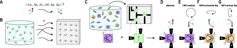
Two challenges arise in the DSA of orgami-templated devices. The first is analogous to the problem of absolute orientation (47) (Fig. 1A) in computational geometry: Given two Cartesian coordinate systems, what translation and rotation can transform the first to the second? Such transformations are key in computer vision and robotics, where they can be used to plan the motion of a virtual camera, or a robot arm. The physical analog for DSA asks: How can an asymmetric device in solution be positioned and aligned relative to a global reference frame in the laboratory? The second challenge is to achieve absolute orientation for many devices at once, such that the position and alignment of each device is arbitrary, i.e. independent of other devices (Fig. 1B). DNA origami placement (DOP) (24, 25, 26) is a potential solution to both challenges. In DOP the match between the overall shape of an origami and lithographically patterned binding sites is used both to position the origami in and , and to control its in-plane rotation . The strength of DOP is that thousands of origami can be oriented with high yield and fidelity: 95% of sites have single origami aligned within 10∘ of a desired . The weakness of DOP has been the exclusive use of equilaterial triangles: an equilateral triangle can attach to its binding site in one of six orientations (at any of three equivalent rotations, flipped right-side up or up-side down). Thus DOP of equilateral triangles does not achieve absolute orientation and its use is limited to devices with compatible symmetry, e.g. point-like (8), three-fold, or six-fold.
Consideration of fully asymmetric (C1 symmetric) devices, like bipolar junction transistors, motivates the development of absolute and arbitrary DSA (Fig. 1C), and clarifies conditions for which DOP of high symmetry shapes (like equilateral triangles and rectangles) or other DSA methods (fig. S1) are insufficent. Were DOP of rectangular origami used for the three-device circuit pictured, the origami’s symmetry would allow it to bind in four orientations relative to each binding site: one (Fig. 1D) desired and three (Fig. 1, E to G) undesired. Random binding at each site would result in exponentially low yield: only % of circuits would have all three transistors in the desired orientation. Flow or field alignment of induced dipoles would allow the same four orientations. Field alignment of origami bearing fixed dipoles could break in-plane rotational symmetry but would still allow two orientations (Fig. 1D and F) related by a horizontal flip. Further, such purely global methods cannot simultaneously specify distinct rotations or translations for multiple devices, and could not fabricate the given circuit in a single step; arbitrary orientation promises independent alignment of an unlimited number of devices in a single step. Approaches which fix the ends of linear nanostructures on metal bars or dots (18, 19, 21), or align them to chemical stripes (16), add arbitrary control of position and in-plane rotation, but still cannot distinguish the orientations in Fig. 1, D to G. Nor can methods which fix the corners of rectangles (22). Here we show that absolute orientation can be achieved by DOP with suitably asymmetric DNA origami shapes, and demonstrate two applications in which absolute and arbitrary orientation work together to optimize or integrate optical devices.
DOP can been performed on any planar substrate (e.g. SiO, quartz, silicon nitride [SiN] and diamond-like carbon) whose surface can be differentiated into negatively-charged binding sites (green features throughout paper) which bind negatively-charged DNA origami strongly in the presence of bridging Mg2+ ions, and a neutral background which binds origami weakly (gray backgrounds). Here e-beam patterned binding sites are made negative via silanols which are ionized at the pH (8.3) of the origami binding buffer and the neutral background is a trimethylsilyl monolayer, generated via silanization. DOP is a complex adsorption process which involves both 3D diffusion to the surface, and 2D diffusion of weakly bound origami on the background. Observations of lateral jamming, binding of multiple origami to a single site, and reorientation of origami already bound to sites suggest that DOP is both nonequilibrium and non-Langmuir (26). Thus to simplify development of absolute orientation, we separated the problem into two parts: first, breaking up-down symmetry on unpatterned SiO (e.g. differentiating between the pair of orientations in Fig. 1, D and E and the pair in Fig. 1, F and G) and second, breaking rotational symmetry in the context of DOP (e.g. differentiating between Fig. 1D and Fig. 1E).
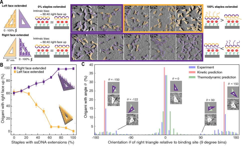
The breaking of up-down symmetry was explored using asymmetric right triangles (Fig. 2A). Synthesized via the SPSA of 200 short DNA staple strands with a long scaffold strand, asymmetric right triangles have left (orange) and right (purple) faces which are easily distinguished by atomic force microscopy (AFM). Our idea was to make one side of the origami non-sticky and hence bias binding, through the addition of single-stranded (ssDNA) extensions to the 5′ ends of staples. To control for geometric details of the right triangle design, and isolate intrinsic bias which might arise from these details instead of ssDNA extensions, two versions were created. In one version, the ends of all staple strands and hence all nicks in the phosphate backbone fell on the origami’s right face; in the other, vice versa. Designed to be flat via twist correction (48), extension-less right triangles of both types exhibited a weak preference to bind unpatterned SiO with their right face up (60:40 right:left, Fig. 2A); thus intrinsic bias was not due to asymmetric flexibility caused by nick position. Bias has been observed in curved single-sheet structures elsewhere (49, 50) suggesting that residual curvature due to imperfect twist correction of the right triangle designs might be responsible for bias here. Strong bias (nearly 100%) was attained by adding 20 nt poly(T) ssDNA extensions to the ends of all 200 staples; origami whose left face was extended bound left-face up, and vice versa (Fig. 1B). Adding poly(A) ssDNA to make all extensions double-stranded and rigid abolished the bias, supporting the idea that on SiO ssDNA extension create bias by acting as entropic brushes which interfere with DNA-SiO binding. However, the symmetry-breaking effect of ssDNA extensions on SiO does not generalize to other surfaces: on mica, where DNA-mica interactions are much stronger than DNA-SiO interactions for the same Mg2+ concentration (26), no bias was observed; on graphene, where - interactions between the unpaired bases and graphene are attractive (51), the bias inverted.
To break rotational symmetry, we began with the DOP of right-face extended triangles (Fig. 2C), used the results to develop a model of binding, and then used the model to design an origami shape which achieved absolute orientation. AFM images of sites binding a single right triangle (73% of 600 sites, fig. S2) were analyzed, and the angle between origami and binding site was measured to the nearest multiple of 4.5∘. Only 34% of origami bound with the desired alignment (∘), too few for reliable absolute orientation. We next asked whether the distribution of states better fit a kinetic or equilibrium model, under the assumption that the binding energy of a given state is linearly proportional to the area of overlap between the origami and binding site; , with its total overlap of origami and binding site, has the highest possible binding energy. The state space was discretized in both and (1 nm increments), and (1∘ increments), encompassing more than 19 million states with positive overlap. For kinetic predictions (Fig. 2C, red), we performed steepest ascent hill climbing using all possible states as initial configurations, and found that (neglecting variations in and ) the state space had three basins of attraction whose maxima (, ∘) corresponded with the three most common experimental states (Fig. 2C, blue). Kinetic abundances predicted by measuring and normalizing basin volumes overestimated experimental abundances with relatively small factors (from 1.1 for ∘ to 2.6 for ∘). Small changes to details of the model (Fig. S3) predicted the existence but not quantitative abundance of minority states (e.g. or 90∘). For thermodynamic predictions (Fig. 2C, green), we calculated expected equilibrium abundances from the partition function, using an energy per unit area overlap derived by constraining the abundance at ∘ to match experiment; thermodynamic abundances underestimated experimental abundances with large factors (from 5.5 for ∘ to 7.3 for ∘). Our data are thus most consistent with a strongly kinetically trapped regime in which origami enter the state space at random (when they collide with a binding site) and simply proceed to a local maxima (fig. S4A) in binding energy.
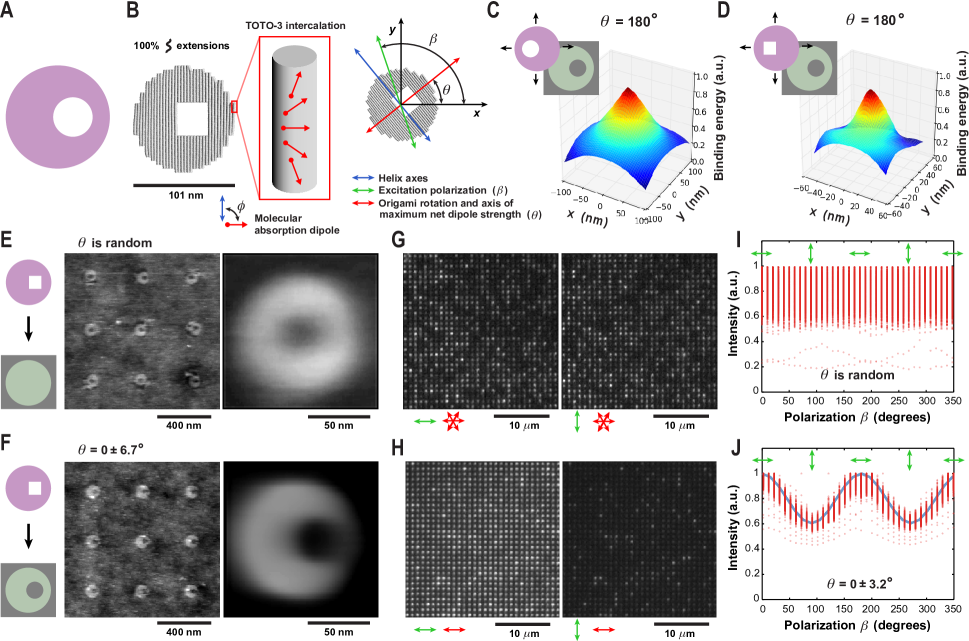
The strong kinetic trapping exhibited by DOP constrains the energy landscapes which can robustly break rotational symmetry: the volume of a single basin of attraction must comprise most of the state space; in the best case the landscape will have a unique global maximum. Exact analysis (52) and general yet simple geometric arguments (53) have shown existence of a unique global maximum for a disk with an offset hole (Fig. 3A), a shape we call a ‘small moon’. Experiments with millimeter-scale models on hydrophobic binding sites (54) confirm that small moons translate and rotate to a unique orientation from initial configurations created by hand using tweezers. Here, we approximated the small moon shape by a DNA origami (Fig. 3B, fig. S5A) with an offset square hole (circumscribed by the ideal hole). Exact mathematical analysis of the energy landscape of the approximate small moon was hindered by its complex jagged outline, so we discretized the landscape as above. Like its idealized counterpart the DNA origami small moon has a unique global maximum in its energy landscape, although the square-shaped hole slightly flattens the landscape in some regions (compare Figs. 3C and D, figs. S4B and C). DOP of small moon origami with ssDNA extensions to break up-down symmetry (fig. S5D) was performed on both disk-shaped control sites (Fig. 3E and fig. S6) and shape-matched sites (Fig. 3F and fig. S7). The average of 498 AFM images of control sites with single origami (83% of 600 total sites) gave an annular shape indicating random orientation; the average of 592 images on shape-matched sites (98.7% of 600 total sites) reconstruct the small moon shape, confirming unique alignment.
By fitting the small moon shape to AFM of small moon origami on shape-matched sites, we found that alignment varied by 6.7∘ (1 SD). This variability includes both real variability due to fabrication error or imperfect assembly, and spurious variability due to the fitting of a model shape to poorly resolved origami; the latter error is difficult to estimate. To get a better estimate of alignment precision, we imaged small moons intercalated post-DOP with the fluorescent dye TOTO-3 (Figs. 3G to J, figs. S8 and S9). For 600-site arrays of small moons on disk-shaped control and shape-matched sites, we measured emission intensity for excitation polarization in 10∘ steps (sampling each twice by rotating the stage from 0∘ to 350∘) and fit the emission to derive distributions for the origami orientation . The reported angle between the molecular absorption dipole of TOTO-3 analogs and the DNA helix axis () ranges from 61∘ to 90∘ (55, 56, 57, 58), but the exact angle is unimportant for measuring variability: it is close enough to 90∘ that averaging over multiple dyes (intercalated at varying rotations due to twist, Fig. 3B) results in a strongly anisotropic net dipole strength in the plane of the origami. Consequently, emission peaks for perpendicular to the helix axes (58), coincident with . The strength of a molecular dipole excited by an electric field along the direction of unit vector is where is the polarization of , and the in-plane dipole angle. According to the dipole approximation (59, 60), emission is proportional to absorption, which is proportional to . Thus experimental intensity can be fit to where is the maximum emission, and is the background (camera noise, reflection). Emission from a collection of molecular dipoles bound to an origami is proportional to , where the net dipole strength***Note that the strength of the net dipole moment is not the same as the net dipole strength. Consider equal and opposite dipoles intercalated 180∘ from each other around the helix. They cancel to yield zero net dipole moment but contribute equally to the net dipole strength, and hence emission under . is given by . Thus the experimental intensity of molecular dipoles with an anisotropic net in-plane dipole strength can be fit to the expression above: if and are defined to lie along the direction of maximum net dipole strength, then is proportional to the difference and is the background plus a contribution proportional to , from the direction of smallest net dipole strength. Emission from control sites (Fig. 3I, fig. S10A and B) individually fit this expression but individual were uniformly distributed (fig. S10C), both confirming random origami orientation and ruling out polarization anisotropy in our setup. As expected, aggregate data could not be fit. In contrast, aggregate data for shape-matched sites (Fig. 3J) fit 0∘ and fits to individual sites (fig. S10D) vary by 3.2∘, our best estimate of alignment precision.
TOTO-3 intercalation of small moons further enabled us to demonstrate arbitrary orientation, prototype the large-scale integration of orientation-dependent devices, and explore variables which can affect the quality of polarization-based devices. However, even when the and are orthogonal to each other there is a small, but reproducible, excitation of the light emitters which we refer to as the bleed-through of the system. We quantified bleed-through for the data in Fig. 3J; after background subtraction we found that emission from origami perpendicular to was 30% of that from origami parallel to β. We quantified bleed-through for the data in Fig. 3J; after background subtraction we found that emission from origami perpendicular to was 30% of that from origami parallel to . In interpreting the source of bleed-through, we consider only the effect of dye alignment and neglect small polarization mixing effects of high numerical aperture on excitation polarization (59). In an ideal device, all dye molecules would align perfectly with : and hence bleed-through would be zero. combines contributions from both placement variability in with incoherence of dye angle relative to the origami. The contribution from placement variability is small, as bleed-through would be only 0.3% were the 3.2∘ variability the only source; 39∘ variability would be required to explain 30% bleed-through. The contribution from incoherent dye alignment within an origami is itself complex: it combines the deterministic rotation of by DNA twist, random wobble (61, 62) from rotational diffusion (reduced here by intercalation and drying), potential alternative binding modes (63), and significant (10.6∘, fig. S11) back-and-forth bending of each helix axis in a DNA origami (2). Here we explain bleed-through simply by a combination of and helix bending, which are the most relevant variables for devices based on intercalators. Attributing all bleed-through to the dipole-helix angle yields 69∘ and adding helix bending increases our estimate of to 70∘; both are consistent with previously measured for TOTO-3 analogs. As with the addition of helix bending, adding other sources of dye alignment incoherence or excitation polarization mixing to the model would increase our estimate of ; thus given our data, 69∘ is a lower bound for . On the other hand, even if 90∘ were achieved and all other sources of alignment incoherence removed, helix bending would still cause 3.5% bleed-through, an unavoidable consequence of randomly intercalating dyes binding to both ∘ and ∘ bent helices. Devices with better-defined alignment relative to DNA origami, such as gold rods (64, 65) or single site-specific rigidly-linked chromophores (66), would exhibit much stronger polarization effects, limited only by the placement variability (i.e. 0.3% bleed-through might be attained).
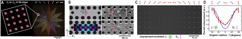
Despite the limitations of intercalating dyes, Fig. 4A shows that arbitrary orientation can integrate 3,456 TOTO-3 labelled small moons with 12 different into a microscopic fluorescent polarimeter, a 100 m device which glows most strongly along the polarization axis of incident light. Microscopic polarimeters constructed using plasmonic antennas have been created in the near-IR (67), and arrays of oriented gold rods have been used for metasurface polarimeters at telecommunication wavelengths (68); the goal of such on-chip instruments is to replace multiple bulky and expensive optical components and to make in situ measurements possible, within devices or transmission lines. Since our polarimeter reports polarization directly, it could be fabricated on microscope slides and used in situ to aid polarized fluorescence microscopy (69): to align excitation polarization grossly by eye without requiring analyzers, to check for polarization bias, or as a calibration standard for fluorescence anisotropy of biomolecules. Operating wavelength could be tuned via intercalation of different dyes (e.g. YOYO-1, 491 nm excitation; TOTO-1, 514 nm; YOYO-1, 612 nm; TOTO-3, 642 nm), or made broadband by using a mixture. Based on the 3.2∘ variability we observe, fitting the orientation of 3,456 origami would allow the angle between excitation polarization and surface features to be measured with a precision of 0.05∘ (SEM). Our polarimeter is unable to measure -polarization, but DOP of 3D origami could add this capability. And while our polarimeter is not a metasurface (70), it provides a roadmap for how DOP could push metal-rod metasurfaces from the near-IR, where the rods are fabricated lithographically, to the visible, via oriented arrays of smaller colloidal gold rods (64, 65).
Hybrid nanophotonic devices (71) combine light emitters or scatterers with microfabricated optical resonators to obtain devices ranging from biosensors (72) to light sources for on-chip quantum information processing (73). The performance (e.g. sensitivity of a detector, or intensity of a light source) of such devices hinges on the strength of the coupling between the emitter and resonator. In particular, emission intensity is proportional to the cavity Purcell enhancement , which is typically a sensitive function of the position of the emitter , and the orientation of the emission dipole relative to the cavity electric field (74). To maximize coupling, the emitter should be positioned in a peak of a resonant mode, with aligned to the polarization of at . Fabricating resonators with simultaneously positioned and aligned emitters has been a difficult challenge (75). Most approaches for positioning involve randomly growing or depositing emitters on a surface, selecting emitters with microscopy, and tediously fabricating resonators around them (73, 76, 74, 77). Some emitters can be grown at predetermined sites within resonators (78), but in general, deterministic approaches for positioning emitters rely on scanning probe microscopy (79, 80). Neither “select and post-process” nor scanning probe approaches can scale to large numbers of devices, or provide deterministic alignment. Conversely, methods for achieving deterministic alignment of molecular or vacancy-based emitters (81, 82, 83, 84, 85) do not address positioning. Previously (8), we used DOP to achieve the large-scale positioning of molecular emitters within L3 photonic crystal cavities (PCCs); TOTO-3 intercalated small moons allowed us to extend that work to control the alignment of in the cavity (Fig. 4, B to D). To optimize emission from the PCCs, we created a 136 array of identical resonators (fig. S13 and S14) with small moons positioned in the center of a -polarized peak in , and varied in 13 steps from 90∘ to -90∘across the width of the array. Emission intensity roughly followed the expected relationship, and a 4.5-fold increase was observed for which maximally align TOTO-3 dipoles with . Potential reasons for disagreement between experimental intensity at 0∘ with FDTD simulation of a single dipole are similar to those for bleed-through above: TOTO-3 dyes are spread out over the 100 nm diameter disk of the small moons rather than in the exact center of the cavity, contributes to a net dipole strength parallel to , and alignment error. Beyond emitter-in-cavity devices, our ability to simultaneously position and orient molecular and nanoparticle components should find wide use in nanophotonics. The collective behavior of multiple emitter systems is highly sensitive to inter-emitter distance and relative dipole orientation, suggesting that our technique will be ideal for studying and engineering fundamental phenomena such as superradiance (86), and other coherence effects (87). Positioning and orientation of molecular emitters within optical nanoantennas will allow antenna performance to be optimized (88); similar control over metal nanoparticle dipoles will enable optical nanocircuit elements to be programmed with series, parallel or intermediate behavior (89, 90).
We have engineered the energy landscape of DNA origami shapes on binding sites to realize absolute and arbitrary orientation, enabling DSA to independently specify all degrees of freedom and thus break all translational and rotational symmetries for arbitrary numbers of C1-symmetric molecular devices. Perhaps surpisingly, we achieved this by combining broken up-down symmetry with a mirror symmetric (D1, bilateral) shape—the small moon; a fully asymmetric (C1) shape was neither necessary nor sufficient—the C1-symmetric right triangle suffered from kinetic trapping.†††A system with multiple local maxima and a single global maximum could break rotational symmetry in the limit of slow annealing to zero temperature. We have yet to find a practical way to anneal DOP, but a combination of heat and monovalent cations has been used to mobilize and crystallize origami kinetically trapped on mica (91). Yet the devices we have presented do not demonstrate the full power of the small moons—the two-fold degeneracy of transition dipoles means that D2 symmetric shapes, e.g. an elongate rectangle or oval, could have been used. No isolated optical device, or coupled array of optical devices yet designed seem to require full symmetry-breaking: 2D chiral scatterers (92) (C4) require up-down symmetry to be broken but not rotational, U-shaped resonators (D1) for certain nonlinear metasurface holograms (93) require that complete rotational symmetry be broken but not up-down. Within electronics, no molecular device with the C1 symmetry of a bipolar junction transistor has been achieved: molecular diodes (94, 95) (D1) can tolerate flips about their mirror plane and crossed-CNT FETs (3) (D2) can tolerate two flips and 180∘ rotation. On the other hand, proposed planar optical and electronic circuits (96) of even just a few symmetric components can almost invariably take advantage of absolute and arbitrary orientation to avoid tortuous paths for interconnect. In part, applications for DSA of molecular components have been constrained by what has been possible. Now that molecular orientation can be controlled, we anticipate that new asymmetric devices and architectures will be explored.
Acknowledgments We acknowledge funding from Office of Naval Research Award N000141410702, U.S. National Science Foundation grant Nos. 1636364 and 1317694 (Expedition in Computing, Molecular Programming Project, http://molecular-programming.org), Air Force Office of Scientific Research FA9550-16-1-0019 (A.M), the Natural Sciences and Engineering Research Council of Canada (D.K.), a Banting Fellowship (C.T.), the Orr Family Foundation, and the Abedin Institute. Fabrication was done at Caltech’s Kavli Nanoscience Institute.
Corresponding authors. Email: ashwing@caltech.edu (A.G.) or pwkr@dna.caltech.edu (P.W.K.R).
Author Contributions A.G. and P.W.K.R. conceived the project. A.G. performed origami synthesis, nanofabrication, AFM, SEM, and fluorescence microscopy. C.T and D.K formalized proof for the deathstar origami design. A.G. and C.T. wrote the simulation code for surface reorientation model. All authors contributed to data interpretation and manuscript preparation.
Supplementary Materials Materials and Methods, Figs. S1 to S14, References (XX–YY), DNA sequences, Design Files, and Simulation Software.
References and Notes
- (1) N. C. Seeman. DNA in a material world. Nature, 421:427–431, 2003.
- (2) P. W. K. Rothemund. Folding DNA to create nanoscale shapes and patterns. Nature, 440(7082):297–302, 2006.
- (3) H. T. Maune, S.-P. Han, R. D. Barish, M. Bockrath, W. A. Goddard III, et al. Self-assembly of carbon nanotubes into two-dimensional geometries using DNA origami templates. Nat. Nanotechnol., 5(1):61–66, 2010.
- (4) Zhao Zhao, Yan Liu, and Hao Yan. DNA origami templated self-assembly of discrete length single wall carbon nanotubes. Org. Biomol. Chem., 11:596–598, 2013.
- (5) Anshuman Mangalum, Masudur Rahman, and Michael L. Norton. Site-specific immobilization of single-walled carbon nanotubes onto single and one-dimensional DNA origami. Journal of the American Chemical Society, 135(7):2451–2454, 2013.
- (6) G. P. Acuna, F. M. Möller, P. Holzmeister, S. Beater, B. Lalkens, et al. Fluorescence enhancement at docking sites of DNA-directed self-assembled nanoantennas. Science, 338(6106):506–510, 2012.
- (7) R. Schreiber, J. Do, E.-M. Roller, T. Zhang, V. J. Schüller, et al. Hierarchical assembly of metal nanoparticles, quantum dots and organic dyes using DNA origami scaffolds. Nature Nanotechnology, 9(1):74–78, 2014.
- (8) Ashwin Gopinath, Evan Miyazono, Andrei Faraon, and Paul W. K. Rothemund. Engineering and mapping nanocavity emission via precision placement of DNA origami. Nature, 535:401–405, 2016.
- (9) S. H. Ko, K. Du, and J. A. Liddle. Quantum-dot fluorescence lifetime engineering with DNA origami constructs. Angewandte Chemie International Edition, 52(4):1193–1197, 2013.
- (10) Jakob Bach Knudsen, Lei Liu, Anne Louise Bank Kodal, Mikael Madsen, Qiang Li, et al. Routing of individual polymers in designed patterns. Nature Nanotechnology, 10:892–898, 2015.
- (11) J. Alexander Liddle, Yi Cui, and Paul Alivisatos. Lithographically directed self-assembly of nanostructures. J. Vac. Sci. Technol. B, 22:3409–3414, 2004.
- (12) Mohamed Asbahi, Shafigh Mehraeen, Fuke Wang, Nikolai Yakovlev, Karen S. L. Chong, et al. Large area directed self-assembly of sub-10 nm particles with single particle positioning resolution. Nano Letters, 15(9):6066–6070, 2015.
- (13) Sang Ouk Kim, Harun H. Solak, Mark P. Stoykovich, Nicola J. Ferrier, Juan J. de Pablo, et al. Epitaxial self-assembly of block copolymers on lithographically defined nanopatterned substrates. Nature, 424:411–414, 2003.
- (14) Jae-Byum Chang, Hong Kyoon Choi, Adam F. Hannon, Alfredo Alexander-Katz, Caroline A. Ross, et al. Design rules for self-assembled block copolymer patterns using tiled templates. Nature Communications, 5:3305, 2014.
- (15) Chin Li Cheung, Julio A. Camarero, Bruce W. Woods, Tianwei Lin, John E. Johnson, et al. Fabrication of assembled virus nanostructures on templates of chemoselective linkers formed by scanning probe nanolithography. Journal of the American Chemical Society, 125(23):6848–6849, 2003.
- (16) Rafael A. Vega, Daniel Maspoch, Khalid Salaita, and Chad A. Mirkin. Nanoarrays of single virus particles. Angewandte Chemie International Edition, 44(37):6013–6015, 2005.
- (17) Yuhuang Wang, Daniel Maspoch, Shengli Zou, George C. Schatz, Richard E. Smalley, et al. Controlling the shape, orientation, and linkage of carbon nanotube features with nano affinity templates. Proceedings of the National Academy of Sciences, 103:2026–31, 2006.
- (18) T. D. Yuzvinsky, A. M. Fennimore, A. Kis, and A. Zettl. Controlled placement of highly aligned carbon nanotubes for the manufacture of arrays of nanoscale torsional actuators. Nanotechnology, 17(2):434, 2006.
- (19) Baoquan Ding, Hao Wu, Wei Xu, Zhao Zhao, Yan Liu, et al. Interconnecting gold islands with DNA origami nanotubes. Nano Letters, 10(12):5065–5069, 12 2010.
- (20) Anthony C. Pearson, Elisabeth Pound, Adam T. Woolley, Matthew R. Linford, John N. Harb, et al. Chemical alignment of DNA origami to block copolymer patterned arrays of 5 nm gold nanoparticles. Nano Lett., 11(5):1981–1987, 2011.
- (21) Risheng Wang, Matteo Palma, Erika Penzo, and Shalom J. Wind. Lithographically directed assembly of one-dimensional DNA nanostructures via bivalent binding interactions. Nano Research, 6:409–417, 2013.
- (22) Piero Morales, Liqian Wang, Abhichart Krissanaprasit, Claudia Dalmastri, Mario Caruso, et al. Suspending DNA origami between four gold nanodots. Small, 12(2):169–173, 2016.
- (23) Aren E. Gerdon, Seung Soo Oh, Kuangwen Hsieh, Yonggang Ke, Hao Yan, et al. Controlled Delivery of DNA Origami on Patterned Surfaces. Small, 5(17):1942–1946, 2009.
- (24) R. J. Kershner, L. D. Bozano, C. M. Micheel, A. M. Hung, A. R. Fornof, et al. Placement and orientation of individual DNA shapes on lithographically patterned surfaces. Nat. Nanotechnol., 4(9):557–561, 2009.
- (25) Erika Penzo, Risheng Wang, Matteo Palma, and Shalom J. Wind. Selective Placement of DNA Origami on Substrates Patterned by Nanoimprint Lithography. J. Vac. Sci. Technol. B, 29(6):06F205, 2011.
- (26) A. Gopinath and P. W. K. Rothemund. Optimized assembly and covalent coupling of single-molecule DNA origami nanoarrays. ACS Nano, 8(12):12030–12040, 2014.
- (27) Yuegang Zhang, Aileen Chang, Jien Cao, Qian Wang, Woong Kim, et al. Electric-field-directed growth of aligned single-walled carbon nanotubes. Applied Physics Letters, 79(19):3155–3157, 2001.
- (28) Miguel A. Correa-Duarte, Marek Grzelczak, Verónica Salgueiriño-Maceira, Michael Giersig, Luis M. Liz-Marzán, et al. Alignment of carbon nanotubes under low magnetic fields through attachment of magnetic nanoparticles. The Journal of Physical Chemistry B, 109(41):19060–19063, 2005.
- (29) Peter A. Smith, Christopher D. Nordquist, Thomas N. Jackson, Theresa S. Mayer, Benjamin R. Martin, et al. Electric-field assisted assembly and alignment of metallic nanowires. Applied Physics Letters, 77(9):1399–1401, 2000.
- (30) Monica Tanase, Laura Ann Bauer, Anne Hultgren, Daniel M. Silevitch, Li Sun, et al. Magnetic alignment of fluorescent nanowires. Nano Letters, 1:155–158, 2001.
- (31) Ahmet Faik Demirörs, Patrick M. Johnson, Carlos M. van Kats, Alfons van Blaaderen, and Arnout Imhof. Directed self-assembly of colloidal dumbbells with an electric field. Langmuir, 26(18):14466–14471, 2010.
- (32) Li Zhang, Jake J. Abbott, Lixin Dong, Bradley E. Kratochvil, Dominik Bell, et al. Artificial bacterial flagella: Fabrication and magnetic control. Applied Physics Letters, 94(6):064107, 2009.
- (33) Kathrin E. Peyer, Li Zhang, and Bradley J. Nelson. Bio-inspired magnetic swimming microrobots for biomedical applications. Nanoscale, 5:1259–1272, 2013.
- (34) Anton Kuzyk, Bernard Yurke, J. Jussi Toppari, Veikko Linko, and Päivi Törmä. Dielectrophoretic trapping of DNA origami. Small, 4(4):447–450, 2008.
- (35) Boxuan Shen, Veikko Linko, Hendrik Dietz, and J. Jussi Toppari. Dielectrophoretic trapping of multilayer DNA origami nanostructures and DNA origami-induced local destruction of silicon dioxide. Electrophoresis, 36(2):255–262, 2015.
- (36) Anja Henning-Knechtel, Matthew Wiens, Mathias Lakatos, Andreas Heerwig, Frieder Ostermaier, et al. Dielectrophoresis of gold nanoparticles conjugated to DNA origami structures. Beilstein Journal of Nanotechnology, 7:948–956, 2016.
- (37) Sam Emaminejad, Mehdi Javanmard, Chaitanya Gupta, Shuai Chang, Ronald W. Davis, et al. Tunable control of antibody immobilization using electric field. Proceedings of the National Academy of Sciences, 112(7):1995–1999, 2015.
- (38) D. Bensimon, A. J. Simon, V. Croquette, and A. Bensimon. Stretching DNA with a receding meniscus: Experiments and models. Phys. Rev. Lett., 74:4754–4757, 1995.
- (39) Junping Jing, Jason Reed, John Huang, Xinghua Hu, Virginia Clarke, et al. Automated high resolution optical mapping using arrayed, fluid-fixed DNA molecules. Proc. Natl. Acad. Sci. USA, 95:8046–8051, 1998.
- (40) Bo Li, Wei Han, Myunghwan Byun, Lei Zhu, Qingze Zou, et al. Macroscopic highly aligned DNA nanowires created by controlled evaporative self-assembly. ACS Nano, 7:4326–4333, 2013.
- (41) Bezu Teschome, Stefan Facsko, Kurt V. Gothelf, and Adrian Keller. Alignment of gold nanoparticle-decorated DNA origami nanotubes: Substrate prepatterning versus molecular combing. Langmuir, 31(46):12823–12829, 2015.
- (42) Dunwei Wang, Ryan Tu, Li Zhang, and Hongjie Dai. Deterministic one-to-one synthesis of germanium nanowires and individual gold nanoseed patterning for aligned nanowire arrays. Angewandte Chemie International Edition, 44(19):2925–2929, 2005.
- (43) Jiaxing Huang, Rong Fan, Stephen Connor, and Peidong Yang. One-step patterning of aligned nanowire arrays by programmed dip coating. Angewandte Chemie International Edition, 46(14):2414–2417, 2007.
- (44) Yu Huang, Xiangfeng Duan, Qingqiao Wei, and Charles M. Lieber. Directed assembly of one-dimensional nanostructures into functional networks. Science, 291(5504):630–633, 2001.
- (45) Bo Li, Chuchu Zhang, Beibei Jiang, Wei Han, and Zhiqun Lin. Flow-enabled self-assembly of large-scale aligned nanowires. Angewandte Chemie International Edition, 54(14):4250–4254, 2015.
- (46) Jingjiao Guan and L. James Lee. Generating highly ordered DNA nanostrand arrays. Proc. Natl. Acad. Sci. U. S. A., 102:18321–18325, 2005.
- (47) Berthold K.P. Horn. Closed-form solution of absolute orientation using unit quaternions. J. Opt. Sci. Am. A, 4:629–642, 1987.
- (48) H. Dietz, S.M. Douglas, and W.M. Shih. Folding DNA into twisted and curved nanoscale shapes. Science, 325:725–730, 2009.
- (49) Bryan Wei, Mingjie Dai, Cameron Myhrvold, Yonggang Ke, Ralf Jungmann, et al. Design space for complex DNA structures. Journal of the American Chemical Society, 135(48):18080–18088, 2013.
- (50) Alexandria N. Marchi, Ishtiaq Saaem, Briana N. Vogen, Stanley Brown, and Thomas H. LaBean. Toward larger DNA origami. Nano letters, 14(10):5740–5747, 2014.
- (51) By Sudhir Husale, Sangeeta Sahoo, Aleksandra Radenovic, Floriano Traversi, Paolo Annibale, et al. ssDNA binding reveals the atomic structure of graphene. Langmuir, 26(23):18078–18082, 2010.
- (52) Xiaorong Xiong, Sheng-Hsiung Liang, and K.F. Böhringer. Geometric binding site design for surface-tension driven self-assembly. In 2004 IEEE International Conference on Robotics and Automation, 2004. Proceedings. ICRA ’04., volume 2, pages 1141–1148. 2004.
- (53) Ashwin Gopinath, David Kirkpatrick, Paul Rothemund, and Chris Thachuk. Progressive alignment of shapes. In Proceedings of the 28th Canadian Conference on Computational Geometry, pages 230–236. 2016.
- (54) Sheng-Hsiung Liang, Xiaorong Xiong, and Karl F. Böhringer. Towards optimal designs for self-alignment in surface tension driven micro-assembly. In 17th IEEE International Conference on Micro Electro Mechanical Systems,(MEMS 2004)., pages 9–12. 2004.
- (55) H. Peter Spielmann, David E. Wemmer, and Jens Peter Jacobsen. Solution structure of a DNA complex with the fluorescent bis-intercalator TOTO determined by NMR spectroscopy. Biochemistry, 34(27):8542–8553, 1995.
- (56) Juleon M. Schins, Alexandra Agronskaia, Bart G. de Grooth, and Jan Greve. Orientation of the chromophore dipoles in the TOTO-DNA system. Cytometry, 37:230–237, 1999.
- (57) Martin L. Bennink, Orlando D. Schärer, Roland Kanaar, Kumiko Sakata-Sogawa, Juleon M. Schins, et al. Single-molecule manipulation of double-stranded DNA using optical tweezers: interaction studies of DNA with RecA and YOYO-1. Cytometry, 36(3):200–208, 1999.
- (58) Fredrik Persson, Fredrik Westerlund, Jonas O. Tegenfeldt, and Anders Kristensen. Local conformation of confined DNA studied using emission polarization anisotropy. Small, 5(2):190–193, 2009.
- (59) Taekjip Ha, Ted A. Laurence, Daniel S. Chemla, and Shimon Weiss. Polarization spectroscopy of single fluorescent molecules. The Journal of Physical Chemistry B, 103(33):6839–6850, 1999.
- (60) B. Sick, B. Hecht, and L. Novotny. Orientational imaging of single molecules by annular illumination. Phys. Rev. Lett., 85:4482–4485, 2000.
- (61) Adam S. Backer, Maurice Y. Lee, and W. E. Moerner. Enhanced DNA imaging using super-resolution microscopy and simultaneous single-molecule orientation measurements. Optica, 3(6):659–666, 2016.
- (62) Cesar Augusto Valades Cruz, Haitham Ahmed Shaban, Alla Kress, Nicolas Bertaux, Serge Monneret, et al. Quantitative nanoscale imaging of orientational order in biological filaments by polarized superresolution microscopy. Proceedings of the National Academy of Sciences, 113(7):E820–E828, 2016.
- (63) N. Milanovich, M. Suh, R. Jankowiak, G. J. Small, and J. M. Hayes. Binding of TO-PRO-3 and TOTO-3 to DNA: fluorescence and hole-burning studies. The Journal of Physical Chemistry, 100(21):9181–9186, 1996.
- (64) Suchetan Pal, Zhengtao Deng, Haining Wang, Shengli Zou, Yan Liu, et al. DNA-directed self-assembly of anisotropic plasmonic nanostructures. Journal of the American Chemical Society, 133(44):17606–17609, 2011.
- (65) A. Kuzyk, R. Schreiber, H. Zhang, A. O. Govorov, T. Liedl, et al. Reconfigurable 3D plasmonic metamolecules. Nature Materials, 13:862–866, 2014.
- (66) Mykhailo Vybornyi, Alina L. Nussbaumer, Simon M. Langenegger, and Robert Häner. Assembling multiporphyrin stacks inside the DNA double helix. Bioconjugate Chemistry, 25(10):1785–1793, 2014.
- (67) Farzaneh Afshinmanesh, Justin S. White, Wenshan Cai, and Mark L. Brongersma. Measurement of the polarization state of light using an integrated plasmonic polarimeter. Nanophotonics, 1:125–129, 2012.
- (68) J. P. Balthasar Mueller, Kristjan Leosson, and Federico Capasso. Ultracompact metasurface in-line polarimeter. Optica, 3(1):42–47, 2016.
- (69) Shalin B. Mehta, Molly McQuilken, Patrick J. La Riviere, Patricia Occhipinti, Amitabh Verma, et al. Dissection of molecular assembly dynamics by tracking orientation and position of single molecules in live cells. Proceedings of the National Academy of Sciences, 113(42):E6352–E6361, 2016.
- (70) Nanfang Yu and Federico Capasso. Flat optics with designer metasurfaces. Nature Materials, 13:139–150, 2014.
- (71) O. Benson. Assembly of hybrid photonic architectures from nanophotonic constituents. Nature, 480(7376):193–199, 2011.
- (72) A. M. Armani, R. P. Kulkarni, S. E. Fraser, R. C. Flagan, and K. J. Vahala. Label-free, single-molecule detection with optical microcavities. Science, 317(5839):783–787, 2007.
- (73) K. Hennessy, A. Badolato, M. Winger, D. Gerace, M. Atatüre, et al. Quantum nature of a strongly coupled single quantum dot–cavity system. Nature, 445(7130):896–899, 2007.
- (74) Janine Riedrich-Möller, Carsten Arend, Christoph Pauly, Frank Mücklich, Martin Fischer, et al. Deterministic coupling of a single silicon-vacancy color center to a photonic crystal cavity in diamond. Nano letters, 14(9):5281–5287, 2014.
- (75) Clotilde Lethiec, Julien Laverdant, Henri Vallon, Clémentine Javaux, Benoît Dubertret, et al. Measurement of three-dimensional dipole orientation of a single fluorescent nanoemitter by emission polarization analysis. Phys. Rev. X, 4:021037, 2014.
- (76) I.J. Luxmoore, R. Toro, O. Del Pozo-Zamudio, N.A. Wasley, E.A. Chekhovich, et al. III–V quantum light source and cavity-QED on silicon. Scientific Reports, 3:1239, 2013.
- (77) L. Sapienza, M. Davanco, A. Badolato, and K. Srinivasan. Nanoscale optical positioning of single quantum dots for bright and pure single-photon emission. Nature Communications, 6:7833, 2015.
- (78) A. Lyasota, S. Borghardt, C. Jarlov, B. Dwir, P. Gallo, et al. Integration of multiple site-controlled pyramidal quantum dot systems with photonic-crystal membrane cavities. Journal of Crystal Growth, 414:192–195, 2015.
- (79) M. Barth, S. Schietinger, S. Fischer, J. Becker, N. Nüsse, et al. Nanoassembled plasmonic-photonic hybrid cavity for tailored light-matter coupling. Nano letters, 10(3):891–895, 2010.
- (80) D. Englund, B. Shields, K. Rivoire, F. Hatami, J. Vučković, et al. Deterministic coupling of a single nitrogen vacancy center to a photonic crystal cavity. Nano Letters, 10(10):3922–3926, 2010.
- (81) R.J. Pfab, J. Zimmermann, C. Hettich, I. Gerhardt, A. Renn, et al. Aligned terrylene molecules in a spin-coated ultrathin crystalline film of p-terphenyl. Chemical Physics Letters, 387:490–495, 2004.
- (82) C. Toninelli, K. Early, J. Bremi, A. Renn, S. Götzinger, et al. Near-infrared single-photons from aligned molecules in ultrathin crystalline films at room temperature. Opt. Express, 18(7):6577–6582, 2010.
- (83) Claudio Polisseni, Kyle D. Major, Sebastien Boissier, Samuele Grandi, Alex S. Clark, et al. Stable, single-photon emitter in a thin organic crystal for application to quantum-photonic devices. Opt. Express, 24(5):5615–5627, 2016.
- (84) M. Lesik, J.-P. Tetienne, A. Tallaire, J. Achard, V. Mille, et al. Perfect preferential orientation of nitrogen-vacancy defects in a synthetic diamond sample. Applied Physics Letters, 104(11):113107, 2014.
- (85) Julia Michl, Tokuyuki Teraji, Sebastian Zaiser, Ingmar Jakobi, Gerald Waldherr, et al. Perfect alignment and preferential orientation of nitrogen-vacancy centers during chemical vapor deposition diamond growth on (111) surfaces. Applied Physics Letters, 104(10):102407, 2014.
- (86) Sang-Hyun Lim, Thomas G. Bjorklund, Frank C. Spano, and Christopher J. Bardeen. Exciton delocalization and superradiance in tetracene thin films and nanoaggregates. Phys. Rev. Lett., 92:107402, Mar 2004.
- (87) C. Hettich, C. Schmitt, J. Zitzmann, S. Kühn, I. Gerhardt, et al. Nanometer resolution and coherent optical dipole coupling of two individual molecules. Science, 298(5592):385–389, 2002.
- (88) Lukas Novotny. From near-field optics to optical antennas. Physics Today, 64(7):47–52, 2011.
- (89) Andrea Alù, Alessandro Salandrino, and Nader Engheta. Parallel, series, and intermediate interconnections of optical nanocircuit elements. 2. nanocircuit and physical interpretation. J. Opt. Soc. Am. B, 24(12):3014–3022, 2007.
- (90) Andrea Alù and Nader Engheta. Tuning the scattering response of optical nanoantennas with nanocircuit loads. Nature Photonics, 2:307–310, 2008.
- (91) Sungwook Woo and Paul W.K. Rothemund. Self-assembly of two-dimensional DNA origami lattices using cation-controlled surface diffusion. Nature Communications, 5:4889, 2014.
- (92) M. Decker, M. W. Klein, M. Wegener, and S. Linden. Circular dichroism of planar chiral magnetic metamaterials. Opt. Lett., 32(7):856–858, 2007.
- (93) Weimin Ye, Franziska Zeuner, Xin Li, Bernhard Reineke, Shan He, et al. Spin and wavelength multiplexed nonlinear metasurface holography. Nature Communications, 7:11930, 2016.
- (94) Arieh Aviram and Mark A. Ratner. Molecular rectifiers. Chemical Physics Letters, 29:277–283, 1974.
- (95) Colin Van Dyck and Mark A. Ratner. Molecular rectifiers: A new design based on asymmetric anchoring moieties. Nano Letters, 15(3):1577–1584, 2015.
- (96) Max M. Shulaker, Gage Hills, Nishant Patil, Hai Wei, Hong-Yu Chen, et al. Carbon nanotube computer. Nature, 501:526–530, 2013.
- (97) E. Stahl, T.G. Martin, F. Praetorius, and H. Dietz. Facile and scalable preparation of pure and dense DNA origami solutions. Angew. Chem. Int. Edit., 53(47):12735–12740, 2014.
- (98) Alan Shaw, Erik Benson, and Björn Högberg. Purification of functionalized DNA origami nanostructures. ACS Nano, 9(5):4968–4975, 2015.
- (99) A. Noy. Handbook of Molecular Force Spectroscopy. Springer, New York, 2007.
- (100) Yong-Xing Zhao, Alan Shaw, Xianghui Zeng, Erik Benson, Andreas M. Nyström, et al. DNA origami delivery system for cancer therapy with tunable release properties. ACS Nano, 6(10):8684–8691, 2012.
- (101) Haorong Chen, Ruixin Li, Shiming Li, Joakim Andréasson, and Jong Hyun Choi. Conformational effects of UV light on DNA origami. Journal of the American Chemical Society, 139(4):1380–1383, 2017. PMID: 28094518.
- (102) Yonggang Ke, Gaëtan Bellot, Niels V. Voigt, Elena Fradkov, and William M. Shih. Two design strategies for enhancement of multilayer–DNA–origami folding: underwinding for specific intercalator rescue and staple-break positioning. Chemical science, 3:2587–2597, 2012.
- (103) Michael C.P. Wang and Byron D. Gates. Directed assembly of nanowires. Materials Today, 12(5):34–43, 2009.
- (104) Jean Michel Arbona, Jean-Pierre Aimé, and Juan Elezgaray. Modeling the mechanical properties of DNA nanostructures. Phys. Rev. E, 86:051912, Nov 2012.