A Review on Image- and Network-based Brain Data Analysis Techniques for Alzheimer’s Disease Diagnosis Reveals a Gap in Developing Predictive Methods for Prognosis
Abstract
Unveiling pathological brain changes associated with Alzheimer’s disease (AD) is a challenging task especially that people do not show symptoms of dementia until it is late. Over the past years, neuroimaging techniques paved the way for computer-based diagnosis and prognosis to facilitate the automation of medical decision support and help clinicians identify cognitively intact subjects that are at high-risk of developing AD. As a progressive neurodegenerative disorder, researchers investigated how AD affects the brain using different approaches: 1) image-based methods where mainly neuroimaging modalities are used to provide early AD biomarkers, and 2) network-based methods which focus on functional and structural brain connectivities to give insights into how AD alters brain wiring. In this study, we reviewed neuroimaging-based technical methods developed for AD and mild-cognitive impairment (MCI) classification and prediction tasks, selected by screening all MICCAI proceedings published between 2010 and 2016. We included papers that fit into image-based or network-based categories. The majority of papers focused on classifying MCI vs. AD brain states, which has enabled the discovery of discriminative or altered brain regions and connections. However, very few works aimed to predict MCI progression based on early neuroimaging-based observations. Despite the high importance of reliably identifying which early MCI patient will convert to AD, remain stable or reverse to normal over months/years, predictive models are still lagging behind.
1 Introduction
Alzheimer’s disease (AD), which is the most common form of dementia, is still today an incurable degenerative disease. It affects 5-8% of all people above 60 years of age, increasing to around 40% of people older than 90% [1]. AD is also known as an irreversible, progressive disorder that destroys neurons which leads to deficits in cognitive functions such as memory and thinking skills. Clinical diagnosis can be supported by biomarkers that detect the presence or absence of the disease. However, identifying such biomarkers, especially in a very early stage, remains challenging as brain changes due to AD occur even before amnestic symptoms appear [2]. The number of people diagnosed with dementia in the UK is expected to rise to over 2 million by 2051 with an estimated cost at between £17 billion and £18 billion a year (Dementia UK report111https://www.alzheimers.org.uk/about-us/policy-and-influencing/dementia-uk-report). Hence, identifying Alzheimer’s disease (AD) earlier before the neurodegeneration is too severe and where treatment is not currently available, might aid in preventing AD onset. Specifically, patients initially diagnosed with mild cognitive impairment (MCI) are known to be a clinically heterogeneous group with different patterns of brain atrophy [3], of which some cases will not progress to AD [4]. To examine the borders between MCI and AD, Magnetic Resonance Imaging (MRI) was extensively used as a non-invasive imaging modality to track changes in brain images of MCI patients as they remain stable, progress to AD, or reverse to normal. Brain dementia MRI data are rapidly growing with emerging international research initiatives aiming to massively collect large high-quality brain images with structural, diffusion and functional imaging modalities, e.g., the public ADNI (Alzheimer’s Disease Neuroimaging Initiative) dataset [5]. However, despite the large body of publications on AD and major advances in neuroimaging technologies, brain image analysis and machine-learning methods, dementia research has not progressed as desired. Fundamentally, there are two major reasons for this.
First, the majority of methods developed for investigating AD stages have focused on learning how to classify AD vs. MCI or normal control (NC) subjects [6, 7, 8, 9, 10, 11, 12, 13, 14, 15, 16, 17, 18, 19, 20, 21, 22, 23, 24]. A conventional classification method would help identify features discriminating between MCI and AD groups; however, it would not allow to identify MCI patients with longer-term followup who will convert to AD after the first MR acquisition timepoint (i.e., baseline). Recently, a challenge on computer-aided diagnosis (CAD) of dementia based on structural MRI, namely CAD-Dementia [4], was launched to evaluate the performance of 29 algorithms from 15 research teams in classifying NC/MCI/AD using a public dataset. However, such dementia challenges have not focused on finding very early biomarkers of prodromal AD, characteristic of the presymptomatic MCI phase of the disease preceding severe cognitive decline, which is a major issue for current international research on AD.
Second, although advanced machine-learning and medical imaging analysis methods for dementia CAD have demonstrated high performance in the literature [4], they are not publicly shared for comparability, reproducibility, and generalizability to unseen data [25]. Although the data for the CAD-dementia challenge is available, the developed methods were not made available for researchers to test on other datasets. A notable exception based on multivariate analysis [26] overlooks the richness and efficiency of recently published machine-learning and data analysis methods for brain disease diagnosis and prognosis [27]. In the following sections, we provide in-depth analysis of AD-related classification and evolution prediction methods from various neuroimaging modalities and identify the gaps in the state-of-the-art.
2 Selection criteria
The analyzed papers in this review were identified from MICCAI conference from 2010 until 2016. We conducted our search using different combinations of the following key words: functional, structural, fMRI, DTI, magnetic resonance imaging, network, brain, connectivity, diffusion, Alzheimer’s disease, mild cognitive impairment, classification, prediction, diagnosis, AD, MCI, biomarker, dementia. We identified 28 papers based on the given search criteria. It is noteworthy that works developed for segmentation tasks and those not focusing primarily on AD/MCI classification or prediction were excluded from this review. The included papers are displayed in Fig. 1. We grouped them into two categories: ‘image-based methods’ and ‘network-based methods’.
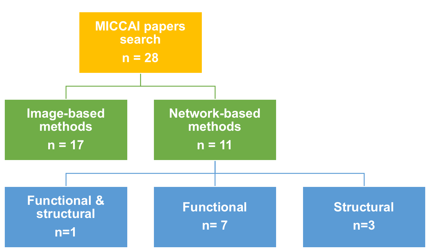
3 Image-based methods
We identified 17 papers that use MR images for dementia state classification. These mainly used hippocampal atrophy and gray matter volume for classifying NC, MCI, and AD brain states. This can be explained by the fact that AD is related to gray matter loss [28] and the shape of subcortical structures (particularly the hippocampus) [29]. To predict clinical decline at the MCI stage and progression to AD, [24] created p-maps from the differences in the shape of the hippocampus between NC and AD subjects and showed increased rates by identifying local regions of interest (ROIs) within the hippocampus using statistical shape models. In a different work, [30] used (left and right) caudate nucleus, and putamen as additional features to hippocampal features to present a system for AD classification using a self-smoothing operator. Other papers suggested the combination of grading measure with HC volume. Specifically, [6] proposed a new method to robustly detect the hippocampal atrophy patterns based on a nonlocal means estimation framework. Combined with HC volume, the grading measure (i.e, the atrophy degree in AD context) led to a success classification rate of 90% between NC and AD subjects.
[7] used two different modalities (MRI + PET) where each subject is represented by two 93-dimensional feature vectors that represent gray matter volume and the average intensity of PET images of 93 ROIs. A novel multi-task learning based feature selection method was proposed to preserve the complementary information conveyed by the two modalities and reached 94% accuracy in distinguishing between AD and NC subjects. In the same context, [9] proposed a manifold regularized multi-task learning framework to jointly select features from multi-modality (MRI + FDG-PET) data as well as [10] and [31, 12, 13]. Similarly, [8] used the same features in addition to three CSF biomarkers and introduced a deep learning method that discovers the non-linear correlations among features which improves the AD, MCI and MCI-C diagnosis accuracy. In [32], a novel Multifold Bayesian Kernelization (MBK) method was proposed to analyze multi-modal MRI biomarkers including average cerebral metabolic rate from PET data, gray matter volume, solidity and convexity features for AD and MCI classification. Another study [11] introduced a different approach to improve AD/NC and p-MCI/s-MCI classification. Basically, it learns a maximum margin representation using multiple atlases jointly with the classification model which resulted in 90% accuracy for AD/NC classification and 73% for p-MCI/s-MCI.
We also identified two landmark papers [33, 34], which devised machine learning frameworks for predicting dementia evolution at later timepoints. [34] proposed a novel canonical feature selection method to fuse information from different imaging modalities (MRI+PET). Specifically, original features (gray matter volume and average intensity of PET images) are projected into a common space. Hence, they become more comparable and easier to depict their relationship in order to predict clinical scores of Alzheimer’s disease. Using the same features, [33] applied a low rank subspace clustering to cluster the data, then used a low rank matrix completion framework to identify pMCI patients and their time of conversion.
4 Network based methods
In this section, we identified 12 papers where the majority (6 papers) used functional networks (derived from fMRI) for dementia diagnosis and prognosis. The remaining studies mainly used structural brain networks (derived from MRI) or fused both networks to enhance AD classification accuracy.
4.1 Functional network
It has been reported that the neurodegenerative process of AD reflects disturbed functional connectivity between brain regions [35, 36]. These alterations are usually measured using resting-state functional MRI (rs-fMRI). Multiple methods have been used for AD diagnosis. For instance, [18] proposed a constrained sparse linear regression model associated with the least absolute shrinkage and selection operator (LASSO) that generates topologically consistent functional connectivity networks from rs-fMRI, thereby improving NC/MCI classification compared with traditional correlation-based methods [36, 37]. [19] introduced a sparse multivariate autoregressive (MAR) modeling to infer effective connectivity from rs-fMRI data and demonstrated its superiority compared to correlation based functional connectivity approaches.
[23] proposed a novel weighted sparse group representation method for brain network modeling, which integrates link strength, group structure as well as sparsity in a unified framework. This models the interactions among multiple brain regions unlike pairwise Pearson correlation and showed superior results in the task of MCI and NC classification. Since simply relying on pairwise functional connectivity between brain regions overlooks how their relationship might be affected at a higher-order level by AD, many studies introduced a functional connectivity hyper-network (FCHN) to infer additional information for AD classifcation. [20] constructed a FCHN using sparse representation modeling where three sets of brain regions are exracted to be fed into a multi-kernel SVM classifier and evaluated on a real MCI dataset. [38] also used a high-order functional connectivity network (HOFC) for MCI classification but generated multiple HOFC networks using hierarchical clustering to further ensemble them with a selective feature fusion method. This approach produced better classification performance than simple use of a single HOFC network.
Dynamic FC. While many studies assumed stationarity on the functional networks over time [39, 40], recent studies in neuroscience have shown that functional organization changes spontaneously over time [41]. For instance, [21] introduced a novel method to model functional dynamics in rs-fMRI for MCI identification. Specifically, a deep network was designed to unravel the non-linear relationships among ROIs in a hierarchical manner and achieved a high classification performance.
4.2 Structural network
Recent findings showed that AD induces a disrupted topology in the structural network characterized by an early damage to synapses and a degeneration of axons [42]. These alterations are basically investigated using MRI with different proposed techniques. In [14], a graph matching framework was devised to match (i) a source graph, where each node encodes a vector that describes regional gray matter volume or cortical thickness features, and (ii) a target graph that includes class label and clinical scores. This approach estimates a target vector for each sample without neglecting its relation with other samples. [15] proposed a view-aligned hypergraph learning (VAHL) method using multi-modality data (MRI, PET, and CSF) for AD/MCI diagnosis where each view corresponds to a specific modality or a combination of several ones. This method can explicitly model the coherence among the views which led to a boost of 4.6% in classification accuracy. [43] proposed a two-stage (query prediction + ranking) medical image retrieval technique with application to MCI diagnosis assistance. This framework was evaluated using three imaging modalities: T1-weighted imaging (T1-w), Diffusion Tensor Imaging (DTI) and Arterial Spin Labeling (ASL).
5 Functional and structural networks
Relying on either structural or functional brain networks may overlook the complementary information that can be leveraged to improve diagnosis and prognosis. For this purpose, [17] integrated two imaging modalities (DTI and fMRI) using a multi-kernel support vector machine (SVM) to improve classification performance where DTI images are parcellated into 90 regions. Then, different structural networks are generated, each conveying a different biophysical property of the brain (e.g., fibers count, fractional anisotropy and mean diffusivity). Additionally, functional connectivity matrices were constructed based on Pearson correlation coefficient to encode the connectivity strength between a pair of ROIs. Furthermore, [44] proposed a centralized hypergraph learning method to model the relationship among subjects using multiple MRIs. Specifically, four MRI sequences were used including T1-weighted MRI (T1), Diffusion Tensor Imaging (DTI), Resting-State functional MRI (RS-fMRI) and Arterial Spin Labeling (ASL) perfusion imaging. This allows to extract supplementary information captured by different neuroimaging data, thereby enhancing the quality of MCI diagnosis.
6 Results and discussion
In this paper, we identified and reviewed 28 works on AD diagnosis and prognosis published in MICCAI proceedings from 2010-2016. Table. 1 displays the different identified papers, while revealing five major gaps that need to be addressed to move dementia research field forward. First, all identified MICCAI papers focused on AD/MCI/NC classification, except for two papers [33, 34], which proposed machine-learning frameworks to predict MCI conversion to AD. Undeniably, accurate discrimination between AD and MCI subjects is an important task to solve as it helps devise more individualized and patient-tailored treatment strategies [45]. However, an accurate prognosis for MCI patients is far more important for providing the optimal treatment and management of the disorder in very early stage. Indeed, early biomarkers identification might help reduce MCI to AD conversion rate. Therefore, predictive models need to be developed to fill this gap and propel the field of MCI prognosis forward.
Such lack of studies could be due to the scarcity of spatiotemporal neuroimaging data where each patient is scanned multiple times. One way to tackle this is by adopting good practices in data analysis and sharing which can promote reliability and collaboration [25]. Second, the classification performance of the proposed technical methods for dementia largely varied with multiple peaks and drops from 2010-2016. This can give insights into the heterogeneity and variability of the disease within subjects and how challenging it is to find an accurate method that works for all cases. In fact, no single approach can be sufficient as each has complementary merits and limitations. Third, comparing these methods is very difficult since they used different approaches and datasets, it is somewhat hard to tell which one performs better if they are not compared against the same baseline methods and evaluated on the same dataset. Fourth, all network-based analysis methods overlooked how dementia states affect the relationship between cortical regions in morphology in both stability, conversion, or reversal MCI evolution scenarios. To fill this gap and noting that several studies [27, 46] reported that morphological features of the brain, such as cortical thickness, can be affected in neurological disorders, one can use the recently proposed morphological brain networks for dementia diagnosis [47, 48, 49, 50]. Last, none of these works proposed a technique for predicting the full trajectory of brain shape changes as MCI progresses towards AD, remains stable, or reverses to normal. Besides, the absence of network-based predictive models is remarkable (Table 1). As such, the use of advanced network and shape analysis methods, using machine learning, could prove fruitful for both classification [47, 48, 49, 50] and prediction tasks.
| Data | Image-based MICCAI papers | Network-based MICCAI papers |
|---|---|---|
| AD/NC | [30, 6, 7, 8, 9, 10, 11, 12, 13] | [14, 15, 16] |
| 96.25%—90%—94.37%—95.9%—95.03%—95.18%—90.69%— 92.1% —96.1% | 92.17%—93.10%—94.05% | |
| NC/MCI | [30, 7, 8, 9, 10, 12, 13] | [17, 18, 14, 19, 20, 21, 22, 15, 23, 16] |
| 91.25%—78.8%—85%—79.27%—79.52%—79.9% —80.3%— | 96.59%—86.49%—81.57%—91.89%—94.6%—81.08%—84.85 %—80.00%—81.8 %—88.59% | |
| cMCI/sMCI | [24, 51, 52, 8, 9, 10, 11, 12, 53] | [15] |
| AUC(0.67) —69.4%—66%—75.8%—68.94%—72.02%—73.69%—80.7%—96.7%— | 79% | |
| Prediction | [32] | |
| NC: 86% — cMCI: 60.61% — sMCI: 66.96% — AD:81.76% | ||
| MCI conversion prediction using baseline MRI | [33] | |
| pMCI to AD: 76% | ||
| MCI conversion prediction using MRI | [34] | |
| 18 months earlier: 76.53%; 12 months earlier: 79.83%; |
7 Conclusion
In this review paper, we examined neuroimaging-based methods for dementia diagnosis and prognosis published in MICCAI 2010-2016 proceedings. The majority of reviewed studies focused on NC, MCI and AD classification tasks using image-based methods or network-based methods including structural and functional brain networks. We noted that very few works developed frameworks to predict MCI conversion to AD at later observations. While the ultimate goal of classification is to provide a computer-aided diagnosis for better clinical decisions, predicting future progression of early demented brains from a baseline observation (i.e., a single timepoint) remains a priority as it might help delay conversion from MCI to AD when early treatment is addressed to the patient. Undoubtedly, predictive intelligence for early dementia diagnosis is still lagging behind, holding various untapped potentials for translational medicine.
References
- [1] : (World Alzheimer Report 2015; https://www.alz.co.uk/research/world-report-2015)
- [2] Buckner, R.L.: Memory and executive function in aging and ad: multiple factors that cause decline and reserve factors that compensate. Neuron 44 (2004) 195–208
- [3] Misra, C., Fan, Y., Davatzikos, C.: Baseline and longitudinal patterns of brain atrophy in MCI patients, and their use in prediction of short-term conversion to AD: Results from ADNI. NeuroImage 44 (2009) 1415 – 1422
- [4] Bron, E.E., Smits, M., van der Flier, W.M., Vrenken, H., Barkhof, F., Scheltens, P., Papma, J.M., Steketee, R.M., Orellana, C.M., Meijboom, R., Pinto, M., Meireles, J.R., Garrett, C., Bastos-Leite, A.J., Abdulkadir, A., Ronneberger, O., Amoroso, N., Bellotti, R., Cardenas-Pena, D., Alvarez-Meza, A.M., Dolph, C.V., Iftekharuddin, K.M., Eskildsen, S.F., Coupe, P., Fonov, V.S., Franke, K., Gaser, C., Ledig, C., Guerrero, R., Tong, T., Gray, K.R., Moradi, E., Tohka, J., Routier, A., Durrleman, S., Sarica, A., Fatta, G.D., Sensi, F., Chincarini, A., Smith, G.M., Stoyanov, Z.V., Sorensen, L., Nielsen, M., Tangaro, S., Inglese, P., Wachinger, C., Reuter, M., van Swieten, J.C., Niessen, W.J., Klein, S.: Standardized evaluation of algorithms for computer-aided diagnosis of dementia based on structural MRI: The CADDementia challenge. NeuroImage 111 (2015) 562 – 579
- [5] Jack Jr, C.R., Bernstein, M.A., Fox, N.C., Thompson, P., Alexander, G., Harvey, D., Borowski, B., Britson, P.J., L. Whitwell, J., Ward, C., et al.: The Alzheimer’s disease neuroimaging initiative (ADNI): MRI methods. Journal of Magnetic Resonance Imaging: An Official Journal of the International Society for Magnetic Resonance in Medicine 27 (2008) 685–691
- [6] Coupé, P., Eskildsen, S.F., Manjon, J.V., Fonov, V., Collins, D.L.: Simultaneous segmentation and grading of hippocampus for patient classification with Alzheimer’s Disease. MICCAI 6893 (2011) 149–157
- [7] Liu, F., Wee, C., Chen, H., Shen, D.: Inter modality relationship constrained multi-task feature selection for AD/MCI classification. MICCAI 8149 (2013) 308–315
- [8] Suk, H.I., Shen, D.: Deep learning-based feature representation for AD/MCI classification. MICCAI 8150 (2013) 583–590
- [9] Jie, B., Zhang, D., Shen, B.C.: Manifold regularized muti-task feature selection for multi-modality classification in AD. MICCAI 8149 (2013) 275–283
- [10] Suk, H.I., Shen, D.: Clustering-induced multi task learning for ad/mci classification. MICCAI 8675 (2014) 393–400
- [11] Min, R., Cheng, J., Price, T., Wu, G., Shen, D.: Maximum-margin based representation learning from multiple atlases for Alzheimer’s Disease classification. MICCAI 8675 (2014) 212–219
- [12] An, L., Adeli, E., Liu, M., Zhang, J., Shen, D.: Semi supervised hierarchical multi modal feature and sample selection for Alzheimer’s Disease diagnosis. MICCAI 9901 (2016) 79–87
- [13] Peng, J., An, L., Zhu, X., Jin, Y., Shen, D.: Structured sparse kernel learning for imaging genetics based Alzheimer’s Disease diagnosis. MICCAI 9901 (2016) 70–78
- [14] Liu, F., Suk, H.I., Wee, C.Y., Chen, H., Shen, D.: High-order graph matching based feature selection for Alzheimer’s Disease identification. MICCAI 8150 (2013) 311–318
- [15] Liu, M., Zhang, J., Yap, P.T., Shen, D.: Diagnosis of Alzheimer’s Disease using view aligned hyergraph learning with incomplete multi-modality data. MICCAI 9900 (2016) 308–316
- [16] Liu, M., Du, J., Jie, B., Zhang, D.: Ordinal patterns for connectivity networks in brain disease diagnosis. MICCAI 9900 (2016) 1–9
- [17] Wee, C.Y., Yap, P.T., Zhang, D., Denny, K., Wang, L., Shen, D.: Identification of individuals with MCI via multimodality connectivity networks. MICCAI 6892 (2011) 277–284
- [18] Wee, C.Y., Yap, P., Zhang, D., Wang, L., Shen, D.: Constrained sparse functional connectivity networks for MCI classification. MICCAI 7511 (2012) 212–219
- [19] Wee, C., Li, Y., Jie, B., Peng, Z., Shen, D.: Identification of MCI using optimal sparse MAR modeled effective connectivity networks. MICCAI 8150 (2013) 319–327
- [20] Jie, B., Shen, D., Zhang, D.: Brain connectivity hyper-network for MCI classification. MICCAI 8674 (2014) 724–732
- [21] Shen, H.S.S.L.D.: A hybrid of deep network and hidden Markov model for MCI classification with Resting-State fMRI. MICCAI 9349 (2015) 573???580
- [22] Chen, X., Zhang, H., Gao, Y., Wee, C.Y., Li, G., Shen, D., the Alzheimer’s Disease Neuroimaging Initiative: High-order resting-state functional connectivity network for MCI classification. Human Brain Mapping 37 (2016) 3282–3296
- [23] Yu, R., Zhang, H., An, L., Chen, X., Wei, Z., Shen, D.: Correlation weighted sparse group representation for brain network construction in MCI classification. MICCAI 9900 (2016) 37–45
- [24] Leung, K.K., Shen, K., Barnes, J., Ridgway, G.R., Clarkson, M.J., Fripp, J., Salvado, O., Meriaudeau, F., Fox, N.C., Bourgeat, P., Ourselin, S.: Increasing power to predict Mild Cognitive Impairment Conversion to Alzheimer’s Disease using hippocampal atrophy rate and statistical shape models. Sringer 6362 (2010) 125–132
- [25] Nichols, T.E., Das, S., Eickhoff, S.B., Evans, A.C., Glatard, T., Hanke, M., Kriegeskorte, N., Milham, M.P., Poldrack, R.A., Poline, J.B., et al.: Best practices in data analysis and sharing in neuroimaging using mri. Nature Neuroscience 20 (2017) 299
- [26] Sabuncu, M.R., Konukoglu, E., Initiative, A.N., et al.: Clinical prediction from structural brain MRI scans: a large-scale empirical study. Neuroinformatics 13 (2015) 31–46
- [27] Brown, C., Hamarneh, G.: Machine learning on human connectome data from MRI. arXiv:1611.08699v1 (2016)
- [28] Karas, G., Burton, E., Rombouts, S., Van Schijndel, R., O’Brien, J., Scheltens, P., McKeith, I., Williams, D., Ballard, C., Barkhof, F.: A comprehensive study of gray matter loss in patients with alzheimer’s disease using optimized voxel-based morphometry. Neuroimage 18 (2003) 895–907
- [29] Du, A., Schuff, N., Amend, D., Laakso, M., Hsu, Y., Jagust, W., Yaffe, K., Kramer, J., Reed, B., Norman, D., et al.: Magnetic resonance imaging of the entorhinal cortex and hippocampus in mild cognitive impairment and alzheimer’s disease. Journal of Neurology, Neurosurgery & Psychiatry 71 (2001) 441–447
- [30] Iglesias, J.E., Jiang, J., Liu, C.Y., Tu, Z., the Alzheimers Disease Neuroimaging Initiative: Classification of Alzheimer’s Disease using a self-smoothing operator. MICCAI 6893 (2011) 58–65
- [31] Zhu, X., Suk, H.I., Shen, D.: Multi-modality canonical feature selection for alzheimer’s disease diagnosis. In: International Conference on Medical Image Computing and Computer-Assisted Intervention, Springer (2014) 162–169
- [32] Liu, S., Song, Y., Cai, W., Pujol, S., Kikinis, R., Wang, X., Feng, D.: Multifold bayesian kernelization in alzheimer’s diagnosis. MICCAI 8150 (2013) 303–310
- [33] Thung, K., Yap, P., Adeli-M, E., Shen, D.: Joint Diagnosis and Conversion Time Prediction of Progressive Mild Cognitive Impairment (pMCI) Using Low-Rank Subspace Clustering and Matrix Completion. MICCAI 9351 (2015) 527–534
- [34] Zhu, X., Suk, H.I., Lee, S.W., Shen, D.: Canonical feature selection for joint regression and multi-class identification in Alzheimer’s disease diagnosis. Brain Imaging and Behavior 10 (2016) 818–828
- [35] Fransson, P.: Spontaneous low-frequency bold signal fluctuations: An fmri investigation of the resting-state default mode of brain function hypothesis. Human brain mapping 26 (2005) 15–29
- [36] Wang, K., Liang, M., Wang, L., Tian, L., Zhang, X., Li, K., Jiang, T.: Altered functional connectivity in early alzheimer’s disease: A resting-state fmri study. Human brain mapping 28 (2007) 967–978
- [37] Wee, C.Y., Yap, P.T., Denny, K., Browndyke, J.N., Potter, G.G., Welsh-Bohmer, K.A., Wang, L., Shen, D.: Resting-state multi-spectrum functional connectivity networks for identification of mci patients. PloS one 7 (2012) e37828
- [38] Chen, X., Zhang, H., Shen, D.: Ensemble hierarchical high-order functional connectivity networks for MCI classification. MICCAI (2016) 18–25
- [39] Li, S., Eloyan, A., Joel, S., Mostofsky, S., Pekar, J., Bassett, S.S., Caffo, B.: Analysis of group ica-based connectivity measures from fmri: application to alzheimer’s disease. PloS one 7 (2012) e49340
- [40] Wee, C.Y., Yap, P.T., Zhang, D., Wang, L., Shen, D.: Group-constrained sparse fmri connectivity modeling for mild cognitive impairment identification. Brain Structure and Function 219 (2014) 641–656
- [41] Hutchison, R.M., Womelsdorf, T., Allen, E.A., Bandettini, P.A., Calhoun, V.D., Corbetta, M., Della Penna, S., Duyn, J.H., Glover, G.H., Gonzalez-Castillo, J., et al.: Dynamic functional connectivity: promise, issues, and interpretations. Neuroimage 80 (2013) 360–378
- [42] Serrano-Pozo, A., Frosch, M.P., Masliah, E., Hyman, B.T.: Neuropathological alterations in alzheimer disease. Cold Spring Harbor perspectives in medicine 1 (2011) a006189
- [43] Gao, Y., Adeli-M, E., Kim, M., Giannakopoulos, P., Haller, S., Shen, D.: Medical image retrieval using multi-graph learning for MCI diagnostic assistance. MICCAI 9350 (2015) 86–93
- [44] Gao, Y., Wee, C.Y., Kim, M., Giannakopoulos, P., Montandon, M.L., Haller, S., Shen, D.: Mci identification by joint learning on multiple mri data. MICCAI 9350 (2015)
- [45] Ithapu, V.K., Singh, V., Okonkwo, O.C., Chappell, R.J., Dowling, N.M., Johnson, S.C., Initiative, A.D.N., et al.: Imaging-based enrichment criteria using deep learning algorithms for efficient clinical trials in mild cognitive impairment. Alzheimer’s & Dementia 11 (2015) 1489–1499
- [46] Querbes, O., Aubry, F., Pariente, J., Lotterie, J., Demonet, J., Duret, V., Puel, M., Berry, I., Fort, J., Celsis, P., The Alzheimer’s Disease Neuroimaging Initiative: Early diagnosis of Alzheimer’s disease using cortical thickness: impact of cognitive reserve. Brain 132 (2009) 2036
- [47] Lisowska, A., Rekik, I.: Pairing-based ensemble classifier learning using convolutional brain multiplexes & multi-view brain networks for early dementia diagnosis. MICCAI-CNI (Connectomics in Neuroimaging) workshop 1 (2017)
- [48] Mahjoub, I., Mahjoub, M.A., Rekik, I.: Brain multiplexes reveal morphological connectional biomarkers fingerprinting late brain dementia states. Scientific reports 8 (2018) 4103
- [49] Lisowska, A., Rekik, I.: Joint pairing and structured mapping of convolutional brain morphological multiplexes for early dementia diagnosis. Brain Connectivity (2018)
- [50] Raeper, R., Lisowska, A., Rekik, I.: Joint correlational and discriminative ensemble classifier learning for dementia stratification using shallow brain multiplexes. MICCAI (2018)
- [51] Cheng, B., Zhang, D., Shen, D.: Domain transfer learning for MCI conversion prediction. MICCAI 7510 (2012) 82–90
- [52] Singh, N., Wang, A.Y., Sankaranarayanan, P., Fletcher, P.T., Joshi, S.: Genetic, functional and structural imaging biomarkers for early detection of conversion from MCI to AD. MICCAI 7510 (2012) 132–140
- [53] Zhang, J., Shi, J., Stonnington, C., Li, Q., Gutman, B.A., Chen, K., Reiman, E.M., Richard??Caselli, Thompson, P.M., Ye, J., Wang, Y.: Hyperbolic space sparse coding with its application on prediction of AD in MCI. MICCAI 9900 (2016) 326–334