Three-dimensional nuclear spin positioning using coherent radio-frequency control
Abstract
Distance measurements via the dipolar interaction are fundamental to the application of nuclear magnetic resonance (NMR) to molecular structure determination, but they only provide information on the absolute distance and polar angle between spins. In this Letter, we present a protocol to also retrieve the azimuth angle . Our method relies on measuring the nuclear precession phase after application of a control pulse with a calibrated external radio-frequency coil. We experimentally demonstrate three-dimensional positioning of individual 13C nuclear spins in a diamond host crystal relative to the central electronic spin of a single nitrogen-vacancy center. The ability to pinpoint three-dimensional nuclear locations is central for realizing a nanoscale NMR technique that can image the structure of single molecules with atomic resolution.
Nuclear magnetic resonance (NMR) and electron paramagnetic resonance (EPR) spectroscopy are among the most important analytical methods in structural biology and the chemical sciences. By combining local chemical information of atoms with pair-wise distance constraints, it becomes possible to reconstruct three-dimensional structures or structural changes of proteins and other biomolecules. While conventional NMR typically analyzes large ensembles of molecules, considerable effort has recently been expended on advancing NMR detection to the level of individual molecules Poggio and Degen (2010); Wrachtrup and Finkler (2016); Schwartz et al. (2017). If successfully extended to the atomic scale, NMR could enable direct imaging of three-dimensional molecular structures, with many applications in structural biology and the nanosciences. A promising platform for this task are diamond chips containing near-surface nitrogen-vacancy (NV) centers whose electronic spins can be exploited as sensitive local NMR probes Staudacher et al. (2013); Mamin et al. (2013).
Structural imaging of single molecules involves determining the three-dimensional coordinates and elemental species of the constituent nuclei. In NV-NMR, information on the spatial position can be gained from the dipolar part of the hyperfine interaction between the nuclei and the central electronic spin Taminiau et al. (2012); Kolkowitz et al. (2012); Zhao et al. (2012). Because of the axial symmetry of the dipolar interaction, however, only the absolute distance and the polar (inter-spin) angle can be inferred from a NMR spectroscopy measurement. Although the axial symmetry can be broken by a static Zhao et al. (2012) or dynamic Zopes et al. (2018) transverse magnetic field, determination of the azimuth angle , required for reconstructing the full three-dimensional distance vector , has remained challenging Laraoui et al. (2015); Wang et al. (2016); Sasaki et al. (2018).
In this Letter, we demonstrate a simple and precise method for retrieving the azimuth of the inter-spin vector, allowing us to perform full three-dimensional nuclear distance measurements. Our technique relies on measuring the nuclear precession phase after application of a radio-frequency (rf) pulse by an external micro-coil. We determine at low and high magnetic fields, and for polarized as well as unpolarized nuclear spins. We exemplify our method by mapping the three-dimensional locations of 13C nuclei for distances up to and angular uncertainties below .
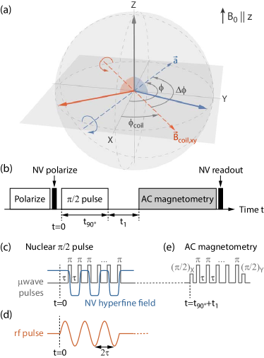
Our scheme for measuring the azimuth angle is introduced in Fig. 1(a-d): starting from a polarized nuclear state, we perform a rotation of the nuclear spin. The rotation is generated either by modulating the hyperfine field of the NV center using microwave pulses (Fig. 1c), or by applying a rf pulse with an external coil (Fig. 1d). Subsequently, we let the nuclear spin precess in the equatorial plane of the Bloch sphere and detect the frequency and phase of the precession by an AC magnetometry measurement with the NV center Taylor et al. (2008); Lange et al. (2011); Kotler et al. (2011) (Fig. 1e).
Crucially, the starting phase of the nuclear precession at is set by the axis of the rotation, which is determined by the spatial direction of the rf field in the laboratory frame of reference. When driving the nuclear rotation via the hyperfine interaction, the rf field direction is given by , where is the secular part of the hyperfine tensor, and are the parallel and transverse hyperfine coupling parameters Taminiau et al. (2014); Boss et al. (2016), and is the nuclear gyromagnetic ratio (Fig. 1a, blue). Conversely, if the external coil is used to generate the rf field, the rotation axis is given by the in-plane component of the coil field (Fig. 1a, red). By comparing the phases of the precession signals, we directly obtain the relative angle between the unknown orientation of the hyperfine vector and the calibrated orientation of the external coil field.
We experimentally determine the angles of three 13C nuclear spins from three different NV centers in two single-crystal diamond chips. We optically polarize and read out the NV spin by short laser pulses () and detect the fluorescence intensity in a confocal microscope arrangement. Microwave pulses at are used to actuate the electronic spin transition. To polarize the nuclear spins, we transfer polarization from the optically aligned NV center using dynamic nuclear polarization with a repetitive initialization sequence Taminiau et al. (2014); Cujia et al. (2018). AC magnetometry is performed by a periodic sequence of microwave pulses with XY8 phase cycling Gullion et al. (1990) enclosed by two pulses that are phase-shifted by Taylor et al. (2008); Boss et al. (2017). We use a permanent magnet to apply bias fields of and for low field and high field experiments, respectively, aligned to within of the NV quantization axis.
The key component of our experiment is the external rf coil, whose field orientation serves as the spatial reference for the angle measurement. Two generations of micro-coils are used: the first coil has a 3-dB-bandwidth of (deduced from the step response recorded with the NV center) and is used for low field experiments. The second coil reaches a bandwidth of . Both rf coils produce fields of and are operated with currents of up to . Crucial for our experiments is a precise knowledge of the direction and temporal shape of the coil magnetic field. We determine the three-dimensional vector of the coil magnetic field using two other nearby NV centers with different crystallographic orientations with an uncertainty of less than in all three spatial components Steinert et al. (2010); Zopes et al. (2018). We align our (,,) laboratory reference frame to the ([],[],[111]) crystallographic axes of the single crystal diamond chips (up to an inversion symmetry about the origin). To calibrate the dynamic response of the coil, we perform in situ measurements of the rf field using time-resolved optically-detected magnetic resonance (ODMR) spectroscopy (Fig. 2(a,e)). We acquire ODMR spectra in snapshots of (a) or (e) over the duration of the rf pulse, and determine the pulse profile by fitting the peak positions of the resonance curves.
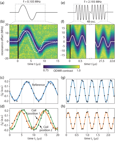
In Fig. 2(c,d), we show a first set of measurements for nuclear spin 13C1 carried out at low magnetic field, . The hyperfine coupling parameters of this nuclear spin are , calibrated by a separate correlation spectroscopy measurement Boss et al. (2016). Fig. 2(c) shows the reference measurement of the nuclear spin precession after application of the pulse using the hyperfine field. Fig. 2(d) plots the corresponding precession signal after applying the rotation with the rf coil. We observe a clear phase shift between the two signals, indicating that the hyperfine field and the coil field point in different spatial directions. We verify that the phase shift changes if we vary the direction of by moving the rf coil to a different position (green data in Fig. 2(d)).
For ideal rf pulses and exact timings, the observed phase shift corresponds to the difference between the azimuth angles of the hyperfine and coil magnetic fields, allowing us to directly deduce . However, due to the limited bandwidth of the rf circuit and the finite length of feed lines, the actual rf pulses tend to be delayed and distorted, leading to a phase offset. In addition, the AC magnetometry measurement is very sensitive to timing errors and resonance offsets in the microwave modulation, causing additional uncertainty in the phase measurement. To compensate for these issues, we determine by fitting the experimental data with a Levenberg-Marquardt algorithm using a density matrix simulation Johansson et al. (2013) as fit function and as fit parameter. We propagate the two-spin density matrix through the full sequence shown in Fig. 1(b) using piece-wise constant Hamiltonians for the nuclear spin propagation, taking the calibrated vector field and temporal shape of Fig. 2(a) as well as the hyperfine parameters (,) as inputs. By calculating the nuclear spin evolution in the laboratory frame of reference, the simulation captures the Bloch-Siegert shift Abragam and the -component of the rf field. In addition, we directly retrieve the absolute laboratory frame azimuth rather than the relative between and .
We start the analysis by fitting the simulation to the reference measurement (Fig. 2(c)), which allows us to determine with an uncertainty smaller than . As defines the nuclear precession frequency, this calibration is of paramount importance for a precise estimate of . Afterwards we determine with a second fit to the measurements with the rf pulse (Fig. 2(d)) while keeping fixed. All fit results are shown by solid lines in Fig. 2(c,d). We find an azimuth location of . We have previously determined the three-dimensional coordinates of the same nuclear spin using a different positioning method Zopes et al. (2018), where , in good agreement with the present result. The accuracy of our experiment is presently limited by the calibration uncertainty of the coil field angle () and by the statistical fit error of the precession phase (). Additional sources of uncertainty, e.g., a misalignment of or the influence of the local chemical environment are not included in the analysis, but are expected to be insignificant for our study. The estimated three-dimensional location for this (13C1) and another nuclear spin (13C2; ) are shown in Fig. 3.
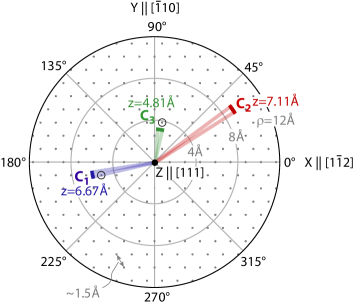
Next, we demonstrate that our azimuth positioning technique can be readily extended to high magnetic fields. High bias fields are desirable in NMR because of a better peak separation and a simplified interpretation of spectra. In addition, in NV-NMR, more efficient dynamical decoupling control and repetitive readout schemes become possible at higher fields Neumann et al. (2010). In Fig. 2(e-h) we show measurements carried out at on a third nuclear spin (13C3) with hyperfine coupling parameters . Here, we find . The three-dimensional location of 13C3 is also indicated in Fig. 3.
The uncertainty at high magnetic field is larger than at low field because of timing errors. At , the nuclear Larmor period is only , such that of timing uncertainty causes a phase uncertainty of about . For the rf pulse in Fig. 2(e), we find a phase delay of , corresponding to an overall timing uncertainty of the ODMR calibration of . Although the measured phase delay is in good agreement with the value predicted from the electrical characteristics of the rf circuit (), it already introduces the largest error to the measurement. For future experiments carried out in the high bias fields of superconducting magnets Aslam et al. (2017) a precise calibration of control fields will therefore become even more critical.
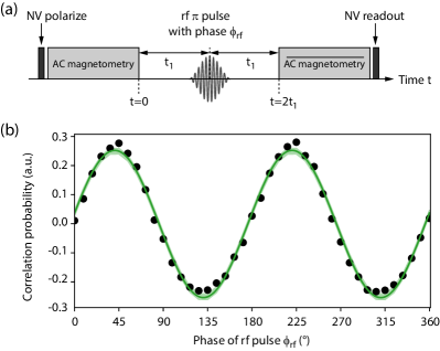
Finally, we discuss a complementary scheme for reconstructing the azimuth angle that does not require pre-polarization of nuclear spins. Instead of recording the nuclear precession signal as a function of , we intersperse a correlation spectroscopy sequence Laraoui et al. (2013); Boss et al. (2016) with a central rf pulse to generate a nuclear spin echo at a fixed time (Fig. 4(a)). By varying the pulse phase from , we modulate the amplitude of the spin echo, leading to an oscillatory signal . We then determine from the phase offset of the oscillation. Fig. 4(b) shows a spin echo oscillation for 13C3 measured at a bias field of . The compatible angles are , in good agreement with the result from Fig. 2(h). Note that the echo method is afflicted by a ambiguity in the angle measurement, because the echo oscillation repeats with modulo . Although the ambiguity could possibly be resolved by applying concomitant rf and microwave rotations or by introducing dc field pulses Zopes et al. (2018), it is unlikely to restrict future experiments on single molecules where relative, rather than absolute, positions are important. In addition, single-molecule NMR experiments can exploit internuclear interactions to further constrain the nuclear positions.
In conclusion, we have introduced a simple method for measuring the inter-spin azimuth , enabling us to perform three-dimensional distance measurements on single nuclear spins. We demonstrate the potential of our technique by mapping the 3D location of individual 13C nuclei in diamond with a precision sufficient for assigning discrete lattice sites. Future experiments will apply 3D distance measurements to molecules deposited on the surface of dedicated diamond NMR sensor chips Mamin et al. (2013); Ohashi et al. (2013); Loretz et al. (2014); Lovchinsky et al. (2016) and provide an avenue to analyze the structure and conformation of single molecules with atomic resolution Sidles (2009).
This work was supported by Swiss National Science Foundation (SNFS) Project Grant No. 200020_175600, the National Center of Competence in Research in Quantum Science and Technology (NCCR QSIT), and the DIAmond Devices Enabled Metrology and Sensing (DIADEMS) program, Grant No. 611143, of the European Commission.
We thank A. Nizovtsev and F. Jelezko for sharing details about the DFT simulation. While finishing this manuscript, we learned about a similar idea put forward by Sasaki and coworkers Sasaki et al. (2018).
†These authors contributed equally to this work.
∗Email: degenc@ethz.ch
References
- Poggio and Degen (2010) M. Poggio and C. L. Degen, Nanotechnology 21, 342001 (2010).
- Wrachtrup and Finkler (2016) J. Wrachtrup and A. Finkler, J. Magn. Reson. 269, 225 (2016).
- Schwartz et al. (2017) I. Schwartz, J. Rosskopf, S. Schmitt, B. Tratzmiller, Q. Chen, L. P. McGuinness, F. Jelezko, and M. B. Plenio, arXiv:1706.07134 (2017).
- Staudacher et al. (2013) T. Staudacher, F. Shi, S. Pezzagna, J. Meijer, J. Du, C. A. Meriles, F. Reinhard, and J. Wrachtrup, Science 339, 561 (2013).
- Mamin et al. (2013) H. J. Mamin, M. Kim, M. H. Sherwood, C. T. Rettner, K. Ohno, D. D. Awschalom, and D. Rugar, Science 339, 557 (2013).
- Taminiau et al. (2012) T. H. Taminiau, J. J. T. Wagenaar, T. V. der Sar, F. Jelezko, V. V. Dobrovitski, and R. Hanson, Phys. Rev. Lett. 109, 137602 (2012).
- Kolkowitz et al. (2012) S. Kolkowitz, Q. P. Unterreithmeier, S. D. Bennett, and M. D. Lukin, Phys. Rev. Lett. 109, 137601 (2012).
- Zhao et al. (2012) N. Zhao, J. Honert, B. Schmid, M. Klas, J. Isoya, M. Markham, D. Twitchen, F. Jelezko, R. Liu, H. Fedder, and J. Wrachtrup, Nature Nano. 7, 657 (2012).
- Zopes et al. (2018) J. Zopes, K. S. Cujia, K. Sasaki, J. M. Boss, K. M. Itoh, and C. L. Degen, arXiv:1806.04883 (2018).
- Laraoui et al. (2015) A. Laraoui, D. Pagliero, and C. A. Meriles, Phys. Rev. B 91, 205410 (2015).
- Wang et al. (2016) Z.-Y. Wang, J. F. Haase, J. Casanova, and M. B. Plenio, Phys. Rev. B 93, 174104 (2016).
- Sasaki et al. (2018) K. Sasaki, K. M. Itoh, and E. Abe, arXiv:1806.00177 (2018).
- Taylor et al. (2008) J. M. Taylor, P. Cappellaro, L. Childress, L. Jiang, D. Budker, P. R. Hemmer, A.Yacoby, R. Walsworth, and M. D. Lukin, Nat. Phys. 4, 810 (2008).
- Lange et al. (2011) G. D. Lange, D. Riste, V. V. Dobrovitski, and R. Hanson, Phys. Rev. Lett. 106, 080802 (2011).
- Kotler et al. (2011) S. Kotler, N. Akerman, Y. Glickman, A. Keselman, and R. Ozeri, Nature 473, 61 (2011).
- Taminiau et al. (2014) T. H. Taminiau, J. Cramer, T. van der Sar, V. V. Dobrovitski, and R. Hanson, Nature Nano. 9, 171 (2014).
- Boss et al. (2016) J. M. Boss, K. Chang, J. Armijo, K. Cujia, T. Rosskopf, J. R. Maze, and C. L. Degen, Phys. Rev. Lett. 116, 197601 (2016).
- Cujia et al. (2018) K. S. Cujia, J. M. Boss, J. Zopes, and C. L. Degen, arXiv:1806.08243 (2018).
- Gullion et al. (1990) T. Gullion, D. B. Baker, and M. S. Conradi, J. Magn. Res. 89, 479 (1990).
- Boss et al. (2017) J. M. Boss, K. S. Cujia, J. Zopes, and C. L. Degen, Science 356, 837 (2017).
- Steinert et al. (2010) S. Steinert, F. Dolde, P. Neumann, A. Aird, B. Naydenov, G. Balasubramanian, F. Jelezko, and J. Wrachtrup, Rev. Sci. Instrum. 81, 43705 (2010).
- Johansson et al. (2013) J. Johansson, P. Nation, and F. Nori, Computer Physics Communications 184, 1234 (2013).
- (23) A. Abragam, (Oxford University Press, Oxford, 1961) .
- Gali et al. (2008) A. Gali, M. Fyta, and E. Kaxiras, Phys. Rev. B 77, 155206 (2008).
- Nizovtsev et al. (2018) A. P. Nizovtsev, S. Y. Kilin, A. L. Pushkarchuk, V. A. Pushkarchuk, S. A. Kuten, O. A. Zhikol, S. Schmitt, T. Unden, and F. Jelezko, New Journal of Physics 20, 023022 (2018).
- Neumann et al. (2010) P. Neumann, J. Beck, M. Steiner, F. Rempp, H. Fedder, P. R. Hemmer, J. Wrachtrup, and F. Jelezko, Science 329, 542 (2010).
- Aslam et al. (2017) N. Aslam, M. Pfender, P. Neumann, R. Reuter, A. Zappe, F. F. de Oliveira, A. Denisenko, H. Sumiya, S. Onoda, J. Isoya, and J. Wrachtrup, Science 357 (2017), 10.1126/science.aam8697.
- Laraoui et al. (2013) A. Laraoui, F. Dolde, C. Burk, F. Reinhard, J. Wrachtrup, and C. A. Meriles, Nature Commun. 4, 1651 (2013).
- Zopes et al. (2017) J. Zopes, K. Sasaki, K. S. Cujia, J. M. Boss, K. Chang, T. F. Segawa, K. M. Itoh, and C. L. Degen, arXiv:1705.07968 (2017).
- Ohashi et al. (2013) K. Ohashi, T. Rosskopf, H. Watanabe, M. Loretz, Y. Tao, R. Hauert, S. Tomizawa, T. Ishikawa, J. Ishi-hayase, S. Shikata, C. L. Degen, and K. M. Itoh, Nano Letters 13, 4733 (2013).
- Loretz et al. (2014) M. Loretz, S. Pezzagna, J. Meijer, and C. L. Degen, Appl. Phys. Lett. 104, 033102 (2014).
- Lovchinsky et al. (2016) I. Lovchinsky, A. O. Sushkov, E. Urbach, N. P. de Leon, S. Choi, K. de Greve, R. Evans, R. Gertner, E. Bersin, C. Muller, L. McGuinness, F. Jelezko, R. L. Walsworth, H. Park, and M. D. Lukin, Science 351, 836 (2016).
- Sidles (2009) J. A. Sidles, Proc. Natl. Acad. Sci. USA 106, 2477 (2009).