A tunable time-resolved spontaneous Raman spectroscopy setup for probing ultrafast collective excitation and quasiparticle dynamics in quantum materials
Abstract
We present a flexible and efficient ultrafast time-resolved spontaneous Raman spectroscopy setup to study collective excitation and quasi-particle dynamics in quantum matter. The setup has a broad energy tuning range extending from the visible to near infrared spectral regions for both the pump excitation and Raman probe pulses. Additionally, the balance between energy and time-resolution can be controlled. A high light collecting efficiency is realized by high numerical aperture collection optics and a high-throughput flexible spectrometer. We demonstrate the functionality of the setup with a study of the zone-center longitudinal optical phonon and hole continuum dynamics in silicon, and discuss the role of the Raman tensor in time-resolved Raman scattering. In addition, we show evidence for unequal phonon softening rates at different high symmetry points in the Brillouin zone of silicon by means of detecting pump-induced changes in the two-phonon overtone spectrum. Demagnetization dynamics in the helimagnet Cu2OSeO3 is studied by observing softening and broadening of a magnon after photo-excitation, underlining the unique power of measuring transient dynamics in the frequency domain, and the feasibility to study phase transitions in quantum matter.
I Introduction
The last decade has seen a surge of experiments where light is exploited as a strong external stimulus to manipulate quantum matter. basov2017 ; zhang2014 ; gandolfi2017 The light-matter interaction can be a fully coherent process as in the case of the Floquet state in photon-dressed materials, gedik2013 or drive quantum matter into a strongly non-thermodynamic state by disturbance of the balance between electronic, orbital, spin, and lattice degrees of freedom. Seminal cases of the latter phenomenon include the photo-induced insulator-metal transition in VO2, cavalleri2001 optical control of colossal magnetoresistivity in manganites, rini2007 and transient signatures of photo-induced superconductivity in a stripe-ordered cuprate. fausti2011
Integral in the description of the emanating dynamics in the optically driven non-equilibrium state is the creation and annihilation of electronic quasiparticles, collective excitations associated with the various degrees of freedom, and their interaction. Their transient annihilation and creation may for instance induce symmetry changes across photo-induced phase transitions, boschini2018 and dictates nonequilibrium temperatures, maldonado2017 while their interaction underlies the coupling, and therefore the equilibration between the various degrees of freedom. turgut2016 A scala of partially complimentary pump-probe techniques have been developed to study quasiparticle and collective excitation dynamics, and their interaction. For instance, the coupling between low-energy collective excitations, and high-energy electronic excitations may be mapped into the time-domain, and quantified, through all-optical coherent fluctuation spectroscopy. mansart2013 ; mann2015 A direct view on momentum-dependent electron and hole quasiparticle population dynamics is provided by means of the matured technique of time-resolved angle-resolved photoemission spectroscopy. sobota2012 ; boschini2018 The situation for direct probing of collective excitation populations is perhaps more ominous. abbamonte2013 Rather novel photon-probes of dynamics of collective excitations such as phonons, magnons, and orbital excitations are time-resolved diffuse x-ray scattering, trigo2013fourier and time-resolved resonant inelastic x-ray scattering. dean2016ultrafast Plasmon dynamics in quantum materials has for instance succesfully been probed with femtosecond electron energy loss spectroscopy. carbone2009 ; piazza2014 However, a quantification of occupation numbers and corresponding effective temperatures with these techniques still seems a stretch.
Time-resolved Raman scattering is a reasonably well-established probe for vibrational dynamics in organic materials, iwata1993 ; hamaguchi1994 ; uesugi1997 ; sahoo2011 ; kruglik2011 and phonon population dynamics in semiconductor materials kash1985 ; oberli1987 ; zhu2017 and carbon allotropes. song2008 ; yan2009 ; kang2010 ; yang2017novel With this technique the transient evolution of the spontaneous Raman spectrum after photo-excitation is studied. faustibook The pump excitation mechanism only needs to induce incoherent dynamics. There are thus no special prerequisites to the pulse duration, as is the case with coherent pump-probe techniques. mansart2013 ; mann2015 In the time-domain spectra transient changes in excitation energies, line-widths, and scattering intensities of Raman-active excitations can be obtained. This has the advantage over coherent excitation techniques that one can follow the true time evolution of the system through its incoherent response, rather than the time evolution of coherent excitations. Through detailed balance of the anti-Stokes to Stokes scattering intensity ratio the temporal evolution of selected effective mode temperatures can be directly determined,faustibook ; compaan1984 as in contrast to techniques relying on a comparison of the dynamical response with the thermodynamic temperature evolution. This makes time-resolved spontaneous Raman spectroscopy a truly unique technique in the study of ultrafast processes, and for instance allows testing of the validity, and limitations of widely applied multiple effective temperature models. waldecker2016 ; maldonado2017 An interesting situation is expected on the shortest time-scales, where truly non-thermal dynamics is present and the fluctuation dissipation theorem may not hold anymore. Phase transitions can be optically induced when a quantum material is driven far out-of-equilibrium. The associated symmetry changes across the phase transition can be deduced from the changes in selection rules of the transient Raman spectrum. fausti2009 Since the transient excitation dynamics is directly measured in the frequency domain, the renormalization of excitation energies and linewidths across the phase transition (or partial melting of a phase) may be measured with large accuracy. This should be compared and contrasted with time-domain techniques which rely on the detection of a frequency chirp in the coherent excitation dynamics. misochko2004
Major factors to the limited use of time-resolved spontaneous Raman spectroscopy in the study of ultrafast dynamics in quantum matter are energy-time resolution limitations, the relevant excitation energy scales, basov2011 and low inelastic light scattering cross sections. In time-resolved Raman spectroscopy the time resolution and spectral resolving power are Fourier transform related. This asks for a proper choice of design parameters as we recapitulate in this article. The Fourier transform limit can in principle be overcome with the femtosecond stimulated variant of the technique. kukura2007femtosecond A recent success with time-resolved femtosecond stimulated Raman spectroscopy (tr-FSRS) is the observation of a modification of the Heisenberg exchange in an antiferromagnet on the femtosecond timescale. batignani2015probing In FSRS the stimulated gain and loss intensities both have contributions from anti-Stokes and Stokes processes. The gain and loss ratio therefore cannot be easily interpreted in terms of an occupation number or equivalent temperature. harbola2013 Time-resolved spontaneous Raman therefore stays an appealing technique to study quasiparticle and collective excitation population dynamics, and optimization of signal detection remains a central issue. Recent advances in high repetition rate amplified laser systems, tunable light sources, and detection techniques can nowadays bring the sensitivity of time-resolved spontaneous Raman spectroscopy close to that encountered in a continuous wave laser based steady-state spontaneous Raman scattering experiment, even without relying on resonances. A resurgence of the technique as a tool to probe quasiparticle population dynamics, photo-induced symmetry changes, and energy and momentum-transfer in quantum materials is thus foreseen.
In this paper we present a flexible and efficient ultrafast time-resolved spontaneous Raman spectroscopy setup to study collective excitation and quasiparticle dynamics in quantum materials. The setup has a broad energy tuning range extending from the visible to near infrared spectral regions for both the pump and probe pulse energies. It thereby allows to selectively excite materials, and to probe under off- and on-resonant Raman conditions. The balance between energy and time-resolution can be controlled, allowing few picosecond time-resolution studies with an energy resolution down to cm-1 or femtosecond studies with cm-1 energy resolution. A high light collecting efficiency is realized by high numerical aperture collection optics, and a high-throughput flexible spectrometer. We demonstrate the functionality of the setup with three different case studies. We first focus on the zone-center longitudinal optical phonon (LO-phonon), and hole continuum dynamics in photo-excited silicon, and discuss the role of the Raman tensor by simultaneously tracking the Stokes and anti-Stokes spectra. Changes in the Raman tensor resulting from photo-induced symmetry changes have been discussed previously. fausti2009 Here we show that changes in the Raman tensor can additionally appear due to resonance effects. This can be used as an alternative probe to track electronic population dynamics. Higher order Raman processes can serve as an all-optical momentum dependent probe. We show evidence for dissimilar electron-phonon scattering rates at different high symmetry points in the Brillouin zone of silicon by detecting pump-induced changes in the two-phonon overtone spectrum. This measurement underscores the unique resolving power of measuring transient changes in excitation energies in the frequency domain. Demagnetization dynamics of helimagnetic order in the chiral insulator Cu2OSeO3 is studied by observing softening and broadening of a magnon excitation after photo-excitation. We thereby illustrate the technique’s feasibility to probe photo-induced phenomena in quantum materials, such as melting of phases, and photo-inducing phase transitions. With the high stability and sensitivity of the described setup new avenues in ultrafast dynamical studies of quantum matter are foreseen. These include the tracking of order parameter evolution and detection of symmetry changes across photo-induced phase transitions, the qualitative determination of energy and angular momentum transfer rates in the relaxation of non-equilibrium states, and time- and momentum-resolved scattering.
II The time-resolved spontaneous Raman spectroscopy setup
II.1 Design considerations
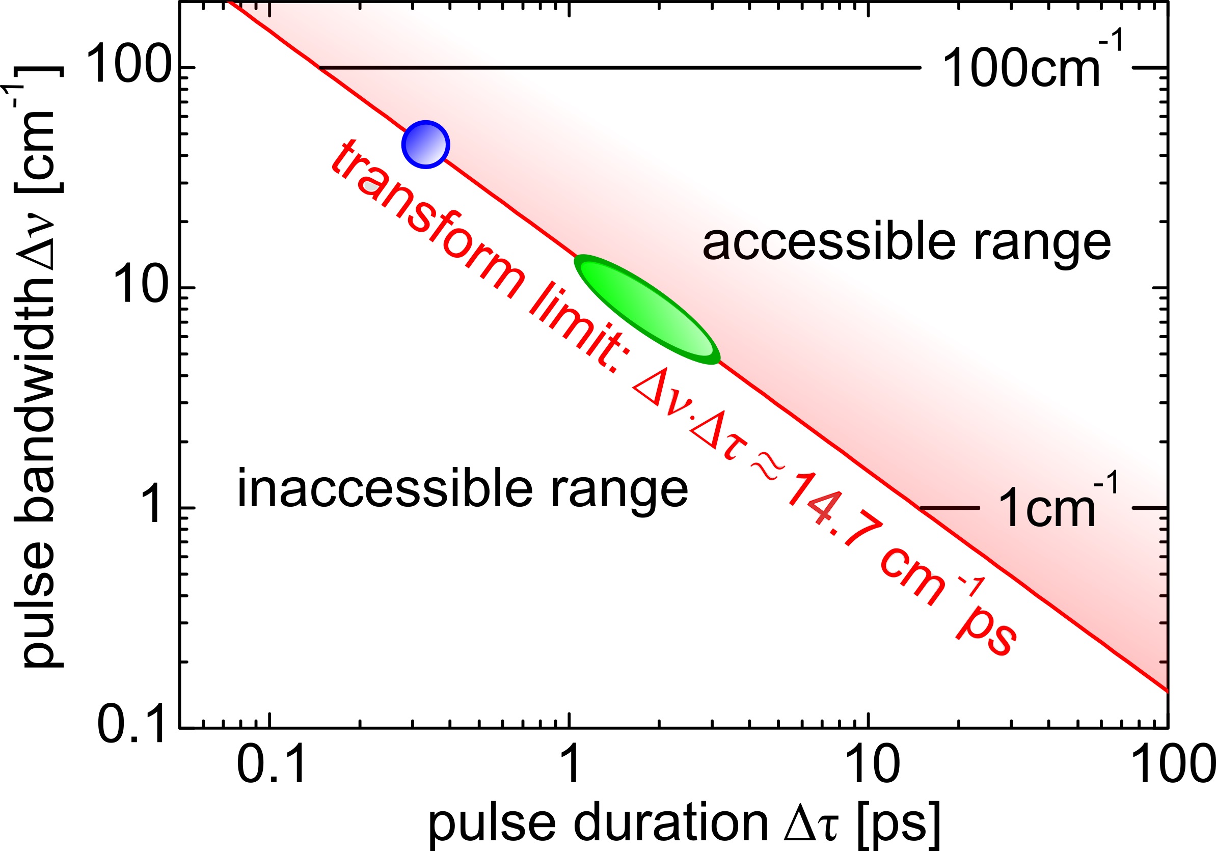
Typical design parameters for a time-resolved spontaneous Raman setup include energy resolution, time-resolution, the laser linewidth, and laser repetition rate. faustibook Other design factors include tunability of the pump excitation and Raman probe light, and suitable schemes to filter the Raman scattered light from the elastically scattered light. The energy- and time-resolution parameters are of special importance in time-resolved spontaneous Raman spectroscopy. For a transform-limited Gaussian pulse the bandwidth-time relation is cm-1ps, with the frequency (energy) bandwidth of a pulse, and the pulse duration. Here and refer to the full width at half maximum (FWHM). A trade-off in time and energy resolution is thus inherent to the technique, and ideally a setup should allow control of energy and temporal resolution. The experimentally accessible region, limited by the Fourier transform relation, is indicated in Fig. 1. In materials with only a few Raman-active modes, such as IV and III-V semiconductors, kash1985 ; kttsen2006 or well-separated high-energy modes in for instance graphite allotrope materials, song2008 ; yan2009 ; kang2010 a pulse bandwidth of cm-1 (FWHM) suffices to resolve individual modes, and thus sub-ps time-resolution can be realized (see Fig. 1). This energy resolution however generally isn’t sufficient to study dynamics of collective excitations in quantum materials such as magnons fleuryloudon1968 and phonons, or the more complex types such as electromagnons, rovillain2012 Cooper-pair breaking, saichu2009 and charge-density wave modes sugai2006 since these modes generally are low energy excitations with E meV. This energy scale corresponds to Raman shifts of cm-1 and lower, and thus requires a higher spectral resolving power. This holds especially in the lowest energy region ( cm-1) where for instance soft modes in ferroelectrics are observed.shigenari2015raman In this region stray light from spectrally broad laser pulses can hinder the observation of the modes of interest. Another case where high resolving power is necessary are materials of low crystallographic symmetry where more interesting modes of electronic and magnetic origin might overlap with a multitude of phonon modes.
In spontaneous Raman spectroscopy the inelastically scattered light needs to be separated from the Raman excitation light. For higher energy modes such as vibrational excitations in carbon-based materials this can for instance be realized with notch filters. When tunability in probe wavelength is of importance a triple subtractive Raman spectrometer is still favored. In time-resolved spontaneous Raman spectroscopy an additional complication appears since the pump-excitation, and the pump-induced Raman spectrum need to be rejected. Different filtering schemes have been applied for this. faustibook In a degenerate pump-probe experiment polarization optics are used to reject the pump-induced elastic, and inelastic scattered light. The downside is that only the parallel Raman polarization geometry can be studied. fausti2009 In addition, unwanted polarization leakage also ends up in the Raman spectrum, and the lowest energy excitations are difficult to access. In a two-color setup however, the pump-induced elastic (and inelastic) scattering can be conveniently rejected by spectral filtering. uesugi1997 ; faustibook
Amplified laser systems allow for tunable two-color experiments, as opposed to MHz-oscillator experiments which are limited to the fundamental and double wavelength for the pump and probe beams. In addition, with an amplifier the problem of average heating is avoided. For low repetition rate systems the average laser power can however bring challenges. The Raman intensity scales with average laser power. For detectable average laser powers with low repetition rate systems the pulse peak intensities can thus easily get too large, which may lead to nonlinear effects, and sample damage. The ideal situation between these laser system limits thus exists in the form of high repetition rate amplifiers. uesugi1997 ; faustibook
II.2 System overview
The time-resolved Raman system consist of three main parts: 1.) the amplified laser system and optical parametric amplifiers to generate pulses for selective pumping, and narrow-bandwidth pulses for Raman probing, 2.) the table optics for pulse cleaning, polarization control and the delay line 3.) the confocal Raman microscopy interface, the high efficiency spectrometer and charge-coupled device detector. A layout of the setup is shown in Fig. 2.
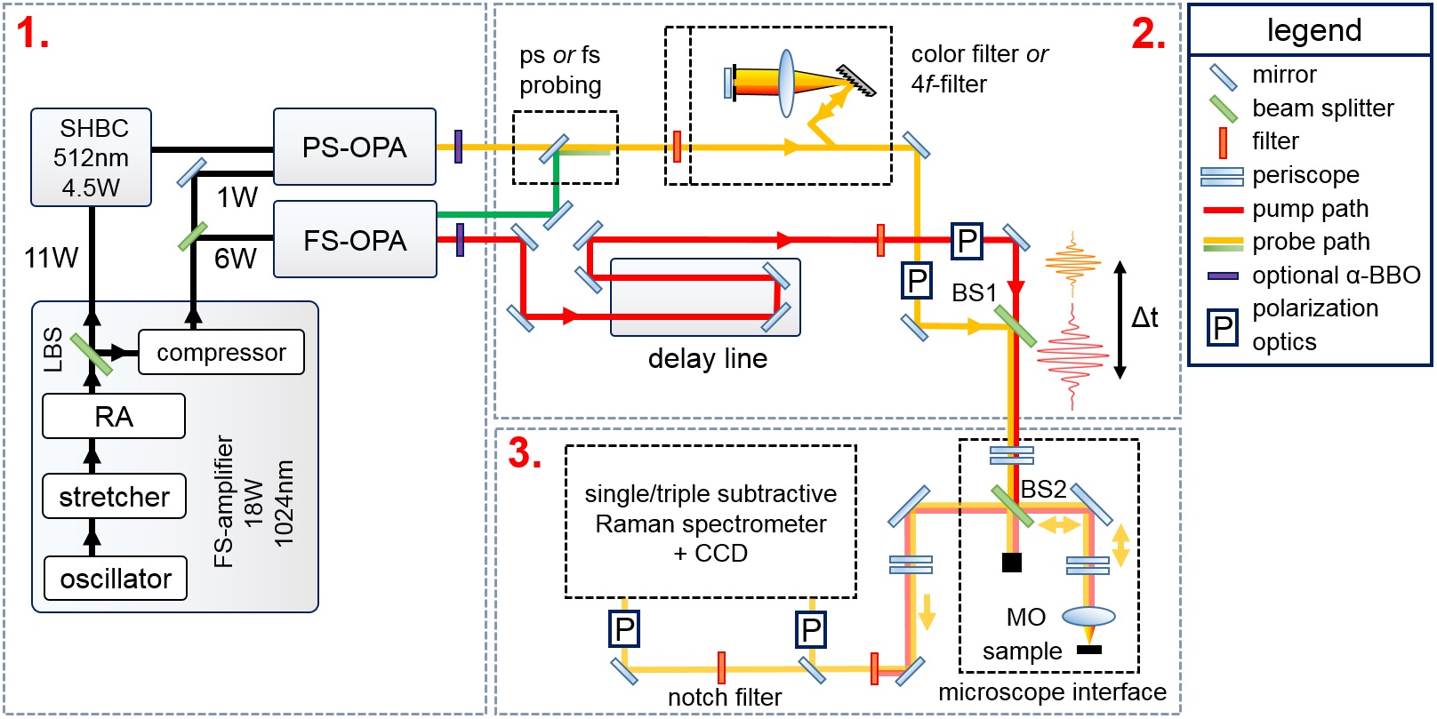
II.2.1 Amplified laser system & selective pump excitation and tunable Raman probe
A chirped pulse amplification based laser system (LightConversion Pharos) was selected for the setup. The Pharos laser system consists of a Kerr-lensing mode locked oscillator module, a regenerative amplifier (RA), and stretcher-compressor units which are all embedded in a compact module. The laser uses diode-pumped Yb:KGW (Ytterbium-doped potassium gadolinium tungstate) as the active medium. The emitted pulses have = nm central wavelength. An internal pulse picker allows to set the repetition rate up to = kHz. After the regenerative amplification stage the nm stretched pulse beam is split into two beams. One beam is directly emitted as an W output of non-compressed highly chirped ps long pulses. The other beam is compressed, resulting in a W average power beam of ps duration pulses.
The uncompressed W beam is routed to a second harmonic bandwidth compressor (LightConversion SHBC). raoult1998 Inside the SHBC, a 1:1 beamsplitter divides the beam. A high-intensity grating gives the split beams an opposite chirp, after which the beams are overlapped on an -BBO crystal. The SHBC converts the nm pulses of full-width at half maximum (FWHM) cm-1, into nm transform limited pulses with cm-1 FWHM and ps temporal width. The SHBC output can directly be used as Raman probe light, or used to pump a three stage white-light seeded picosecond optical parametric amplifier (LightConversion PS-OPA). The white light seed is generated with W of ps nm compressed pulses. The PS-OPA can continuously tune the Raman probe wavelength from nm to nm. The signal and idler of the PS-OPA output can be externally doubled to tune the probe wavelength in the range - nm.
A total power of P = W of the ps nm compressed beam is routed to a double-pass white-light seeded optical parametric amplifier (LightConversion Orpheus FS-OPA) to generate pump pulses. The signal wavelength is continuously tunable from nm to nm, where the idler runs from nm to nm. An external -BBO crystal can be used to extend the pump wavelength range from to nm.
The time-resolved system operates in the green oval region in the (,)-plane of Fig. 1 when probing is realized with the SHBC or PS-OPA output. High time-resolution, with lower energy resolving powers can be realized by only working with the FS-OPA. The FS-OPA’s signal output is in this case still used for pumping. The residual output of the OPA’s ps, = nm pump beam is used for probing. For the FS-OPA based operation mode the system works in the blue sphere in the (,)-plane.
II.2.2 Table optics
Narrow-band laser line filters are placed in the pump beam to remove unwanted spectral components which may otherwise lead to spurious signals in the Raman spectra. The polarization state of the pump beam can be controlled by a Berek compensator. For the probe beam the unwanted spectral components are either removed by laser line filters or a folded grating-based pulse shaper, kawashima1995femtosecond where the latter results in the cleanest pulse, as required for probing small Raman shifts ( cm-1). In addition, the pulse shaper allows to narrow down the spectral pulse bandwidth at least to about cm-1 FWHM at the expense of time-resolution and average probing power. A Berek compensator is used to control the polarization state of the pump beam.
The temporal delay between the pump and probe pulse is controlled via a mechanical delay line of cm length (Physikalische Instrumente M-531.EC) placed in the pump beam path. The step size resolution is m, corresponding to a time-resolution of approximately fs. The time-resolution is thus effectively determined by the pump-probe pulse cross-correlation. A silver-coated retro-reflector is mounted on the delay line. The pump and probe beams are made collinear before the Raman microscopy interface with beam combiner BS1.
II.2.3 Raman interface, spectrometer and CCD-detector
The collinearly propagating pump and probe beams enter the Raman microscopy interface via a periscope. The collinear pump and probe beams are reflected from beam splitter BS2 (R/T = 0.2/0.8) into a high numerical aperture microscope objective MO (Olympus LMPLFLN-series long working distance objectives are used, where the NA=, , working distance WD= mm is the standard choice). The backscattered light is collected by the microscope objective, transmitted through beam splitter BS2, and focused on the spectrometer entrance slit.
The maximum pump pulse energy lies around nJ at the sample position for nm pump excitation. The maximum probe pulse energy lies around nJ at the sample position for nm probe excitation when the grating-based pulse shaper is used. Since the Raman scattering efficiency scales with incident laser intensity, a large average incident probe power is preferred. For diffraction-limited pump-probe spot sizes this however results in a few challenges. Pulse fluences get too large, and samples damage easily. This can be solved by defocussing the microscope objective. The divergence of the scattered light in this case is corrected with a telescope system placed after BS2. This allows working with spot sizes of about m to m diameter. The samples can be placed in a Janis ST500 coldfinger cryostat with small working distance (about mm).
A successful implementation of time-resolved Raman spectroscopy significantly hinges on the efficiency of the spectrometer. Raman-scattered light is an order weaker compared to the incident excitation light. This necessitates a high stray light rejection. We use a cascade of three Czerny-Turner richardsongrating imaging spectrographs (TriVista 555, S&I GmbH) which are operated in subtractive mode (high stray light rejection by the first two spectrometers). Different sets of holographic, and ruled gratings allow to maximize the scattered light detection efficiency. Silver-coated optical elements ensure a high throughput. The last stage (spectrometer stage) has a second entrance port. This allows to bypass the subtractive stage of the spectrometer. In this case a notch filter is used to block the elastically scattered light. The use of a notch filter can provide up to a factor increase in throughput efficiency with respect to the subtractive filtering. A color filter may additionally be used to suppress the scattered pump light.
The used imaging spectrometer lerner2006 contains toroidal mirrors which corrects for the astigmatism present in a spectrometer containing spherical mirrors. In the toroidal case a smaller image size in the lateral direction is thus produced on the charge-coupled device (CCD) chip. This has the advantage that less CCD-rows need to be binned to integrate the scattered light spectrum. This significantly reduces the appearance of occasional spikes from cosmic events in the Raman spectrum, and reduces the CCD-readout noise with respect to an ordinary spectrometer. The low spherical aberration of the spectrometer makes that one can resort to the use of pseudo-confocal microscopy. williams1994 In this case the entrance slit rejects out-of-focus light in the horizontal direction, whereas the CCD-binning is used to digitally reject out-of-focus light in the vertical direction from the Raman spectrum. By resorting to pseudo-confocality the use of an opto-mechanical confocal system is avoided. In the latter case a secondary focus point through a mechanical pinhole before the spectrometer is used to obtain confocality. This would however lead to reflection losses for the scattered light from additional lenses. The CCD-detector is a low-etaloning Pylon-100:BR-eXcelon, pixels CCD, with a pixel size of m2. The quantum-efficiency lies above QE>90% for the wavelength range - nm.
III Experimental results
III.1 Optical phonon population and hole continuum dynamics in silicon
Time-resolved Raman spectroscopy allows to address population dynamics of individual low-energy excitation modes after photo-excitation. Through detailed balance of the Stokes and anti-Stokes signals the population number of different modes can be determined, and calculated into effective mode temperatures.compaan1984 This direct way of measuring transient temperature evolution in the time-domain allows to test the validity and limitations of multiple effective temperature models,waldecker2016 ; maldonado2017 which on the shortest time-scales may not hold anymore due to the presence of truly non-thermal occupation statistics. Such effective temperature models are not only widely applied to describe ultrafast dynamics in simple materials, waldecker2016 ; maldonado2017 but also in quantum matter where different spin, orbital, electronic and lattice degrees of freedom are present with their respective excitations. averitt2001 ; ogasawara2005prl
Raman tensor dynamics is a less treated aspect of time-resolved Raman scattering. It is however of crucial importance in the correct determination of effective mode temperatures. compaan1984 The anti-Stokes intensity scales as IAS , were is the mode occupation number, and the squared Raman tensor. The Stokes-intensity scales as IS and is thus less sensitive to transient population dynamics. This can be taken as a motivation to ”neglect” dynamics of the Stokes-spectrum. However, from these relations it directly becomes clear that assigning transient anti-Stokes dynamics solely to transient population changes may not necessarily be true since the Raman tensor may also show photo-induced changes. This is expected to be of special importance under resonant probing conditions. We’ve recently demonstrated in silicon that for a correct transient mode temperature determination indeed a dynamical modification of the Raman tensor needs to be taken into account. zhu2017 The quench of in silicon was discussed to originate from the photo-induced hole density. Below we reiterate our results on the LO-phonon population and hole continuum dynamics in silicon for similar measurement conditions. A dynamical modification of the Raman tensor is expected to play a role in many other materials such as //-oxides, carbon allotropes ferrari2013raman like graphene or Bucky-tubes, and transition metal dichalcogenides. lee2017
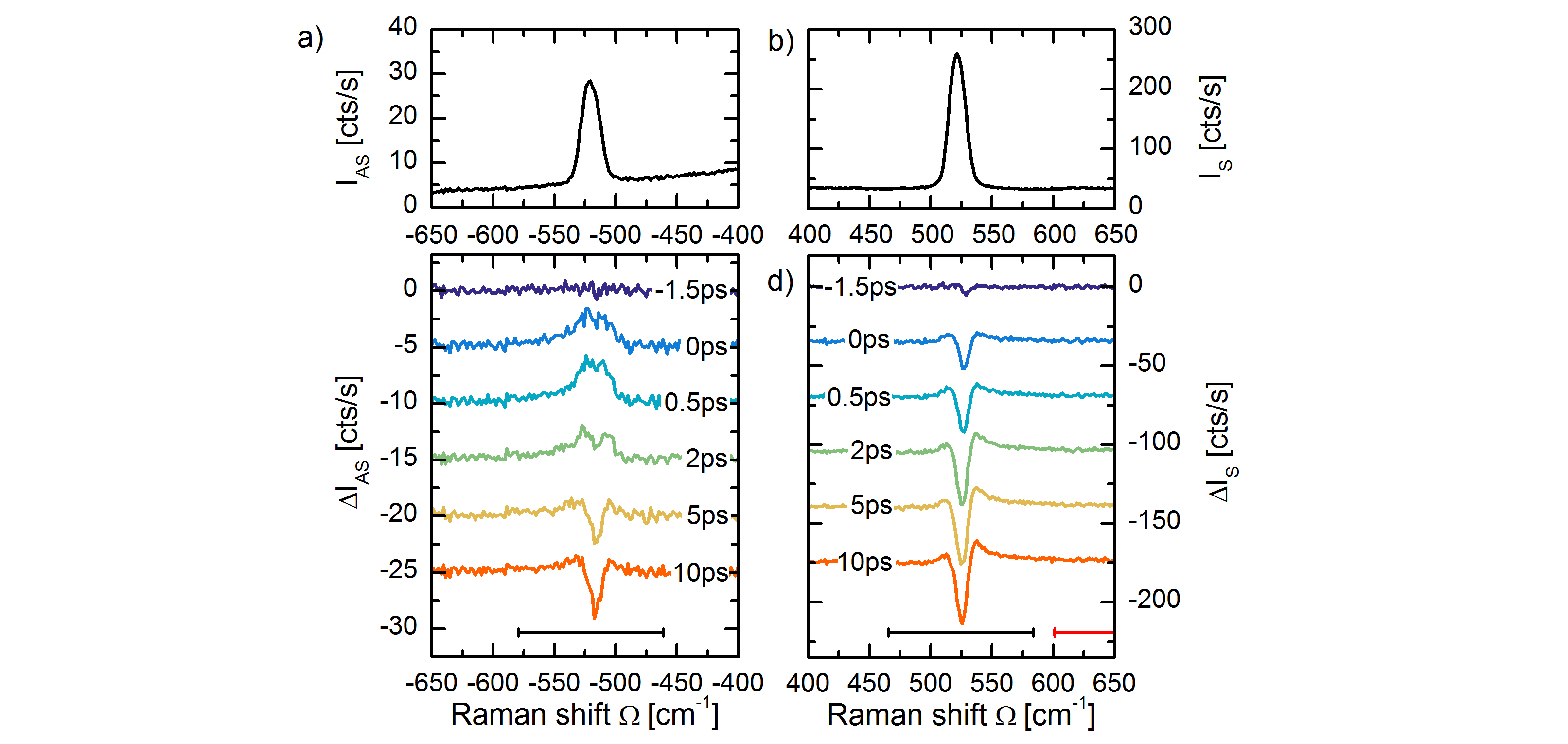
An intrinsic (100) oriented silicon wafer (resistivity m), at room temperature, is excited above the indirect band-gap with nm pulses of ps duration. The Raman probe excitation is narrow-band ( cm-1 FWHM) pulsed light of nm central wavelength, and ps temporal duration. The pump and probe powers were set to Ppump = mW and Pprobe = mW for the pump and probe beam respectively. With a pump spot size of m diameter, the photo-excitation density is about cm-3. Both pump and probe beams are aligned with the [110] crystallographic directions. The backscattered light is collected with the single-stage spectrometer, where a notch-filter (Kaiser Optical Systems Inc. SuperNotch-Plus nm) is used to block the elastically scattered light.
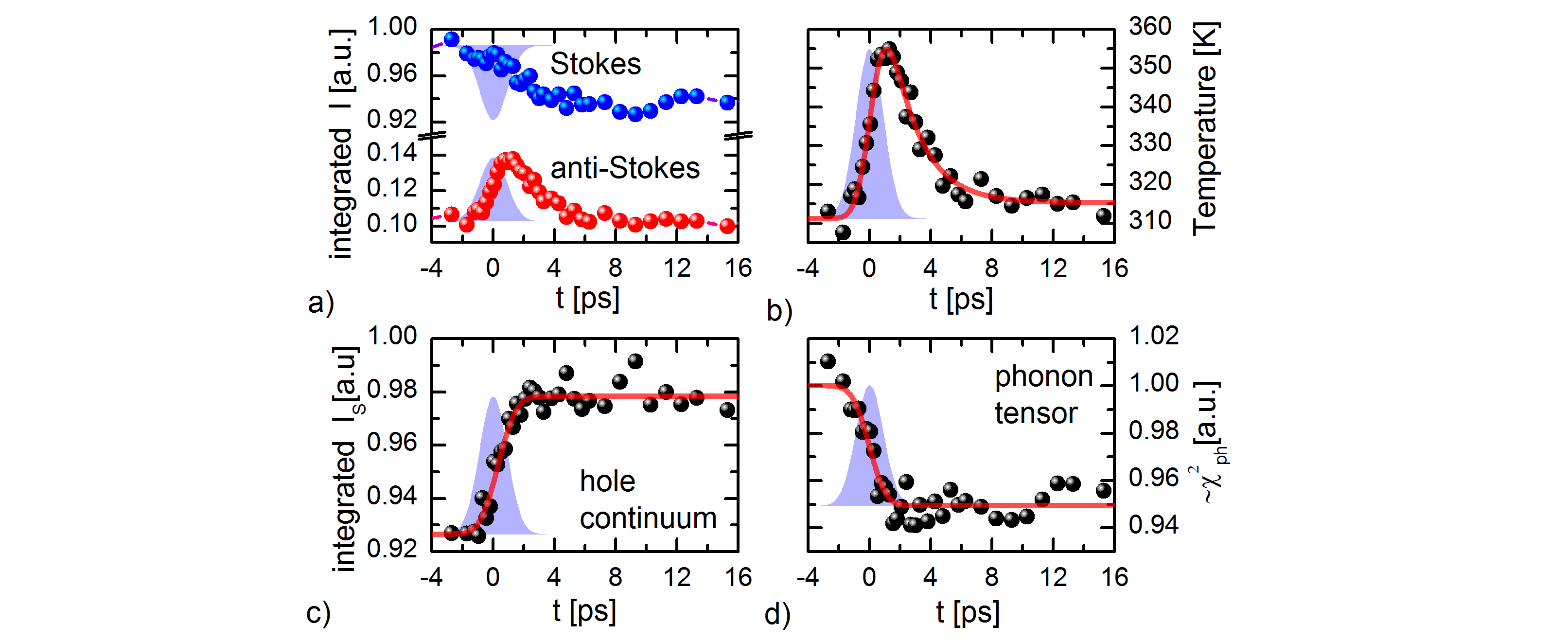
Figure 3a and b show the anti-Stokes IAS and Stokes IS spectra of silicon (spectra at t = ps). The spectra consist of the zone-center longitudinal optical (LO) phonon at cm-1, and an electronic continuum originating from hole scattering. hart1970 ; cerdeira1973 The silicon peak has a (Gaussian) half width at half maximum of cm-1. This is larger than the intrinsic half-width of cm-1 at room temperature, hart1970 since Gaussian ps-pulses were used to collect the Raman spectrum. In Fig. 3c and d the differential anti-Stokes scattering intensity IAS(t) = IAS(t) I, and Stokes scattering intensity IS(t) = IS(t) I is shown for various representative delay-times. At ps to ps a scattering increase is observed on the anti-Stokes side. This corresponds to the creation of a transient optical phonon population. However, on the Stokes side a negative differential feature is observed, which evidently cannot originate from a transient phonon population. In addition a small positive shoulder in the IS(t) spectra is observed. The integrated phonon scattering intensities are shown in Fig. 4a. The phonon response is integrated over the spectral regions [ - ] cm-1 (indicated with black bars) for the Stokes and anti-Stokes side respectively. A linear background was subtracted to take into account the increased hole continuum scattering over the integration region. The cross-correlation of the pump and probe pulse is plotted in faded blue for comparison.
The Stokes intensity IS, and anti-Stokes intensity IAS are given as compaan1984 :
| (1) |
and
| (2) |
Here ( , ) gives the Raman tensor for the anti-Stokes and Stokes resonance, is the probe excitation frequency, and is the occupation number for the mode with energy . The factors C( , ) contain the incident intensity, and optical constants (absorption, transmission and refractive index) at the incident and scattered frequencies. compaan1984 We however neglect the frequency dependence of and , which will lead to a small error of % in the absolute phonon temperature determination. compaan1984 Note1 The anti-Stokes to Stokes scattering ratio can be used to calculate the phonon temperature, or equivalently the particle occupation number , through the ratio:
| (3) |
In Fig. 4b the transient temperature, determined according to Eq. 3 is plotted. An average heating to K is measured before time-zero. After photo-excitation the temperature rises by T K to K within the time-resolution, followed by a decay with a time-scale of ps. The rapid rise, and consecutive decay is consistent with the transient creation of an optical phonon population through electron-phonon coupling, which thereafter decays into acoustic phonons through anharmonic coupling. zhu2017 ; klemens1966 After ps the temperature reaches quasi-equilibrium at an increased temperature of T K, which does not recover within the measured time window.
Figure 4c shows the transient increase of hole continuum scattering in the region +[ - ] cm-1 (indicated with the red bar). The transient scattering signal is well-fitted with a step function convoluted with a cross-correlation function of the pump and probe pulse. The photo-induced electron-hole-density has other effects on the Raman spectrum. Since the phonon Raman transition probability is proportional to the density of electrons in the valence band, and holes in the conduction band, the photo-induced electron-hole density is expected to alter . gillet2013 ; sangalli2016 From the Stokes intensity IS and the calculated population , the time-evolution of is calculated according to Eq. 1, and shown in Fig. 4d. A step-wise tensor quench % is observed, underlining the electronic origin of the transient decrease of the Stokes intensity IS. The transient hole density in addition leads to an increase in the Fano-asymmetry, as evidenced by the ingrowing positive shoulder in the IS(t) spectra. The origin of the tensor quench and the transient Fano-asymmetry are discussed in more detail in Ref. zhu2017, .
III.2 Time- and momentum resolved scattering in silicon
Electron-phonon, and phonon-phonon interaction strengths, or more generally the coupling between any type of quasiparticles, depends on the momentum of the interacting quasiparticles. birmanbook The characteristic timescales associated with optical phonon creation, and relaxation through anharmonic coupling, are thus momentum-dependent. maldonado2017 ; tandon2015 Pump-probe techniques spanning from x-ray to the optical range allow for time- and momentum-resolved probing. Whereas the momentum-dependence is implicit for pump-probe techniques in the x-ray range, such as diffuse scattering, trigo2013fourier diffraction, johnson2017 or resonant inelastic x-ray scattering, dean2016ultrafast this may not directly be obvious for optical pump-probe techniques as these are momentum transfer probes. Optical inelastic scattering techniques, notably femtosecond stimulated Raman spectroscopy, batignani2015probing and spontaneous Raman spectroscopy however can be utilized to disentangle momentum-dependent dynamics through higher-order scattering processes. In a two-particle process, two excitations of opposite momenta and are created (or annihilated). cardonabook Two-particle scattering processes appear in the inelastic light scattering spectrum as bands of the frequency dependence of the combined density of states of the excitation pair, weighted by a scattering efficiency distribution. johnson1964 The multi-particle scattering response thus forms a mapping of momentum-space to the energy domain, and thereby allows for the study of momentum-resolved particle dynamics. After careful analysis of the line-shape even momentum-relaxation dynamics should be in principle feasible to detect. lockwood1987 With time-resolved spontaneous Raman spectroscopy we succeeded to detect photo-induced phonon softening of different high-symmetry Brillouin Zone-points (BZ-points) in the two-phonon spectrum of silicon. We find evidence that the softening at the different points occurs with dissimilar time-scales, which is indicative of dissimilar electron-phonon scattering rates.
Probing of the two-phonon spectrum on a (100) oriented silicon wafer was performed at room temperature. A nm Raman probe excitation was used, with the probe polarization along the [110] axis. The two-phonon spectrum is observed between cm-1 and cm-1 as seen in Fig. 5a (dark blue line). The two-phonon spectrum consists of a summation of TO-phonon overtones from four high-symmetry Brillouin Zone (BZ) points. temple1973 Previous reports have assigned the characteristic spectral features based on the analysis of critical points in the phonon density-of-states. johnson1964 ; temple1973 ; gillet2017ab The sharp increase at cm-1 is related to scattering from the X-point, the shoulder at cm-1 to the W-point, and the shoulder at cm-1 to scattering from the L-point. The tail extending from cm-1 to cm-1 is the overtone from the zone center, i. e. the -point. The different regions are marked with bars.
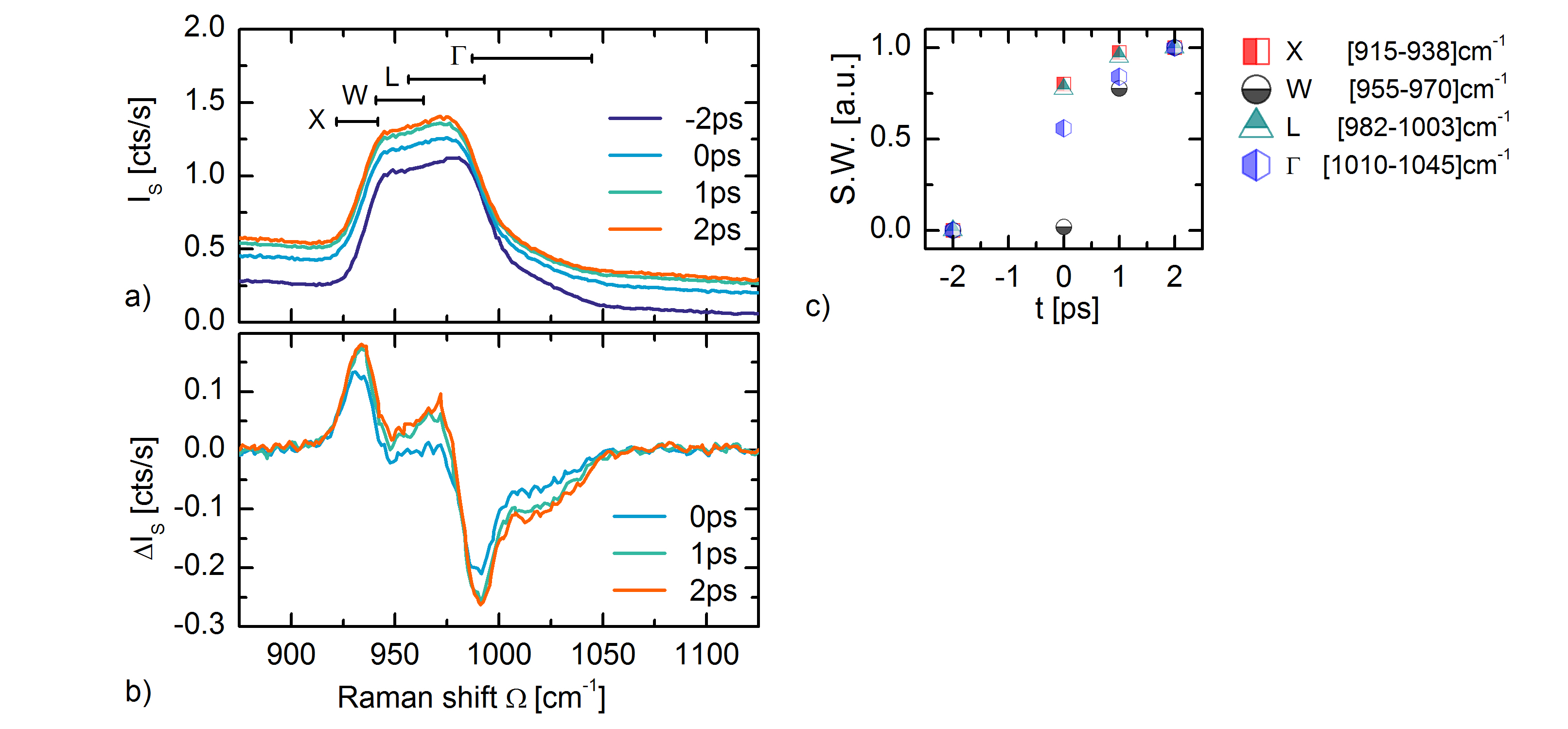
The sample is excited above the indirect band-gap with nm pulses of ps duration, and pump polarization along the [110] axis. The photo-induced electron-hole density is cm-3. Figure 5a shows the transient Stokes spectra IS(t) of the two-phonon peak for various pump-probe delays. A photo-induced redshift of the two-phonon spectrum is observed, in addition to an increased hole continuum scattering background, as discussed above. Figure 5b shows the differential spectra IS(t) = IS(t) IS() for the respective time-delays, where the hole continuum scattering increase is subtracted (the tail of a Voight profile is fitted to the increase in electronic scattering in the IS(t) spectra). The IS(t) / I-2ps spectra are integrated over four different wavelength regions and plotted normalized to the IS / I-2ps value at ps. This gives the spectral weight S.W. as a function of time delay as shown in Fig. 5c.
The two-phonon Stokes signal has a I dependence (cf. Eq. 1). Both the Raman tensor and the population factor transiently change upon photo-excitation as discussed above, where the population term can only increase. The tensor dynamics thus appears to dominate the transient two-phonon response. This is also evidenced by the large redshift cm-1. The redshift is understood as a photo-induced phonon softening at the different high symmetry points in the Brillouin Zone. Extracting time-constants for the different BZ-points is not possible due the limited amount of time-delay points. However, from the IS(t) spectra in Fig. 5b, and the spectral weight S.W. in Fig. 5c, it appears that the phonon softening at the X- and L-point has a faster ingrowth than at the W- and -point. This hints to dissimilar electron-phonon coupling constants for the different points. The quantitative determination of time-constants for the four BZ-points asks for a higher time-resolution. In spontaneous Raman spectroscopy this would however result in a loss of frequency resolving power, and thereby momentum-resolving power. This would again lead to a complication in disentangling time-and momentum resolved scattering, however now because of frequency resolution limitations. A qualitative disentanglement of momentum-dependent scattering in silicon thus forms an interesting study case for the time-resolved femtosecond stimulated Raman scattering variant.
III.3 Photo-induced melting of helimagnetic order in the chiral magnet Cu2OSeO3
The understanding of out-of-equilibrium phenomena in magnetic materials is a highly active part of condensed matter research. kirilyuk2010 Recent scientific advances with a high potential towards applications include all-optical magnetization switching, savoini2012 and picosecond optical writing and read-out of magnetic bits. stupakiewicz2017ultrafast With increasingly complex magnetic phases and dynamical phenomena being studied, there is a high demand for novel all-optical probes. Magnetization dynamics in finite magnetization phases, such as ferromagnetic and ferrimagnetic order, or a more exotic phase such as the skyrmion lattice, ogawa2015ultrafast can be probed by optical techniques which are sensitive to the order parameter M which describes the (macroscopic) magnetization. Linear magneto-optical effects, for instance the Faraday or magneto-optical Kerr effect, measure a polarization rotation proportional to the magnetization. pershan1967magneto The dynamical variant allows to detect photo-induced incoherent and coherent dynamics of the order parameter M. (ogasawara2005prl, ; ogawa2015ultrafast, ) All-optical detection of antiferromagnetic order dynamics already becomes more challenging. The antiferromagnetic order parameter L is proportional to the difference in magnetic sublattice magnetizations; - . Optical probing of L is realized by second-order magneto-optical effects, such as magnetic linear birefringence and second harmonic generation. Ultrafast variants have been used to study photo-induced dynamics in antiferromagnets. bossini2014controlling ; fiebig2008ultrafast For more complex net-zero magnetization order, such as cycloidal, helical, or spin wave order it is more challenging to probe the magnetic order parameter. One suggestion is tracking the thermalization of a magnon mode with THz-time-domain spectroscopy after photo-excitation, as was realized in the spin-cycloid material TbMnO3. bowlan2016 The Raman-activity of a magnon mode would however permit all-optical detection, as opposed to detection through its dipole activity. Here we show that time-resolved Raman spectroscopy allows to all-optically probe melting of helimagnetic order after photo-excitation in the chiral magnet Cu2OSeO3. seki2012 This is realized by tracking the softening and broadening of a Raman-active magnon mode in the time-domain.
Cu2OSeO3 has a long-range magnetic ordering temperature of TN K. seki2012 The magnetic ground state consists of helimagnetically aligned effective = spin clusters, which by itself consist of three-up-one-down = spins on the Cu2+() sites. janson2014quantum ; ozerov2014 The spin cluster ordering results in high energy meV magnons, from which the magnons at the -point are observable by spontaneous Raman spectroscopy. gnezdilov2010 A [111] oriented Cu2OSeO3 sample, at bias temperature K in the helimagnetic phase was studied. The SHBC output at nm, with cm-1 FWHM was used as Raman probe ( mW probe power at sample position). In Fig.6a part of the Stokes spectrum is shown. The M-mode at cm-1 corresponds to a = magnon mode, which can be understood as a spin-flip excitation within the three-up-one-down spin cluster. romhanyi2014 The modes labeled ”P” correspond to phonons, or regions of not fully resolved overlapping phonons.
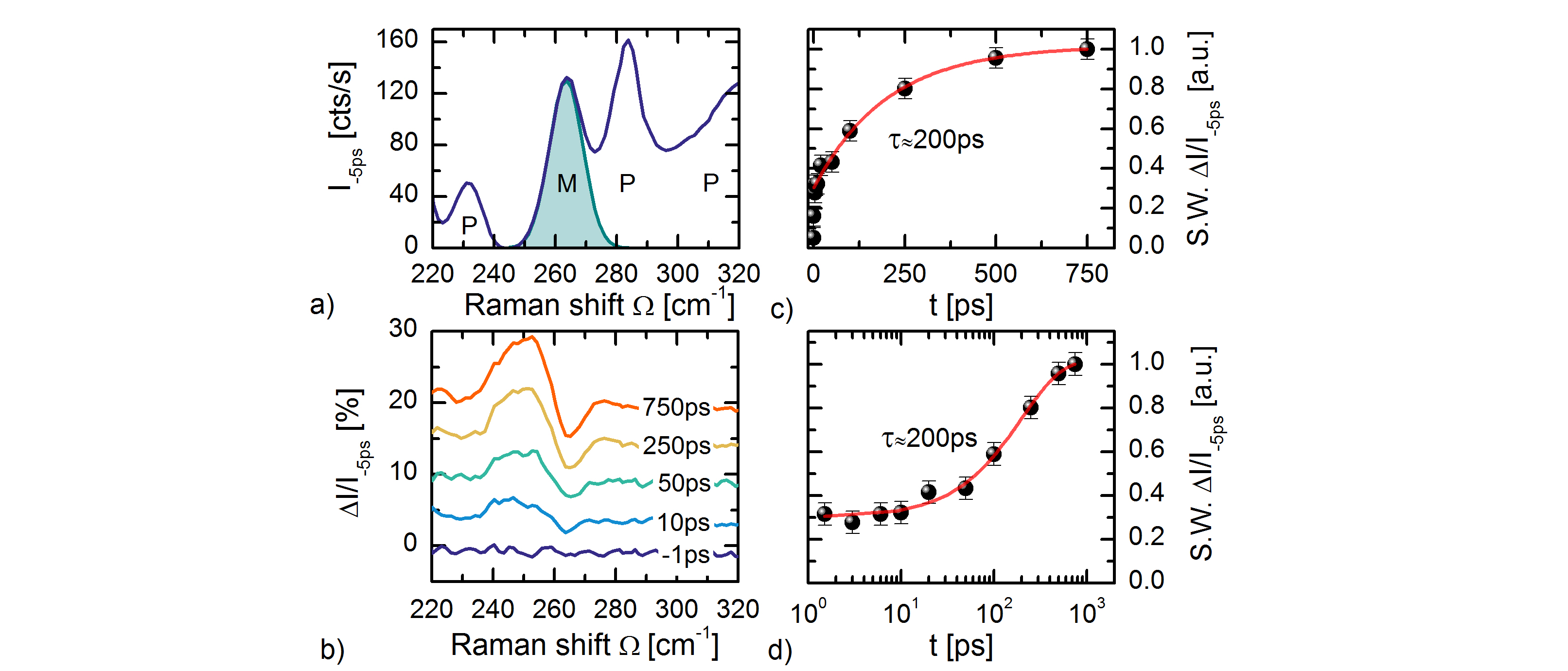
Local crystal field excitations versteeg2016 were excited with ps, = nm pump pulses (with mW pump power, and fluence F mJ/cm2), after which the thermalization dynamics is measured in the transient Raman spectra. In Fig. 6b the scaled differential spectra IS(t) / I-5ps are shown for representative time-delays, where IS(t) = IS(t) IS(). A derivative-like lineshape is observed in the IS(t) / I-5ps spectra, resulting from a spectral weight shift to lower Raman shift for the magnon excitation. For the low temperatures, and Stokes range of interest ( =- cm-1) the occupation number (,T) . The change in occupation thus also is (,T) . We thereby assign the spectral weight transfer to a change in the Raman tensor . This physically corresponds to a dynamic softening and broadening of the magnon excitation. From fitting of the = peak position in the IS(t) spectra it is found that the dynamic shift ( ps) at late delay times lies below cm-1. By comparison to the temperature dependent position we conclude that the transient magnetic temperature does not rise above K. Under our pump excitation conditions the long-range magnetic order thus gets strongly perturbed, but not fully destroyed. The phonon peak shifts are too small to be resolvable in the IS(t) / I-5ps spectra. The ingrowing spectral weight transfer (S.W.) of the IS(t) / I-5ps signal, corresponding to the temporal evolution of the magnetic order parameter, is shown in Fig. 6c and d in linear and logarithmic timescales. A typical time-scale of ps for spin-lattice thermalization in insulators is observed. kirilyuk2010 This agrees well with the findings of Langner et al. where a spin-lattice thermalization time of ps was observed by means of time-resolved resonant x-ray diffraction. langner2017 The slight discrepancy might originate from the different pump excitation conditions, and the different bias temperatures.
IV Conclusions and outlook
In this paper have presented a flexible and efficient ultrafast time-resolved spontaneous Raman spectroscopy setup, and illustrated its strength and capabilities with different conceptual time-resolved Raman studies of collective excitation and quasi-particle dynamics. One of the strongest feats of time-resolved spontaneous Raman is the determination of transient population numbers, and effective modes temperatures through detailed balance of the anti-Stokes to Stokes intensity ratio. A less studied aspect of the dynamics is the resonance enhancement in the Raman process. This however plays a role, as we demonstrated in the prototype test material silicon. We’ve shown that the photo-induced hole density leads to a quench of the Raman tensor, and in addition to an increase in electronic inelastic scattering. Dynamic changes in the electronic population and structure are of importance to photo-induced phenomena in many classes of quantum matter, such as //-materials, carbon allotropes, and transition metal dichalgocenides. Time-resolved resonant Raman can thereby provide a convenient tool to study both electronic population dynamics, as well as photo-induced changes in the electronic band structure such as band gap renormalization.
Although Raman spectroscopy is a zero-momentum transfer probe, momentum-dependent scattering can nevertheless be resolved in the higher-order Raman response. The overtone spectrum can thus serve as a momentum-and time-resolved scattering probe. As a proof of concept, we’ve studied photo-induced changes in the two-phonon response of silicon, and found evidence for dissimilar phonon softening rates at different high-symmetry points of the Brillouin Zone. Applying this concept to slower phenomena, such as spin-lattice relaxation in -antiferromagnetic materials, seems fortuitous to pursue, and possibly allows to detect momentum-dependent transient population dynamics and momentum relaxation.
Raman-active collective excitations such as magnons, charge-density wave modes, and the Cooper-pair breaking peak, can serve as a time-resolved probe for the order parameter of quantum phases. We illustrated this in the helimagnet Cu2OSeO3, where the photo-induced melting of helimagnetic order can be tracked by the observation of a dynamic softening and broadening of a magnon peak. This principle can also be applied to probe dynamics of other net-zero magnetization order such as cycloidal, spin-density wave, and antiferromagnetic order. On a broader scope this can be applied to photo-induced phase transitions, where the associated change of symmetry can be derived from the transient Raman spectrum. The Raman spectrum of collective low energy excitations can additionally serve as a probe to study energy and angular momentum transfer between the lattice, electronic, orbital and magnetic degrees of freedom in quantum matter.
V Acknowledgements
This project was partially financed by the Deutsche Forschungsgemeinschaft (DFG) through SFB Grossgeräteantrag INST217/782-1, and SFB-1238 (projects A02 and B05). R.B.V. acknowledges funding through the Bonn-Cologne Graduate School of Physics and Astronomy (BCGS).
References
- (1) D. N. Basov, R. D. Averitt, and D. Hsieh, Towards properties on demand in quantum materials, Nat. Mater. 16, 1077 (2017).
- (2) J. Zhang and R. D. Averitt, Dynamics and control in complex transition metal oxides, Annu. Rev. Mater. Res. 44, 19 (2014).
- (3) M. Gandolfi, G. L. Celardo, F. Borgonovi, G. Ferrini, A. Avella, F. Banfi, and C. Giannetti, Emergent ultrafast phenomena in correlated oxides and heterostructures, Phys. Scr. 92, 034004 (2017).
- (4) Y. H. Wang, H. Steinberg, P. Jarillo-Herrero, and N. Gedik, Observation of floquet-bloch states on the surface of a topological insulator, Science, 342, 453 (2013).
- (5) A. Cavalleri, Cs. Tóth, C. W. Siders, J. A. Squier, F. Ráksi, P. Forget, and J. C. Kieffer, Femtosecond structural dynamics in VO2 during an ultrafast solid-solid phase transition, Phys. Rev. Lett. 87, 237401 (2001).
- (6) M. Rini, N. Dean, J. Itatani, Y. Tomioka, Y. Tokura, R. W. Schoenlein, and A. Cavalleri, Control of the electronic phase of a manganite by mode-selective vibrational excitation, Nature 449, 72 (2007).
- (7) D. Fausti, R. I. Tobey, N. Dean, S. Kaiser, A. Dienst, M. C. Hoffmann, S. Pyon, T. Takayama, H. Takagi, and A. Cavalleri, Light-induced superconductivity in a stripe-ordered cuprate, Science 331, 189 (2011).
- (8) F. Boschini, E. H. da Silva Neto, E. Razzoli, M. Zonno, S. Peli, R. P. Day, M. Michiardi, M. Schneider, B. Zwartsenberg, P. Nigge, R. D. Zhong, J.. Schneeloch, G. D. Gu, S. Zhdanovich, A. K. Mills, G. Levy, D. J. Jones, C. Giannetti, and A. Damascelli, Collapse of superconductivity in cuprates via ultrafast quenching of phase coherence, Nat. Mater. 17, 416 (2018).
- (9) P. Maldonado, K. Carva, M. Flammer, and P. M. Oppeneer, Theory of out-of-equilibrium ultrafast relaxation dynamics in metals, Phys. Rev. B 96, 174439 (2017).
- (10) E. Turgut, D. Zusin, D. Legut, K. Carva, R. Knut, J. M. Shaw, C. Chen, Z. Tao, H. T. Nembach, T. J. Silva, S. Mathias, M. Aeschlimann, P. M. Oppeneer, H. C. Kapteyn, M. M. Murnane, and P. Grychtol, Stoner versus Heisenberg: Ultrafast exchange reduction and magnon generation during laser-induced demagnetization, Phys. Rev. B 94, 220408 (2016).
- (11) B. Mansart, J. Lorenzana, A. Mann, A. Odeh, M. Scarongella, M. Chergui, and F. Carbone, Coupling of a high-energy excitation to superconducting quasiparticles in a cuprate from coherent charge fluctuation spectroscopy, Proc. Natl. Acad. Sci. USA 110, 4539 (2013).
- (12) A. Mann, E. Baldini, A. Tramontana, E. Pomjakushina, K. Conder, C. Arrell, F. van Mourik, J. Lorenzana, and F. Carbone. Probing the electron-phonon interaction in correlated systems with coherent lattice fluctuation spectroscopy, Phys. Rev. B 92, 035147 (2015).
- (13) J. A. Sobota, S. Yang, J. G. Analytis, Y. L. Chen, I. R. Fisher, P. S. Kirchmann, and Z.-X. Shen, Ultrafast optical excitation of a persistent surface-state population in the topological insulator , Phys. Rev. Lett. 108, 117403 (2012).
- (14) P. Abbamonte, Condensed-matter physics: Picking up fine vibrations, Nat. Phys. 9, 759 (2013).
- (15) M. Trigo, M. Fuchs, J. Chen, M. P. Jiang, M. Cammarata, S. Fahy, D. M. Fritz, K. Gaffney, S. Ghimire, A. Higginbotham, S. L. Johnson, M. E. Kozina, J. Larsson, H. Lemke, A. M. Lindenberg, G. Ndabashimiye, F. Quirin, K. Sokolowski-Tinten, C. Uher, G. Wang, J. S. Wark, D. Zhu, and D. A. Reis, Fourier-transform inelastic x-ray scattering from time-and momentum-dependent phonon-phonon correlations, Nat. Phys. 9, 790 (2013).
- (16) M. P. M. Dean, Y. Cao, X. Liu, S. Wall, D. Zhu, R. Mankowsky, V. Thampy, X. M. Chen, J. G. Vale, D. Casa, J. Kim, A. H. Said, P. Juhas, R. Alonso-Mori, J. M. Glownia, A. Robert, J. Robinson, M. Sikorski, S. Song, M. Kozina, H. Lemke, L. Patthey, S. Owada, T. Katayama, M. Yabashi, Y. Tanaka, T. Togashi, J. Liu, C. Rayan Serrao, B. J. Kim, L. Huber, C.-L. Chang, D. F. McMorrow, M. Först, and J. P. Hill, Ultrafast energy-and momentum-resolved dynamics of magnetic correlations in the photo-doped Mott insulator Sr2IrO4, Nat. Mater. 15, 601 (2016).
- (17) F. Carbone, B. Barwick, O.-H. Kwon, H. Soon Park, J. Spencer Baskin, and A. H. Zewail, EELS femtosecond resolved in 4D ultrafast electron microscopy, Chem. Phys. Lett. 468, 107 (2009).
- (18) L. Piazza, C. Ma, H. X. Yang, A. Mann, Y . Zhu, J. Q. Li, and F. Carbone, Ultrafast structural and electronic dynamics of the metallic phase in a layered manganite, Struct. Dyn. 1, 014501 (2014).
- (19) K. Iwata, S. Yamaguchi, and H. Hamaguchi, Construction of a transform-limited picosecond time-resolved Raman spectrometer, Rev. Sci. Instrum. 64, 2140 (1993).
- (20) H. Hamaguchi and T. L. Gustafson, Ultrafast time-resolved spontaneous and coherent Raman spectroscopy: the structure and dynamics of photogenerated transient species, Annu. Rev. Phys. Chem. 45, 593 (1994).
- (21) Y. Uesugi, Y. Mizutani, and T. Kitagawa, Developments of widely tunable light sources for picosecond time-resolved resonance Raman spectroscopy, Rev. Sci. Instrum. 68, 4001 (1997).
- (22) S. K. Sahoo, S. Umapathy, and A. W. Parker, Time-resolved resonance Raman spectroscopy: Exploring reactive intermediates, Appl. Spectrosc. 65, 1087 (2011).
- (23) S. G. Kruglik, J.-C. Lambry, J.-L. Martin, M. H. Vos, and M. Negrerie, Sub-picosecond raman spectrometer for time-resolved studies of structural dynamics in heme proteins, J. Raman Spectrosc. 42, 265 (2011).
- (24) J. A. Kash, J. C. Tsang, and J. M. Hvam, Subpicosecond time-resolved Raman spectroscopy of LO phonons in GaAs, Phys. Rev. Lett. 54, 2151 (1985).
- (25) D. Y. Oberli, D. R. Wake, M. V. Klein, J. Klem, T. Henderson, and H. Morkoç, Time-resolved Raman scattering in GaAs quantum wells, Phys. Rev. Lett. 59, 696 (1987).
- (26) J. Zhu, R. B. Versteeg, P. Padmanabhan, and P. H. M. van Loosdrecht, Dynamic asymmetry in the spontaneous Raman scattering of silicon, arXiv:1806.05986 [cond-mat.mtrl-sci] (2018).
- (27) D. Song, F. Wang, G. Dukovic, M. Zheng, E. D. Semke, L. E. Brus, and T. F. Heinz, Direct measurement of the lifetime of optical phonons in single-walled carbon nanotubes, Phys. Rev. Lett. 100, 225503 (2008).
- (28) H. Yan, D. Song, K. F. Mak, I. Chatzakis, J. Maultzsch, and T. F. Heinz, Time-resolved Raman spectroscopy of optical phonons in graphite: Phonon anharmonic coupling and anomalous stiffening, Phys. Rev. B 80, 121403 (2009).
- (29) K. Kang, D. Abdula, D. G. Cahill, and M. Shim, Lifetimes of optical phonons in graphene and graphite by time-resolved incoherent anti-Stokes Raman scattering, Phys. Rev. B 81, 165405 (2010).
- (30) J.-A. Yang, S. Parham, D. Dessau, and D. Reznik, Novel electron-phonon relaxation pathway in graphite revealed by time-resolved Raman scattering and angle-resolved photoemission spectroscopy, Sci. Rep. 7, 40876 (2017).
- (31) D. Fausti and P. H. M. van Loosdrecht, in Chapter 14 of Optical Techniques for Solid-State Materials Characterization, edited by R. P. Prasankumar and A. J. Taylor, (CRC Press, Boca Raton London New York, 2012).
- (32) A. Compaan and H. J. Trodahl, Resonance Raman scattering in Si at elevated temperatures, Phys. Rev. B 29, 793 (1984).
- (33) L. Waldecker, R. Bertoni, R. Ernstorfer, and J. Vorberger, Electron-phonon coupling and energy flow in a simple metal beyond the two-temperature approximation, Phys. Rev. X 6, 021003 (2016).
- (34) O. V. Misochko, M. Hase, K. Ishioka, and M. Kitajima, Observation of an amplitude collapse and revival of chirped coherent phonons in bismuth, Phys. Rev. Lett. 92, 197401 (2004).
- (35) D. Fausti, O. V. Misochko, and P. H. M. van Loosdrecht, Ultrafast photoinduced structure phase transition in antimony single crystals, Phys. Rev. B 80, 161207 (2009).
- (36) R. P. Saichu, I. Mahns, A. Goos, S. Binder, P. May, S. G. Singer, B. Schulz, A. Rusydi, J. Unterhinninghofen, D. Manske, P. Guptasarma, M. S. Williamsen, and M. Rübhausen, Two-component dynamics of the order parameter of high temperature superconductors revealed by time-resolved Raman scattering, Phys. Rev. Lett. 102, 177004 (2009).
- (37) D. N. Basov, R. D. Averitt, D. van der Marel, M. Dressel, and K. Haule, Electrodynamics of correlated electron materials, Rev. Mod. Phys. 83, 471 (2011).
- (38) P. Kukura, D. W. McCamant, and R. A. Mathies, Femtosecond stimulated Raman spectroscopy, Annu. Rev. Phys. Chem. 58, 461 (2007).
- (39) G. Batignani, D. Bossini, N. Di Palo, C. Ferrante, E. Pontecorvo, G. Cerullo, A. Kimel, and T. Scopigno, Probing ultrafast photo-induced dynamics of the exchange energy in a Heisenberg antiferromagnet, Nat. Photon. 9, 506 (2015).
- (40) U. Harbola, S. Umapathy, and S. Mukamel, Loss and gain signals in broadband stimulated-Raman spectra: Theoretical analysis. Phys. Rev. A 88, 011801 (2013).
- (41) K. T. Tsen, J. G. Kiang, D. K. Ferry, and H. Morkoç, Subpicosecond time-resolved Raman studies of LO phonons in GaN: Dependence on photoexcited carrier density, Appl. Phys. Lett. 89, 112111 (2006).
- (42) P. A. Fleury and R. Loudon, Scattering of light by one- and two-magnon excitations, Phys. Rev. 166, 514 (1968).
- (43) P. Rovillain, J. Liu, M. Cazayous, Y. Gallais, M-A. Measson, H. Sakata, and A. Sacuto, Electromagnon and phonon excitations in multiferroic TbMnO3, Phys. Rev. B 86, 014437 (2012).
- (44) S. Sugai, Y. Takayanagi, and N. Hayamizu, Phason and amplitudon in the charge-density-wave phase of one-dimensional charge stripes in , Phys. Rev. Lett. 96, 137003 (2006).
- (45) T. Shigenari, Raman spectra of soft modes in ferroelectric crystals in Ferroelectric Materials-Synthesis and Characterization, edited by A. Peláiz-Barranco, (InTech, 2015)
- (46) F. Raoult, A. C. L. Boscheron, D. Husson, C. Sauteret, A. Modena, V. Malka, F. Dorchies, and A. Migus, Efficient generation of narrow-bandwidth picosecond pulses by frequency doubling of femtosecond chirped pulses, Opt. Lett.23, 1117 (1998).
- (47) H. Kawashima, M. M. Wefers, and K. A. Nelson, Femtosecond pulse shaping, multiple-pulse spectroscopy, and optical control, Annu. Rev. Phys. Chem. 46, 627 (1995).
- (48) C. A. Palmer and E. G. Loewen, Diffraction grating handbook, 7th edition, (Newport Corporation, New York, 2014).
- (49) J. M. Lerner, Imaging spectrometer fundamentals for researchers in the biosciences: a tutorial, Cytometry Part A 69A, 712 (2006).
- (50) K. P. J. Williams, G. D. Pitt, D. N. Batchelder, and B. J. Kip, Confocal Raman microspectroscopy using a stigmatic spectrograph and CCD detector, Appl. Spectrosc. 48, 232 (1994).
- (51) R. D. Averitt, A. I. Lobad, C. Kwon, S. A. Trugman, V. K. Thorsmølle, and A. J. Taylor, Ultrafast conductivity dynamics in colossal magnetoresistance manganites, Phys. Rev. Lett. 87, 017401 (2001).
- (52) T. Ogasawara, K. Ohgushi, Y. Tomioka, K. S. Takahashi, H. Okamoto, M. Kawasaki, and Y. Tokura, General features of photoinduced spin dynamics in ferromagnetic and ferrimagnetic compounds, Phys. Rev. Lett. 94, 087202 (2005).
- (53) A. C Ferrari and D. M. Basko, Raman spectroscopy as a versatile tool for studying the properties of graphene, Nat. Nanotech 8, 235 (2013).
- (54) J.-U. Lee and H. Cheong, Resonance Raman effects in transition metal dichalcogenides, J. Raman Spectrosc. 49, 66 (2018).
- (55) T. R. Hart, R. L. Aggarwal, and B. Lax, Temperature dependence of Raman scattering in silicon, Phys. Rev. B 1, 638 (1970).
- (56) F. Cerdeira, T. A. Fjeldly, and M. Cardona, Effect of free carriers on zone-center vibrational modes in heavily doped -type Si. II. optical modes, Phys. Rev. B 8, 4734 (1973).
- (57) Pump-probe reflectivity measurements under similar conditions show that the optical properties change less than %.
- (58) P. G. Klemens, Anharmonic decay of optical phonons, Phys. Rev. 148, 845 (1966).
- (59) Y. Gillet, M. Giantomassi, and X. Gonze, First-principles study of excitonic effects in Raman intensities, Phys. Rev. B 88, 094305 (2013).
- (60) D. Sangalli, S. Dal Conte, C. Manzoni, G. Cerullo, and A. Marini, Nonequilibrium optical properties in semiconductors from first principles: A combined theoretical and experimental study of bulk silicon, Phys. Rev. B 93, 195205 (2016).
- (61) J. L. Birman and V. M. Agranovich, Theory of Light Scattering in Condensed Matter, (Plenum Press, New York, 1975).
- (62) N. Tandon, J. D. Albrecht, and L. R. Ram-Mohan, Electron-phonon interaction and scattering in Si and Ge: Implications for phonon engineering, J. Appl. Phys. 118, 045713 (2015).
- (63) S. L. Johnson, M. Savoini, P. Beaud, G. Ingold, U. Staub, F. Carbone, L. Castiglioni, M. Hengsberger, and J. Osterwalder, Watching ultrafast responses of structure and magnetism in condensed matter with momentum-resolved probes, Struct. Dyn. 4, 061506 (2017).
- (64) M. Cardona, in Chapter 1 of Light Scattering in Solids, edited by M. Cardona, (Springer-Verlag, Berlin Heidelberg New York, 1975).
- (65) F. A. Johnson and R. Loudon, Critical-point analysis of the phonon spectra of diamond, silicon and germanium, Proc. R. Soc. London, Ser. A 281, 274 (1964).
- (66) D. J. Lockwood and M. G. Cottam, Light scattering from magnons in , Phys. Rev. B 35, 1973 (1987).
- (67) P. A. Temple and C. E. Hathaway, Multiphonon Raman spectrum of silicon, Phys. Rev. B 7, 3685 (1973).
- (68) Y. Gillet, S. Kontur, M. Giantomassi, C. Draxl, and X. Gonze, Ab initio approach to second-order resonant Raman scattering including exciton-phonon interaction, Sci. Rep. 7, 7344 (2017).
- (69) A. Kirilyuk, A. V. Kimel, and T. Rasing, Ultrafast optical manipulation of magnetic order, Rev. Mod. Phys. 82, 2731 (2010).
- (70) M. Savoini, R. Medapalli, B. Koene, A. R. Khorsand, L. Le Guyader, L. Duò, M. Finazzi, A. Tsukamoto, A. Itoh, F. Nolting, A. Kirilyuk, A. V. Kimel, and T. Rasing, Highly efficient all-optical switching of magnetization in GdFeCo microstructures by interference-enhanced absorption of light, Phys. Rev. B 86, 140404 (2012).
- (71) A. Stupakiewicz, K. Szerenos, D. Afanasiev, A. Kirilyuk, and A. V. Kimel, Ultrafast nonthermal photo-magnetic recording in a transparent medium, Nature 542, 71 (2017).
- (72) N. Ogawa, S. Seki, and Y. Tokura, Ultrafast optical excitation of magnetic skyrmions, Sci. Rep. 5, 9552 (2015).
- (73) P. S. Pershan, Magneto-Optical Effects, J. Appl. Phys. 38, 1482 (1967).
- (74) D. Bossini, A. M. Kalashnikova, R. V. Pisarev, T. Rasing, and A. V. Kimel, Controlling coherent and incoherent spin dynamics by steering the photoinduced energy flow, Phys. Rev. B 89, 060405 (2014).
- (75) M. Fiebig, N. P. Duong, T. Satoh, B. B. van Aken, K. Miyano, Y. Tomioka, and Y. Tokura, Ultrafast magnetization dynamics of antiferromagnetic compounds, J. Phys. D 41, 164005 (2008).
- (76) P. Bowlan, S. A. Trugman, X. Wang, Y. M. Dai, S.-W. Cheong, E. D. Bauer, A. J. Taylor, D. A. Yarotski, and R. P. Prasankumar, Directly probing spin dynamics in insulating antiferromagnets using ultrashort terahertz pulses, Phys. Rev. B 94, 184429 (2016).
- (77) S. Seki, X. Z. Yu, S. Ishiwata, and Y. Tokura, Observation of skyrmions in a multiferroic material, Science 336, 198 (2012).
- (78) O. Janson, I. Rousochatzakis, A. A. Tsirlin, M. Belesi, A. A. Leonov, U. K. Rößler, J. Van den Brink, and H. Rosner, The quantum nature of skyrmions and half-skyrmions in , Nat. Commun. 5, 5376 (2014).
- (79) M. Ozerov, J. Romhányi, M. Belesi, H. Berger, J.-Ph. Ansermet, Jeroen van den Brink, J. Wosnitza, S. A. Zvyagin, and I. Rousochatzakis, Establishing the fundamental magnetic interactions in the chiral skyrmionic Mott insulator by terahertz electron spin resonance, Phys. Rev. Lett. 113, 157205 (2014).
- (80) V. P. Gnezdilov, K. V. Lamonova, Y. G. Pashkevich, P. Lemmens, H. Berger, F. Bussy, and S. L. Gnatchenko, Magnetoelectricity in the ferrimagnetic : symmetry analysis and Raman scattering study, Low Temp. Phys. 36, 550 (2010).
- (81) J. Romhányi, J. van den Brink, and I. Rousochatzakis, Entangled tetrahedron ground state and excitations of the magnetoelectric skyrmion material , Phys. Rev. B 90, 140404 (2014).
- (82) R. B. Versteeg, I. Vergara, S. D. Schäfer, D. Bischoff, A. Aqeel, T. T. M. Palstra, M. Grüninger, and P. H. M. van Loosdrecht, Optically probed symmetry breaking in the chiral magnet , Phys. Rev. B 94, 094409 (2016).
- (83) M. C. Langner, S. Roy, S. W. Huang, J. D. Koralek, Y.-D. Chuang, G. L. Dakovski, J. J. Turner, J. S. Robinson, R. N. Coffee, M. P. Minitti, S. Seki, Y. Tokura, and R. W. Schoenlein, Nonlinear ultrafast spin scattering in the skyrmion phase of , Phys. Rev. Lett. 119, 107204 (2017).