Anomalous Antiferromagnetism in Metallic RuO2 Determined by Resonant X-ray Scattering
Abstract
We studied the magnetic ordering of thin films and bulk crystals of rutile RuO2 using resonant X-ray scattering across the Ru L2 absorption edge. Combining polarization analysis and azimuthal-angle dependence of the magnetic Bragg signal, we have established the presence and characteristic of collinear antiferromagnetism in RuO2 with TN 300 K. In addition to revealing a spin-ordered ground state in the simplest ruthenium oxide compound, the persistence of magnetic order even in nanometer-thick films lays the ground for potential applications of RuO2 in antiferromagnetic spintronics.
In electronic systems with localized electrons and an insulating ground state, ordered magnetism arises from strong exchange interactions that are often described within the framework of the Heisenberg model. However, in metals with partly itinerant electrons, it is often more appropriate to interpret magnetic phenomena on the basis of correlation effects between band-like states. A basic understanding of magnetism in ferromagnetic metals has been obtained at a level of mean field approximation and beyond, in the framework of the Hubbard model Moriya (1979). However, a general description of spin order in antiferromagnetic metals remains challenging. The best-known example is probably that of Cr metal, whose incommensurate spin density wave (SDW) is characterized by a wave vector determined by the nesting properties of its Fermi surface Fawcett (1988). Some perovskite chromates, such as CaCrO3 and SrCrO3, have been recently established as antiferromagnetic metals (AFMs) as well, but the roots of the AFM order has remained elusive Komarek et al. (2011); Zhou et al. (2006); Williams et al. (2006); Ortega-San-Martin et al. (2007); Komarek et al. (2008); Streltsov et al. (2008); Bhobe et al. (2011).
The family of ruthenium based perovskite oxides encircles several compounds with a rich phenomenology and distinct electronic ground states. For example, Sr2RuO4 exhibits unconventional superconductivity with triplet pairing below 2 K Maeno et al. (1994) while its close structural relative Sr3Ru2O7 has a metamagnetic ground state Perry et al. (2001). Ca2RuO4 Nakatsuji et al. (1997) and Ca3Ru2O7 Yoshida et al. (2005) are antiferromagnetic insulators in their ground sates, whereas CaRuO3 and SrRuO3 are a paramagnetic and ferromagnetic metal, respectively Longo et al. (1968). The parent compound RuO2 was long assumed to be paramagnetic and metallic. This characterization was primarily based on measurements of bulk magnetization Ryden and Lawson (1970); Guthrie and Bourland (1931). However, both a quadratic and linear temperature dependence of the magnetic susceptibility were observed in these studies, which have motivated further diffraction studies to resolve the microscopic spin structure of RuO2. A recent neutron diffaction study on single crystal RuO2 reported antiferromagnetic order up to at least 300 K with a small room temperature magnetic moment of approximately 0.05 Berlijn et al. (2017). This discovery does not only raise new inquiries into the nature of itinerant antiferromagnetism in RuO2, but also underscores its potential use in antiferromagnetic spintronic devices, which are drawing considerable attention recently Železný et al. (2018); Baltz et al. (2018); Fukami et al. (2016); Núñez et al. (2006); Park et al. (2011); Tshitoyan et al. (2015). Ruthenium oxide possesses the key traits of a spintronic material: a metallic ground state; room temperature antiferromagnetism, with high Néel temperature ( 300 K); and a theoretically proposed collinear AFM structure, which has not been fully resolved, to date. To assess the potential of RuO2 for spintronic applications, it is also essential to establish the presence of magnetic order in thin film materials that can serve as a basis for the fabrication of electronic devices.
Here, we used resonant X-ray Scattering (RXS) measurements at the Ru L2 resonance ( 2.968 keV) to investigate antiferromagentic order in RuO2 thin films. RXS is a photon-in/photon-out and element-specific probe of electronic orders in the Fourier domain, and has been previously used to detect spin ordering in other ruthenate compounds Bohnenbuck et al. (2009, 2008); Zegkinoglou et al. (2005); Nelson et al. (2007). In particular, resonant diffraction is a well-suited probe of magnetism in thin film materials, whose thickness (1-100 nm) is ideally matched with the probing depth of tender X-ray photons (200-500 nm). In this study, we establish the presence of antiferromagnetism and resolve the underlying spin texture in thin films and bulk crystals of RuO2.
Thin film samples of (1 0 0) oriented RuO2 have been synthesized on (0 0 1) SrTiO3 substrates via pulsed laser deposition (PLD) using a KrF excimer laser ( = 248 nm) Stoerzinger et al. (2014). Single crystals are grown by the chemical vapor transport (CVT) method. RuO2 powder was heated to 1300 ∘C at one end of the tube, while maintaining a linear flow of oxygen gas to transport the vaporized material. RuO2 single crystals slowly form in the cooler zone at the opposite end of the tube Lister et al. (2003). The RXS experiments were performed at beam line 4-ID-D of the Advanced Photon Source at Argonne National Laboratory. The scattering measurements were conducted in vacuum to minimize beam attenuation using a windowless vacuum shroud on loan from beamline P09/PETRA \@slowromancapiii@ Strempfer et al. (2013). The polarization analysis of scattered photons was carried out using a Si(111) analyzer crystal.

In Figure 1(A) and (B) we show the longitudinal momentum scans across wavevector QAFM = (1 0 0), which is a structurally forbidden reflection, at the Ru resonance (2.9685 keV) for a typical thin film and bulk crystal, respectively. To further demonstrate the electronic nature of the (1 0 0) reflection from the resonant enhancement of the scattering cross section, Figure 1(C) and (D) show the photon energy dependence of the scattered intensity for the film and bulk crystal, respectively. As shown in Figure 1(C), a double-peak structure is observed for the RXS intensity at fixed wavevector (1 0 0). The first peak at 2.9685 keV reflects the resonant enhancement of the scattering cross section, which arises from electric dipole transitions from 2p1/2 core levels directly into the partially occupied 4d orbitals. The second peak at 2.9716 keV is likely due to transitions into the unoccupied 4d orbitals, similar to those previously observed in Ca2RuO4 and RuSr2GdCu2O8 Bohnenbuck et al. (2009); Zegkinoglou et al. (2005); Fang et al. (2004). As shown in Figure 1(D), the double-peak structure is similarly found in the bulk crystal, however the scattering resonance profile is also more asymmetric. This difference between the film and bulk crystal is due to the self-absorption effect in the bulk case. The film thickness (25 nm) is much shorter than the absorption length (600 nm at 2.9685 keV; see Supplemental Material for details sup ; Chantler (2000, 1995); Haverkort et al. (2010)), and the entire film is probed within the energy range used. However, in the case of bulk crystal, the thickness is much larger than the absorption length, and the probing volume changes significantly with the incident energy. Especially, the higher energy peak in Figure 1(D) is located near the whiteline of the x-ray absorption, and this reduces the probing depth significantly, suppressing the peak intensity. Nevertheless, the equivalence in the ordering wavevector and the similarity of the profiles of diffracted intensity vs. photon energy suggest that the observed resonant reflections for both samples originate from the same phenomenon, namely the magnetic order proposed in the previous neutron scattering study Berlijn et al. (2017).
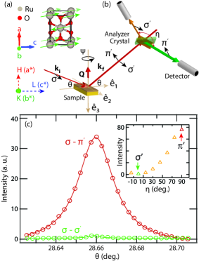
In order to distinguish between magnetic and charge channels for the observed resonant reflection, we carried out photon polarization analysis using a Si(111) analyzer crystal. The polarization of the incoming photon is fixed to and we measured the out-going photon in both and polarization projections of the scattered photons, where and represent, respectively, the polarization component perpendicular and parallel to the scattering plane. Figure 2(A) shows the unit cell of rutile RuO2 with the collinear AFM structure. The fundamental magnetic wavevector is (1 0 0) as we have observed at Ru-L2 edge. Figure 2(B) graphically systematizes the experimental configuration. The selection between - and - is controlled by rotating the analyzer around the scattered wavevector by = 90∘. As shown in Figure 2(C), the intensity of the rocking curve at the (1 0 0) position is dominant in the - channel. The inset in Figure 2(C) shows the integrated intensity of the analyzer scans as a function of , which reaches its maximum in the - channel and near zero in the - channel.
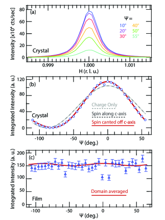
To decode the origin of the resonant reflection at the wave vector (1 0 0), we carried out a detailed symmetry-restricted tensorial analysis of the azimuthal angle dependence of the scatterred intensity (see Supplemental Material for details sup ). In Figure 3(A), we show a series of representative momentum scans across the (1 0 0) reflection for different azimuthal angles . In Figure 3(B), we plot the integrated peak area (extracted from Gaussian fits to the momentum scans) as a function of . A clear modulation with a period of can be visually inferred, with the magnetic scattering intensity being maximized at = 0∘ and minimized at = -90∘, where the azimuthal angle = 0∘ corresponds to the (0 0 1) direction lying in the diffraction plane. A pure two-fold modulation at the (1 0 0) reflection can be equivalently described by a model derived by scattering from quadrupolar-type charge anisotropy or magnetic scattering with moments oriented strictly along the c-axis (see Supplementary Material for further details and a more extended description of the model sup ). The best-fit result for a pure scattering of the type corresponding to these two distinct mechanisms is given in Figure 3(B) as the gray and black dotted lines. In the charge anisotropy picture, this is the only component that is allowed by symmetry of the Ru atoms in the space group. However, in the case that the scattering is of magnetic origin, one can generalize the model to include a slight canting of the moment off of the c-axis. The resulting higher harmonic content in the azimuthal dependence of the scattering intensity arises when more than a single component of the magnetic moment is nonzero. A fit to this generalized model is given by the solid red curve in Figure 3(B), showing a significantly improved agreement to the data, further supporting the magnetic origin of this peak. We caution, however, that with only a single observable reflection at Ru-L2 edge, it is not possible to completely rule out a partial contribution from charge anisotropy. Figure 3(C) reports the azimuthal dependence of the magnetic scattering intensity from the thin film sample. Unlike the case of the bulk crystal, it does not exhibit any significant modulation with . This seeming discrepancy between the thin film and bulk crystal is explained by the twinned nature of the thin films, reflecting the existence of multiple domains. The introduction of an arbitrarily canted moment off of the high-symmetry axis necessitates the consideration of eight distinct species of orthogonal domains sharing a common (1 0 0) epitaxial axis either parallel or antiparallel to the scattering vector. The local scattering intensity from simultaneously probed domains can be related to a global azimuthal angle by a domain-dependent phase shift and rotations by . Taking the best-fit parameters from the bulk case in Figure 3(B), incoherently averaging the contributions and using the known orientation of the film at zero azimuth yields the predicted dependence for the magnetic peak in the thin film as shown in Figure 3(C). The domain-averaging has suppressed lower-frequency components of the azimuthal dependence, leaving a nearly constant intensity with a small residual higher-frequency modulation. The amplitude of the residual modulation lies well below the noise and the prediction is consistent with the observations within experimental uncertainty.
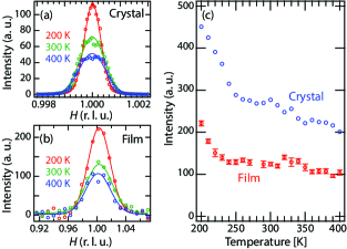
A major aspect and motivation of our investigation is to assess whether magnetic order persists up to room temperature in RuO2 films. Figure 4(A) and (B) show representative longitudinal scans across the magnetic ordering vector at various temperatures, for the bulk crystal and thin film, respectively. For both samples, the magnetic scattering yield decreases when increasing temperature but spin order persists up to at least 400 K. The temperature dependence of the scattered intensity is shown in Figure 4(C) (data are rescaled and offset for clarity). Both samples exhibit a remarkably similar temperature evolution of the magnetic order parameter. The intensity diminishes rapidly, a factor of 2 from 200 K to 250 K, then smoothly tails off above 250 K and up to 400 K. The increase in the line-width of the (1 0 0) reflection for increasing temperatures suggests a progressive reduction in the spin-spin correlation lengths, which however remain finite across the whole temperature range surveyed in this study. In any case, the energy dependence of the magnetic scattering in the thin film sample is almost identical at 200 K and 320 K (see again Figure 1(C)), indicating that the emergence of a second phase below 250 K is unlikely. The observed resonant reflection at Q = (1 0 0) retains its magnetic character both below and above 250 K within the range of measured temperatures.
In summary, we have established the existence of collinear antiferromagnetism in both thin film and bulk rutile RuO2. At odds with previous reports that AFM order is short-ranged [15], our experiments reveal sharp antiferromagnetic diffraction signatures in bulk crystals with a correlation length in excess of 4000 Å in the direction. In the thin film, the correlation length is reduced to about 50 Å in the direction. This far smaller correlation length compared to the bulk crystal can be accounted for by the dimensional confinement along the direction in the thin film. The azimuthal angle dependence analysis suggests a collinear AFM magnetic structure with spin moments having dominant projection along (0 0 1), which agrees well with theoretical predictions from density functional theory Berlijn et al. (2017). The emergence of antiferromagnetism in a highly conducting oxide is rare and unusual, often implying some exotic physics at play. The AFM instability manifested by RuO2 may evoke some analogies with the paradigmatic case of Cr metal, a spin-density-wave antiferromagnet. However, and at variance with the incommensurate AFM ordering of Cr metal, here the magnetic diffraction data rule out any significant incommensurablity of the magnetic wave vector in RuO2. This fact might suggest a possibly different origin of the observed AFM spin textures. The itinerant AFM state of RuO2 is also reminiscent of the anomalous magnetism found in some perovskite chromates (CaCrO3 and SrCrO3), whose origin has long remained unclear. Our experiments in highly conducting RuO2 thus raise fundamental inquiry into the nature of the itinerant antiferromagnetism in this 4 transition metal oxide. From an applied perspective, the presence of room temperature antiferromagnetism in 25-nm films of a metallic oxide underscore RuO2 a potential candiate for spintronic devices. In addition, the evidence of the magnetic moments in RuO2 may prove to be important in catalysis of oxygen evolution reaction Rao et al. (2017) where the spin conservation rule plays an important role in producing oxygen molecules with spin Torun et al. (2013).
This research was supported by NSF through the Massachusetts Institute of Technology Materials Research Science and Engineering Center DMR - 1419807. R. C. acknowledges support from the Alfred P. Sloan Foundation. J. P. acknowledges financial support by the Swiss National Science Foundation Early Postdoc.Mobility fellowship project number P2FRP2171824. The work in the Materials Science Division of Argonne National Laboratory (bulk crystal synthesis) was supported by the U.S. Department of Energy, Office of Science, Basic Energy Sciences, Materials Science and Engineering Division. The work performed at the Advanced Photon Source was supported by the U.S. Department of Energy, Office of Science, and Office of Basic Energy Sciences under Contract No. DE-AC02-06CH11357
References
- Moriya (1979) T. Moriya, Journal of Magnetism and Magnetic Materials 14, 1 (1979), ISSN 0304-8853, URL http://www.sciencedirect.com/science/article/pii/0304885379902014.
- Fawcett (1988) E. Fawcett, Reviews of Modern. Physics 60, 1 (1988), ISSN 0304-8853, URL https://journals.aps.org/rmp/abstract/10.1103/RevModPhys.60.209.
- Komarek et al. (2011) A. C. Komarek, T. Möller, M. Isobe, Y. Drees, H. Ulbrich, M. Azuma, M. T. Fernández-Dáz, A. Senyshyn, M. Hoelzel, G. André, et al., Physical Review B 84, 125114 (2011), URL https://link.aps.org/doi/10.1103/PhysRevB.84.125114.
- Zhou et al. (2006) J.-S. Zhou, C.-Q. Jin, Y.-W. Long, L.-X. Yang, and J. B. Goodenough, Physical Review Letters 96, 046408 (2006), URL https://link.aps.org/doi/10.1103/PhysRevLett.96.046408.
- Williams et al. (2006) A. J. Williams, A. Gillies, J. P. Attfield, G. Heymann, H. Huppertz, M. J. Martínez-Lope, and J. A. Alonso, Physical Review B 73, 104409 (2006), URL https://link.aps.org/doi/10.1103/PhysRevB.73.104409.
- Ortega-San-Martin et al. (2007) L. Ortega-San-Martin, A. J. Williams, J. Rodgers, J. P. Attfield, G. Heymann, and H. Huppertz, Physical Review Letters 99, 255701 (2007), URL https://link.aps.org/doi/10.1103/PhysRevLett.99.255701.
- Komarek et al. (2008) A. C. Komarek, S. V. Streltsov, M. Isobe, T. Möller, M. Hoelzel, A. Senyshyn, D. Trots, M. T. FernÁndez-Díaz, T. Hansen, H. Gotou, et al., Physical Review Letters 101, 167204 (2008), URL https://link.aps.org/doi/10.1103/PhysRevLett.101.167204.
- Streltsov et al. (2008) S. V. Streltsov, M. A. Korotin, V. I. Anisimov, and D. I. Khomskii, Physical Review B 78, 054425 (2008), URL https://link.aps.org/doi/10.1103/PhysRevB.78.054425.
- Bhobe et al. (2011) P. A. Bhobe, A. Chainani, M. Taguchi, R. Eguchi, M. Matsunami, T. Ohtsuki, K. Ishizaka, M. Okawa, M. Oura, Y. Senba, et al., Physical Review B 83, 165132 (2011), URL https://link.aps.org/doi/10.1103/PhysRevB.83.165132.
- Maeno et al. (1994) Y. Maeno, H. Hashimoto, K. Yoshida, S. Nishizaki, T. Fujita, J. G. Bednorz, and F. Lichtenberg, Nature 372, 532 (1994), ISSN 1476-4687, URL https://www.nature.com/articles/372532a0.
- Perry et al. (2001) R. S. Perry, L. M. Galvin, S. A. Grigera, L. Capogna, A. J. Schofield, A. P. Mackenzie, M. Chiao, S. R. Julian, S. I. Ikeda, S. Nakatsuji, et al., Physical Review Letters 86, 2661 (2001), URL https://link.aps.org/doi/10.1103/PhysRevLett.86.2661.
- Nakatsuji et al. (1997) S. Nakatsuji, S. Ikeda, and Y. Maeno, Journal of the Physical Society of Japan 66, 1868 (1997), ISSN 0031-9015, URL https://journals.jps.jp/doi/abs/10.1143/JPSJ.66.1868.
- Yoshida et al. (2005) Y. Yoshida, S.-I. Ikeda, H. Matsuhata, N. Shirakawa, C. H. Lee, and S. Katano, Physical Review B 72, 054412 (2005), URL https://link.aps.org/doi/10.1103/PhysRevB.72.054412.
- Longo et al. (1968) J. M. Longo, P. M. Raccah, and J. B. Goodenough, Journal of Applied Physics 39, 1327 (1968), ISSN 0021-8979, URL https://aip.scitation.org/doi/abs/10.1063/1.1656282.
- Ryden and Lawson (1970) W. D. Ryden and A. W. Lawson, Journal of Chemical Physics 52, 6058 (1970), ISSN 0021-9606, URL https://aip.scitation.org/doi/10.1063/1.1672908.
- Guthrie and Bourland (1931) A. N. Guthrie and L. T. Bourland, Physical Review 37, 303 (1931), URL https://link.aps.org/doi/10.1103/PhysRev.37.303.
- Berlijn et al. (2017) T. Berlijn, P. Snijders, O. Delaire, H.-D. Zhou, T. Maier, H.-B. Cao, S.-X. Chi, M. Matsuda, Y. Wang, M. Koehler, et al., Physical Review Letters 118, 077201 (2017), URL https://link.aps.org/doi/10.1103/PhysRevLett.118.077201.
- Železný et al. (2018) J. Železný, P. Wadley, K. Olejník, A. Hoffmann, and H. Ohno, Nature Physics 14, 220 (2018), ISSN 1745-2481, URL https://www.nature.com/articles/s41567-018-0062-7.
- Baltz et al. (2018) V. Baltz, A. Manchon, M. Tsoi, T. Moriyama, T. Ono, and Y. Tserkovnyak, Reviews of Modern Physics 90, 015005 (2018), URL https://link.aps.org/doi/10.1103/RevModPhys.90.015005.
- Fukami et al. (2016) S. Fukami, C. Zhang, S. DuttaGupta, A. Kurenkov, and H. Ohno, Nature Materials 15, 535 (2016), ISSN 1476-4660, URL https://www.nature.com/articles/nmat4566.
- Núñez et al. (2006) A. S. Núñez, R. A. Duine, P. Haney, and A. H. MacDonald, Physical Review B 73, 214426 (2006), URL https://link.aps.org/doi/10.1103/PhysRevB.73.214426.
- Park et al. (2011) B. G. Park, J. Wunderlich, X. Martí, V. Holý, Y. Kurosaki, M. Yamada, H. Yamamoto, A. Nishide, J. Hayakawa, H. Takahashi, et al., Nature Materials 10, 347 (2011), ISSN 1476-4660, URL https://www.nature.com/articles/nmat2983.
- Tshitoyan et al. (2015) V. Tshitoyan, C. Ciccarelli, A. P. Mihai, M. Ali, A. C. Irvine, T. A. Moore, T. Jungwirth, and A. J. Ferguson, Physical Review B 92, 214406 (2015), URL https://link.aps.org/doi/10.1103/PhysRevB.92.214406.
- Bohnenbuck et al. (2009) B. Bohnenbuck, I. Zegkinoglou, J. Strempfer, C. S. Nelson, H.-H. Wu, C. Schüß-Langeheine, M. Reehuis, E. Schierle, P. Leininger, T. Herrmannsdörfer, et al., Physical Review Letters 102, 037205 (2009), URL https://link.aps.org/doi/10.1103/PhysRevLett.102.037205.
- Bohnenbuck et al. (2008) B. Bohnenbuck, I. Zegkinoglou, J. Strempfer, C. Schüßler-Langeheine, C. S. Nelson, P. Leininger, H.-H. Wu, E. Schierle, J. C. Lang, G. Srajer, et al., Physical Review B 77, 224412 (2008), URL https://journals.aps.org/prb/abstract/10.1103/PhysRevB.77.224412.
- Zegkinoglou et al. (2005) I. Zegkinoglou, J. Strempfer, C. S. Nelson, J. P. Hill, J. Chakhalian, C. Bernhard, J. C. Lang, G. Srajer, H. Fukazawa, S. Nakatsuji, et al., Physical Review Letters 95, 136401 (2005), URL https://link.aps.org/doi/10.1103/PhysRevLett.95.136401.
- Nelson et al. (2007) C. S. Nelson, H. Mo, B. Bohnenbuck, J. Strempfer, N. Kikugawa, S. I. Ikeda, and Y. Yoshida, Physical Review B 75, 212403 (2007), URL https://link.aps.org/doi/10.1103/PhysRevB.75.212403.
- Stoerzinger et al. (2014) K. A. Stoerzinger, L. Qiao, M. D. Biegalski, and Y. Shao-Horn, Journal of Physical Chemistry Letters 5, 1636 (2014), ISSN 1948-7185, URL https://doi.org/10.1021/jz500610u.
- Lister et al. (2003) T. E. Lister, Y. V. Tolmachev, Y. Chu, W. G. Cullen, H. You, R. Yonco, and Z. Nagy, Journal of Electroanalytical Chemistry 554-555, 71 (2003), ISSN 1572-6657, URL http://www.sciencedirect.com/science/article/pii/S0022072803000482.
- Strempfer et al. (2013) J. Strempfer, S. Francoual, D. Reuther, D. K. Shukla, A. Skaugen, H. Schulte-Schrepping, T. Kracht, and H. Franz, Journal of Synchrotron Radiation 20, 541 (2013), ISSN 0909-0495, URL http://scripts.iucr.org/cgi-bin/paper?ie5091.
- Fang et al. (2004) Z. Fang, N. Nagaosa, and K. Terakura, Physical Review B 69, 045116 (2004), URL https://link.aps.org/doi/10.1103/PhysRevB.69.045116.
- (32) See Supplemental Material at [url] for detailed information of the sample growth, characterizations, and theoretical calculations, which includes Refs. [33-35].
- Chantler (2000) C. T. Chantler, Journal of Physical and Chemical Reference Data 29, 597 (2000), ISSN 0047-2689, URL https://aip.scitation.org/doi/abs/10.1063/1.1321055.
- Chantler (1995) C. T. Chantler, Journal of Physical and Chemical Reference Data 24, 71 (1995), ISSN 0047-2689, URL https://aip.scitation.org/doi/10.1063/1.555974.
- Haverkort et al. (2010) M. W. Haverkort, N. Hollmann, I. P. Krug, and A. Tanaka, Physical Review B 82, 094403 (2010), URL https://link.aps.org/doi/10.1103/PhysRevB.82.094403.
- Rao et al. (2017) R. R. Rao, M. J. Kolb, N. B. Halck, A. F. Pedersen, A. Mehta, Y. You, K. A. Stoerzinger, Z. Feng, H. A. Hansen, H. Zhou, et al., Energy & Environmental Science 10, 2626 (2017), URL http://pubs.rsc.org/en/Content/ArticleLanding/2017/EE/C7EE02307C.
- Torun et al. (2013) E. Torun, C. M. Fang, G. A. de Wijs, and R. A. de Groot, The Journal of Physical Chemistry C 117, 6353 (2013), ISSN 1932-7447, URL https://doi.org/10.1021/jp4020367.