Crosslinker mobility weakens transient polymer networks
Abstract
Transient networks comprised of polymers connected by short-lived bonds are a common design theme for both biological and synthetic materials. Transient bonds can provide mechanical rigidity, while still allowing for visco-elastic flows on timescales longer than the bond lifetime. In many biological polymer networks such as the actin cytoskeleton, the short-lived bonds are formed by accessory proteins that diffuse away after unbinding. By contrast, bonds in synthetic networks, such as the pendant groups of telechelic polymers, can only rebind in the same location. Using a recently developed theoretical model of the fracturing of visco-elastic materials, we here investigate the effect of linker mobility on the bond dynamics of a network under stress. We find that although mean field properties such as the average bond linker lifetime are barely affected by bond mobility, networks cross linked by mobile bonds fracture more readily due to ’leaking’ of linkers from crack areas to less stressed regions within the network. We propose a theoretical model to describe the redistribution of mobile linkers, which we validate by simulations. Our work offers insight into a potential trade-off that cells face, between fracture strength versus the modularity and tight dynamic control offered by mobile linkers.
I Introduction
Transient polymer networks are connected by individually short-lived bonds, which collectively result in a long-lived mechanical resistance by distributed load sharing review . This design principle is the basis of viscoelastic materials: the reversible bond dynamics allow the network to flow whilst maintaining mechanical integrity Meng2016c . Furthermore, transient networks are self-healing due to the reversibility of unbinding Yang2013 and the sensitivity of the bond dynamics to a range of external conditions makes these materials stimuli-responsive Ward2008 ; Spruijt2010 ; Gralka2015 . Due to the viscoelastic flow, transient networks are much more deformable than permanent networks Kong2003 . However, viscoelastic materials can resist mechanical stress only for a limited time, after which the system suddenly loses its mechanical percolation, a process which is known as fracturing Gibaud2010 ; Sprakel2011 ; telechelic ; Jasper . Crack initiation of viscoelastic materials occurs stochastically Jasper ; review due to fluctuations in the local density of bound linkers Nava2017 ; Mulla2018a , which eventually results in fracture due to rapid crack propagation Baumberger2006 . In order to understand the fracturing behavior of transient networks, one needs to understand the linker dynamics.
Transient networks can be divided in networks bound by immobile or by mobile linkers. Mobile linkers can be found in many biological systems. A well-studied example is the actin cortex, which consists of a network of actin filaments connected by actin-binding proteins Broedersz2010 . These actin binding proteins can freely diffuse after unbinding. Similarly, cells use integrins Tsunoyama2018 and cadherins TruongQuang2013 for cell-matrix and cell-cell adhesion Schwarz2013 , respectively. These adhesive proteins diffuse in the plane of the membrane after unbinding. By contrast, examples of networks connected by immobile linkers are colloidal gels connected by pendant groups Mueggenburg2007 and adhesive polymer networks Zhang2018 , such as hydrogen-bonded associative polymers Munstedt2011 , telechelic polymers Jasper , ionomers Sun2013 and polyelectrolyes Foyart2016 . A biological example of immobile linkers can be found in fibrin blood clots, where fibrin forms fibers with pendant sticky groups which unbind upon mechanical loading Kurniawan2016 .
Both mobile and immobile linkers result in transient networks, but only mobile linkers allow for linker rebinding in new locations of the network. This linker mobility allows for bond redistribution upon the application of mechanical stress Schmoller2010 ; Majumdar2018 . Due to crack-induced stress localization Griffith;1920 and subsequent force-induced linker unbinding Bell1978d , bond redistribution is most pronounced around cracks. We therefore wonder what is the effect of linker mobility on the mechanical strength of a material?
In order to investigate the effect of bond mobility on network strength, we use a model which we recently developed in the context of crack initiation in visco-elastic materials Mulla2018a . This model includes force-sensitive reversible bonds that are subjected to an external stress. Different from previous transient network models Seifert2000 ; Erdmann2004a ; Dietz2008 ; Novikova2013 ; Bell1978d ; Vaca2015 ; Gralka2015 , this model acknowledges that the applied stress is distributed inhomogeneously over the bonds on basis of their spatial distribution Majmudar2005 ; Guo2014a ; Arevalo2015 . As a result of this inhomogeneous force distribution, cracks stochastically initiate and subsequently propagate due to fluctuations in the local bond density Mulla2018a . However, this model was specific for the case of immobile linkers. We here extend the model, allowing us to include the effect of bond mobility to investigate the influence of bond mobility on the fracture strength of the network.
The main result of our work is that bond mobility hardly affects mean field properties of a bond, such as the average bound lifetime, but significantly reduces the network’s strength. We attribute the reduced network strength to the ’leaking’ of linkers from crack areas to less stressed regions within the network. Intriguingly, mobile linkers are widespread in biology despite the reduced fracture strength compared to networks connected by immobile linkers. We speculate that cells trade fracture strength for the modularity and tight dynamic control offered by mobile linkers.
II Model
II.1 Immobile linkers
In this work, we compare a network connected by mobile linkers to a network connected by immobile linkers. For immobile linkers, we use a model that was introduced in reference Mulla2018a . Here, we briefly summarize its salient features for clarity, and afterwards explain how we extend this model to the case of mobile linkers.
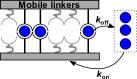
We initialize a one-dimensional (1D) network with equally spaced linkers using periodic boundary conditions, each link having a probability to start in a bound state (figure 1a). Next we model the dynamics of the linkers with a kinetic Monte Carlo scheme Gillespie1976 using the following linker dynamics:
| (1) |
where is the rate of linker binding and the rate of linker unbinding in the absence of force. We normalize time by the on-rate, . The off-rate increases exponentially with the applied force on the linker , in keeping with the Bell model Bell1978d :
| (2) |
where is the force where the off-rate has decreased to . We calculate the force per linker via
| (3) |
where is the stress on the system and is a yet to be defined stress intensity factor per linker. To account for the effect of inhomogeneous force distribution characteristic of polymer networks Majmudar2005 ; Guo2014a ; Arevalo2015 , we assume local loading sharing. Previously, we have shown that local load sharing provides an accurate description of crack initiation in macroscopic viscoelastic materials () Mulla2018a consistent with experiments Gladden2007 ; ABR ; Jasper ; Foyart2016 . Specifically, we assume that the force distribution is dependent on the distance of a linker to its nearest bound linker on both sides. Explicitly, we define a stress intensity factor on a bound linker at site by:
| (4) |
Note that the total force is independent of the bound fraction and normalized by the system size,=. We normalize the applied stress by the linker force sensitivity . After calculating the force on all linkers, we employ a kinetic Monte Carlo step to either bind or unbind a linker stochastically. We repeat this process until all linkers are unbound, and define the time at which the last linker unbinds as the rupture time .
II.2 Mobile linkers
We model mobile linkers by initializing linkers with a probability to start in the bound state. Every linker gets assigned a random location in a network of length . Each bound linker follows the same unbinding rules as explained above for immobile linkers, and each unbound linker binds with a rate . However, crucially, the difference with immobile linkers is that mobile linkers get assigned a new location in the network (figure 1b), whereas immobile linkers always rebind in the same location as where they previously unbound. For mobile linkers, we consider the limit of rapid diffusion after unbinding, and therefore rebinding occurs in a random new location. Throughout the paper, we compare mobile and immobile linkers using the same parameters , and .

III Results
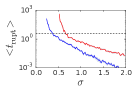
To probe the mechanical strength of transient networks, we study the network lifetime as a function of the applied stress. We run 30 simulations for each set of parameters and record the rupture time as the time where the fraction of closed bonds drops to zero. This way, we compare the rupture time as a function of stress for networks connected by mobile linkers versus immobile linkers, using otherwise identical parameters (figure 1b). We find that the average lifetime decreases with applied stress with two distinct regimes for both types of networks (figure 1c). In the high stress regime, the network lifetime is significantly shorter than the bond turnover time (, dashed line). In this regime, the network is unstable and the lifetime decreases exponentially as applied stress promotes linker unbinding. In the low stress regime, the network is metastable and linkers re-bind many times before rupturing is observed. The average network lifetime again decreases exponentially with stress, but more steeply compared to the high stress regime as not only the linker unbinding speeds up as a function of stress, but also the critical length for crack nucleation decreases (see section IV). Qualitatively, mobile and immobile linkers show a similar biphasic stress-dependence of the network rupture time. Strikingly, however, the mobile networks are weaker for all observed stresses, even though the linker affinity and number of linkers are identical.
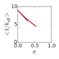
To investigate why linker mobility compromises network strength, we compare the microscopic linker properties at steady state. We first consider the average lifetime of the bound linkers as a function of stress. As shown in Figure 2a, the average bound linker lifetime decreases with stress for both networks, due to force-induced unbinding. Moreover, the average lifetimes are comparable for mobile and immobile linkers. Similarly, we find that the average bond-bond distance is comparable for mobile and immobile linkers ( and respectively, vertical lines in figure 2b).
Why is the network lifetime with mobile linkers drastically smaller than with static linkers, even though the average linker lifetime and bond-bond distance are similar? To investigate this paradox, we need to look beyond the mean field properties as rupture is a stochastic phenomenon, initiated by the emergence and growth of cracks due to local fluctuations Gladden2007 ; ABR ; Jasper ; Foyart2016 ; Mulla2018a . Plotting the distribution of bound linker distances at steady state reveals a crucial difference between immobile and mobile linkers: whereas their mean values are comparable, the bond-bond distance is significantly more widely distributed for mobile than for immobile linkers (figure 2b). We conclude that the reduced strength of networks crosslinked by mobile linkers is due to a more inhomogeneous force distribution over all bonds.

To investigate at what system size the difference between mobile and immobile linkers emerges, we compare networks with sizes ranging from to (figure 3). For both mobile and immobile linkers, increases with for microscopically small systems (up to for mobile linkers or for immobile linkers), as relative fluctuations in the number of bound linkers become smaller. Conversely, for macroscopically large systems ( for mobile linkers or for immobile linkers) the average rupture time decreases with system size according to a power of , as the number of crack nucleation sites increases linearly with the system size. However, an intermediate size regime exists for mobile linkers () where a faster decrease of the rupture time is observed. We hypothesize that this intermediate regime is caused by ’leaking’ of linkers from stressed areas (large ) to the rest of the material.
In the limit of small systems, smaller than the crack length, rebinding of linkers always happens in the vicinity of the unbinding area. In the opposite limit of macroscopic materials, much larger than the crack length, the pool of free linkers is constant in time and therefore uncorrelated from local fluctuations in bound linker density. Thus, the network lifetime decreases solely due to the increased number of crack nucleation sites. For intermediately sized systems, there is an enhanced reduction in network lifetime with increasing system size, because the correlation between local bound linker density and pool of free linkers becomes smaller with system size. Local unbinding of a linker increases the fraction of free linkers that can subsequently rebind in the crack area. For macroscopically large systems however, the fraction of free linkers is relatively constant in time due to the rule of large numbers. In other words, linkers in macroscopic systems effectively ’leak away’ from the crack area into the bulk of the material.

IV
To test the hypothesis that mobile linkers leak away, we modeled the dynamics of a gap area free of linkers within the material of length . The two processes which affect are binding of linkers anywhere within the gap, and unbinding of either of the two linkers at the edge of the gap. We assume that the gap length is significantly longer than the distance of each edge linker to its nearest neighbor. The unbinding rate of either of the linkers at the edge is therefore:
| (5) |
The pre-factor results from the fact that two linkers can unbind, and the exponent is because the force over the gap () is distributed over both edge linkers. We next consider the binding of linkers anywhere within the gap. As the gap size increases, the rate of binding increases as more binding possibilities are present:
| (6) |
where is the rate of binding in the gap, and is the number of bound linkers. The factor for mobile linkers arises from the fact that the pool of free linkers decreases with the fraction of bound linkers. For immobile linkers, rebinding does not depend on the global pool of free linkers, as every linker only rebinds locally. For macroscopic systems, is independent of , namely . We calculate by numerically solving:
| (7) |
where is the total rate of linker binding and is the total rate of linker unbinding within the network. As increases linearly with the gap size, whereas increases exponentially, gaps will always become unstable for large enough as unbinding occurs significantly more rapidly than re-binding Mulla2018a . We are interested in the length at which gaps become unstable and propagate. We approximate by calculating the length at which the rate of unbinding at the edge equals the rate of binding within the center , which we can numerically solve by combining equations 5, 6 and 7.
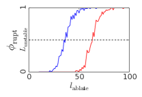
We test our theory quantitatively by measuring by performing simulations where we ablate a gap of controlled length: first we equilibrate a network under stress until , after which we unbind all bound linkers in the positions . Next, we observe the network until (figure 4a). We repeat this procedure for networks per condition and plot the fraction of ruptured networks as a function of (figure 4b). For small , all networks stay intact, whereas networks become unstable and rapidly fracture for large . We extract from simulations by calculating the ablation length at which via linear interpolation. We observe that mobile linkers have a shorter typical ablation length than immobile linkers ( versus , respectively (figure 4b). For both types of networks, increases with as cracks are more likely to propagate under increasing stress (symbols in figure 4c). The theoretical model describes this trend correctly, although there is a systematic under-estimation of the absolute value of by approximately (lines in figure 4c). A fully quantitative calculation of would require solving , which is not possible as we do not have an equation for . Therefore, we have approximated the unstable point by calculating the gap length at which linker unbinding is equally likely as linker binding within the gap. However, is only a good approximation of the unstable length if an unbinding event increases the gap size by an equal amount as a rebinding event would decrease it. This assumption is not fully correct, as a linker unbinding event might only cause a marginal increase in gap length in case the unbinding linker has a neighboring linker that is close-by, whereas a linker rebinding in the middle of the gap halves the gap size .
We next calculated the crack length as a function of the bond affinity for both mobile and immobile linkers at a fixed (figure 4d). We fix rather , because otherwise would increase far more rapidly as a function of and would not be computationally tractable for large . Furthermore, we choose to plot , rather than , as this quantity roughly represents the number of ablated bound linkers. It is therefore more straightforward to interpret than for different values of , as the density of bound linkers varies with . We find that stays approximately constant for mobile linkers upon increasing the bond affinity , whereas it increases for immobile linkers. As a result, the difference in between mobile and immobile linkers is most pronounced for high , whereas the two types of networks become similar at low . Indeed, as seen from equation 6, the only difference between mobile and immobile linkers is the factor , which reduces the rate of binding within a gap. In the limit of a low bond affinity , and therefore immobile and mobile linkers behave identically.


V Discussion
We studied the dynamics of a transient network of reversible bonds under mechanical stress, and have compared immobile linkers, which always rebind in the same place as where they unbound, with mobile linkers, which can rebind anywhere within the network. We found that the mean lifetime of bound linkers in a transient network is unaffected by the mobility of the linker (figure 2a). Yet, networks connected by mobile linkers are significantly weaker than network connected by immobile linkers, with fracturing times that are orders of magnitude lower (figure 1c). We attribute the reduced strength of networks connected by mobile linkers to the redistribution of mobile linkers from areas low in linker density, corresponding to highly stressed areas, to the rest of the material. This effect does not occur for immobile linkers, as they stay in the place from which they unbound.
Our results raise the question of why mobile linkers exist at all in nature Broedersz2010 ; Tsunoyama2018 ; TruongQuang2013 ; Schwarz2013 , as immobile linkers yield stronger networks. An important thing to note in this context is that fracturing in biology is not always detrimental. In fact, fracturing in some cases is even required for biological function. For example local failure of the actin cortex can lead to cell polarization Paluch2005 ; Carvalho2013 ; AbuShah2014 and facilitate a mode of cellular migration which relies on the formation of membrane blebs Paluch2013 . Similarly, destabilization and subsequent rupturing of the polar actomyosin cortex aids proper positioning of the cytokinetic furrow Sedzinski2011a . However, in many other circumstances fracturing of transient networks in biology is related to developmental defects Martin2010 ; Casares2015 and diseases Henderson2009 ; Feng2018 . Therefore, the widespread existence of mobile linkers involved in cellular adhesion Tsunoyama2018 ; TruongQuang2013 ; Schwarz2013 and crosslinking of biopolymer networks Broedersz2010 ; Lin2010 requires explanation.
Both cell-cell and cell-matrix adhesion are mediated by proteins embedded in the membrane, which are either bound to their substrate or diffuse within the plane of the membrane and can therefore be classified as mobile linkers. Examples of such protein families are E-cadherin for cell-cell adhesion and integrin for cell-matrix adhesion Schwarz2013 . The collagen matrix to which integrins adhere have mesh sizes of up to several micrometers Zoumi2002 , whereas the individual collagen fibers are only a few tens of nanometers thick Sell2010 . As a result, the fraction of the membrane area which is in close enough proximity to fibers to allow for binding is very low. A large fraction of the plasma membrane area would have to be covered with immobile linkers in order to have a significant number of integrins interacting with the extracellular matrix. Instead, we speculate that linker mobility allows for diffusion through the membrane to facilitate binding to the sparse fibers. Therefore, linker mobility allows for cellular adhesion whilst requiring only a small fraction of the membrane area.
Biopolymer networks are either connected via stickiness of the fibers, for example in the case of fibrin fibers Kurniawan2016 , or via mobile linkers such as cross linking ions which cross link the intermediate filament vimentin Lin2010 . Different from the case of cellular adhesion, linker mobility does not necessarily increase the connectivity of biopolymer networks: where immobile linkers only require close proximity of two fibers, mobile linkers require the proximity of two fibers and the proximity of a linker. For ionically cross linked intermediate filaments, linkers are abundantly present as the concentration of magnesium ions in the cytosol is on the order of a Sun2009 . Furthermore, as the concentration of intermediate filaments is orders of magnitude lower, in the regime Lai1993 , magnesium is abundant and has a low effective bond affinity . In this regime mobile linkers are as strong as immobile linkers (figure 4d).
Another case of mobile linkers are actin binding proteins which cross link the actin cytoskeleton Broedersz2010 . Many different types of cross linking proteins exist, with an enormous variety in their cross linking properties such as length Wagner2006 , compliance Schwaiger2004 , preferred binding angle Courson2010 , angular flexibility Flexibility , typical lifetime of actin binding Wachsstock1994 and force sensitivity of the unbinding rate Ferrer2008 . Many of these linker properties have been found to affect biopolymer network mechanics Wagner2006 ; Broedersz2010 ; Flexibility ; Gralka2015 ; Didonna2006 . As a result, this variety of mobile linkers allows the cell to have tight dynamic control of the mechanical properties of its actin cytoskeleton, which would be difficult to obtain if the actin filaments were connected by the stickiness of the fibers.
VI Conclusion
To summarize, we have studied the fracturing of transient networks connected by either mobile or immobile linkers. Our main result is that linker mobility weakens networks under stress. We have proposed and verified a theoretical model to explain this effect on basis of the leaking of linkers from crack areas to less stressed areas within the material. Even though linker mobility weakens transient networks, mobile linkers are widespread in biology. We speculate that cellular adhesion proteins are more likely to interact due to linker mobility and the large variety of mobile linkers connecting the actin cytoskeleton allows for a flexible control over the mechanics.
Acknowledgements.
We thank Pieter Rein ten Wolde for fruitful discussions. This work is part of the research program of the Netherlands Organisation for Scientific Research (NWO). We gratefully acknowledge financial support from an ERC Starting Grant (335672-MINICELL).References
- [1] Christian Ligoure and Serge Mora. Fractures in complex fluids: the case of transient networks. Rheologica Acta, 52(2):91–114, feb 2013.
- [2] Fanlong Meng, Robyn H. Pritchard, and Eugene M. Terentjev. Stress Relaxation, Dynamics, and Plasticity of Transient Polymer Networks. Macromolecules, 49(7):2843–2852, apr 2016.
- [3] Ying Yang and Marek W. Urban. Self-healing polymeric materials. Chemical Society Reviews, 42(17):7446, 2013.
- [4] Sabine M. Volkmer Ward, Astrid Weins, Martin R. Pollak, and David A. Weitz. Dynamic viscoelasticity of actin cross-linked with wild-type and disease-causing mutant alpha-actinin-4. Biophysical journal, 95(10):4915–23, nov 2008.
- [5] Evan Spruijt, Joris Sprakel, Marc Lemmers, Martien A Cohen Stuart, and Jasper van der Gucht. Relaxation dynamics at different time scales in electrostatic complexes: time-salt superposition. Physical review letters, 105(20):208301, nov 2010.
- [6] Matti Gralka and Klaus Kroy. Inelastic mechanics: A unifying principle in biomechanics. Biochimica et Biophysica Acta (BBA) - Molecular Cell Research, 1853(11):3025–3037, nov 2015.
- [7] Hyun Joon Kong, Emma Wong, and David J. Mooney. Independent Control of Rigidity and Toughness of Polymeric Hydrogels. Macromolecules, 36(12):4582–4588, jun 2003.
- [8] Thomas Gibaud, Damien Frelat, and Sébastien Manneville. Heterogeneous yielding dynamics in a colloidal gel. Soft Matter, 6(15):3482, 2010.
- [9] Joris Sprakel, Stefan B. Lindström, Thomas E. Kodger, and David A. Weitz. Stress Enhancement in the Delayed Yielding of Colloidal Gels. Physical Review Letters, 106(24):248303, jun 2011.
- [10] J.-F. Berret and Y Séréro. Evidence of Shear-Induced Fluid Fracture in Telechelic Polymer Networks. Physical Review Letters, 87(4):048303, jul 2001.
- [11] Paulina J. Skrzeszewska, Joris Sprakel, Frits a. de Wolf, Remco Fokkink, Martien a. Cohen Stuart, and Jasper van der Gucht. Fracture and Self-Healing in a Well-Defined Self-Assembled Polymer Network. Macromolecules, 43(7):3542–3548, apr 2010.
- [12] Giovanni Nava, Marina Rossi, Silvia Biffi, Francesco Sciortino, and Tommaso Bellini. Fluctuating Elasticity Mode in Transient Molecular Networks. Physical Review Letters, 119(7):078002, aug 2017.
- [13] Yuval Mulla, Giorgio Oliveri, Johannes T. B. Overvelde, and Gijsje H. Koenderink. Crack initiation in viscoelastic materials. pages 1–8, feb 2018.
- [14] Tristan Baumberger, Christiane Caroli, and David Martina. Solvent control of crack dynamics in a reversible hydrogel. Nature Materials, 5(7):552–555, jul 2006.
- [15] Chase P. Broedersz, Martin Depken, Norman Y. Yao, Martin R. Pollak, David a. Weitz, and Frederick C. MacKintosh. Cross-Link-Governed Dynamics of Biopolymer Networks. Physical Review Letters, 105(23):238101, nov 2010.
- [16] Taka A. Tsunoyama, Yusuke Watanabe, Junri Goto, Kazuma Naito, Rinshi S. Kasai, Kenichi G. N. Suzuki, Takahiro K. Fujiwara, and Akihiro Kusumi. Super-long single-molecule tracking reveals dynamic-anchorage-induced integrin function. Nature Chemical Biology, 14(5):497–506, may 2018.
- [17] Binh-An Truong Quang, Madhav Mani, Olga Markova, Thomas Lecuit, and Pierre-François Lenne. Principles of E-Cadherin Supramolecular Organization In Vivo. Current Biology, 23(22):2197–2207, nov 2013.
- [18] Ulrich S. Schwarz and Samuel a. Safran. Physics of adherent cells. Reviews of Modern Physics, 85(3):1327–1381, aug 2013.
- [19] Klara E. Mueggenburg, Xiao-Min Lin, Rodney H. Goldsmith, and Heinrich M. Jaeger. Elastic membranes of close-packed nanoparticle arrays. Nature Materials, 6(9):656–660, sep 2007.
- [20] Zhijie Zhang, Quan Chen, and Ralph H. Colby. Dynamics of associative polymers. Soft Matter, 14(16):2961–2977, 2018.
- [21] Helmut Münstedt. Rheological properties and molecular structure of polymer melts. Soft Matter, 7(6):2273, 2011.
- [22] Tao Lin Sun, Takayuki Kurokawa, Shinya Kuroda, Abu Bin Ihsan, Taigo Akasaki, Koshiro Sato, Md. Anamul Haque, Tasuku Nakajima, and Jian Ping Gong. Physical hydrogels composed of polyampholytes demonstrate high toughness and viscoelasticity. Nature Materials, 12(10):932–937, 2013.
- [23] Guillaume Foyart, Christian Ligoure, Serge Mora, and Laurence Ramos. Rearrangement Zone around a Crack Tip in a Double Self-Assembled Transient Network. ACS Macro Letters, 5(10):1080–1083, oct 2016.
- [24] Nicholas A. Kurniawan, Bart E. Vos, Andreas Biebricher, Gijs J.L. Wuite, Erwin J.G. Peterman, and Gijsje H. Koenderink. Fibrin Networks Support Recurring Mechanical Loads by Adapting their Structure across Multiple Scales. Biophysical Journal, 111(5):1026–1034, sep 2016.
- [25] K M Schmoller, P Fernández, R C Arevalo, D L Blair, and a R Bausch. Cyclic hardening in bundled actin networks. Nature Communications, 1(9):134, dec 2010.
- [26] Sayantan Majumdar, Louis C. Foucard, Alex J. Levine, and Margaret L. Gardel. Mechanical hysteresis in actin networks. Soft Matter, 14(11):2052–2058, 2018.
- [27] A. A. Griffith. The Phenomena of Rupture and Flow in Solids. Philosophical Transactions of the Royal Society A: Mathematical, Physical and Engineering Sciences, 221(582-593):163–198, jan 1921.
- [28] G. Bell. Models for the specific adhesion of cells to cells. Science, 200(4342):618–627, may 1978.
- [29] Udo Seifert. Rupture of Multiple Parallel Molecular Bonds under Dynamic Loading. Physical Review Letters, 84(12):2750–2753, mar 2000.
- [30] T. Erdmann and U. S. Schwarz. Stability of Adhesion Clusters under Constant Force. Physical Review Letters, 92(10):108102, mar 2004.
- [31] Hendrik Dietz and Matthias Rief. Elastic Bond Network Model for Protein Unfolding Mechanics. Physical Review Letters, 100(9):098101, mar 2008.
- [32] Elizaveta A. Novikova and Cornelis Storm. Contractile Fibers and Catch-Bond Clusters: a Biological Force Sensor? Biophysical Journal, 105(6):1336–1345, sep 2013.
- [33] Christian Vaca, Roie Shlomovitz, Yali Yang, Megan T. Valentine, and Alex J. Levine. Bond breaking dynamics in semiflexible networks under load. Soft Matter, 11(24):4899–4911, 2015.
- [34] T. S. Majmudar and R. P. Behringer. Contact force measurements and stress-induced anisotropy in granular materials. Nature, 435(7045):1079–1082, jun 2005.
- [35] Jun Guo, Yuexiu Wang, Frederick Sachs, and Fanjie Meng. Actin stress in cell reprogramming. Proceedings of the National Academy of Sciences, 111(49):E5252–E5261, dec 2014.
- [36] Richard C. Arevalo, Pramukta Kumar, Jeffrey S. Urbach, and Daniel L. Blair. Stress Heterogeneities in Sheared Type-I Collagen Networks Revealed by Boundary Stress Microscopy. PLOS ONE, 10(3):e0118021, mar 2015.
- [37] Daniel T. Gillespie. A general method for numerically simulating the stochastic time evolution of coupled chemical reactions. Journal of Computational Physics, 22(4):403–434, dec 1976.
- [38] Joseph R. Gladden and Andrew Belmonte. Motion of a Viscoelastic Micellar Fluid around a Cylinder: Flow and Fracture. Physical Review Letters, 98(22):224501, may 2007.
- [39] H. Tabuteau, S. Mora, G. Porte, M. Abkarian, and C. Ligoure. Microscopic Mechanisms of the Brittleness of Viscoelastic Fluids. Physical Review Letters, 102(15):155501, apr 2009.
- [40] Ewa Paluch, Matthieu Piel, Jacques Prost, Michel Bornens, and Cécile Sykes. Cortical actomyosin breakage triggers shape oscillations in cells and cell fragments. Biophysical Journal, 89(1):724–733, 2005.
- [41] Kevin Carvalho, Feng-Ching Tsai, Lees Edouard, Voituriez Raphael, Gijsje H. Koenderink, Feng Zhang, Simon J Morley, and Cecile Sykes. Cell-sized liposomes reveal how actomyosin cortical tension drives shape change. Proceedings of the National Academy of Sciences, 110(49):19968–19968, nov 2013.
- [42] E. Abu Shah and K. Keren. Symmetry breaking in reconstituted actin cortices. eLife, 3:e01433–e01433, apr 2014.
- [43] Ewa K. Paluch and Erez Raz. The role and regulation of blebs in cell migration. Current Opinion in Cell Biology, 25(5):582–590, 2013.
- [44] Jakub Sedzinski, Maté Biro, Annelie Oswald, Jean Yves Tinevez, Guillaume Salbreux, and Ewa Paluch. Polar actomyosin contractility destabilizes the position of the cytokinetic furrow. Nature, 476(7361):462–468, 2011.
- [45] Adam C. Martin, Michael Gelbart, Rodrigo Fernandez-Gonzalez, Matthias Kaschube, and Eric F. Wieschaus. Integration of contractile forces during tissue invagination. Journal of Cell Biology, 188(5):735–749, 2010.
- [46] Laura Casares, Romaric Vincent, Dobryna Zalvidea, Noelia Campillo, Daniel Navajas, Marino Arroyo, and Xavier Trepat. Hydraulic fracture during epithelial stretching. Nature Materials, 14(3):343–351, 2015.
- [47] Joel M Henderson, Mariam P Alexander, and Martin R Pollak. Patients with ACTN4 mutations demonstrate distinctive features of glomerular injury. Journal of the American Society of Nephrology : JASN, 20(5):961–8, 2009.
- [48] Di Feng, Jacob Notbohm, Ava Benjamin, Shijie He, Minxian Wang, Lay-Hong Ang, Minaspi Bantawa, Mehdi Bouzid, Emanuela Del Gado, Ramaswamy Krishnan, and Martin R. Pollak. Disease-causing mutation in -actinin-4 promotes podocyte detachment through maladaptation to periodic stretch. Proceedings of the National Academy of Sciences, 115(7):1517–1522, feb 2018.
- [49] Yi Chia Lin, Norman Y. Yao, Chase P. Broedersz, Harald Herrmann, Fred C. MacKintosh, and David A. Weitz. Origins of elasticity in intermediate filament networks. Physical Review Letters, 104(5):1–4, 2010.
- [50] A Zoumi, A Yeh, and B J Tromberg. Imaging cells and extracellular matrix in vivo by using second-harmonic generation and two-photon excited fluorescence. Proceedings of the National Academy of Sciences, 99(17):11014–11019, aug 2002.
- [51] Scott A. Sell, Patricia S. Wolfe, Koyal Garg, Jennifer M. McCool, Isaac A. Rodriguez, and Gary L. Bowlin. The Use of Natural Polymers in Tissue Engineering: A Focus on Electrospun Extracellular Matrix Analogues. Polymers, 2(4):522–553, nov 2010.
- [52] Liyuan Sun, Yuki Kosugi, Emiko Kawakami, Ying-Shan Piao, Tomoyo Hashimoto, and Kiyomitsu Oyanagi. Magnesium concentration in the cerebrospinal fluid of mice and its response to changes in serum magnesium concentration. Magnesium research, 22(4):266–72, dec 2009.
- [53] Yiu-Kay Lai, Wen-Chuan Lee, and Kuang-Den Chen. Vimentin serves as a phosphate sink during the apparent activation of protein kinases by okadaic acid in mammalian cells. Journal of Cellular Biochemistry, 53(2):161–168, oct 1993.
- [54] B Wagner, R Tharmann, I Haase, M Fischer, and a R Bausch. Cytoskeletal polymer networks: the molecular structure of cross-linkers determines macroscopic properties. Proceedings of the National Academy of Sciences of the United States of America, 103(38):13974–13978, 2006.
- [55] Ingo Schwaiger, Angelika Kardinal, Michael Schleicher, Angelika A Noegel, and Matthias Rief. A mechanical unfolding intermediate in an actin-crosslinking protein. Nature Structural & Molecular Biology, 11(1):81–85, jan 2004.
- [56] David S Courson and Ronald S Rock. Actin cross-link assembly and disassembly mechanics for alpha-Actinin and fascin. The Journal of biological chemistry, 285(34):26350–7, aug 2010.
- [57] M L Gardel, F Nakamura, J H Hartwig, J C Crocker, T P Stossel, and D a Weitz. Prestressed F-actin networks cross-linked by hinged filamins replicate mechanical properties of cells. Proceedings of the National Academy of Sciences of the United States of America, 103(6):1762–1767, 2006.
- [58] D.H. Wachsstock, W.H. Schwarz, and T.D. Pollard. Cross-linker dynamics determine the mechanical properties of actin gels. Biophysical Journal, 66(3):801–809, mar 1994.
- [59] Jorge M Ferrer, Hyungsuk Lee, Jiong Chen, Benjamin Pelz, Fumihiko Nakamura, Roger D Kamm, and Matthew J Lang. Measuring molecular rupture forces between single actin filaments and actin-binding proteins. Proceedings of the National Academy of Sciences of the United States of America, 105(34):9221–9226, 2008.
- [60] B. a. Didonna and Alex J. Levine. Filamin cross-linked semiflexible networks: Fragility under strain. Physical Review Letters, 97(6):1–4, 2006.