Athanasios Vlontzosathanasios.vlontzos14@imperial.ac.uk1
\addauthorKrystian Mikolajczykk.mikolajczyk@imperial.ac.uk1
\addinstitution
Dept. of Electrical and Electronic Engineering
Imperial College
London, UK
Deep Segmentation and Registration
Deep Segmentation and Registration in X-Ray Angiography Video
Abstract
In interventional radiology, short video sequences of vein structure in motion are captured in order to help medical personnel identify vascular issues or plan intervention. Semantic segmentation can greatly improve the usefulness of these videos by indicating exact position of vessels and instruments, thus reducing the ambiguity. We propose a real-time segmentation method for these tasks, based on U-Net network trained in a Siamese architecture from automatically generated annotations. We make use of noisy low level binary segmentation and optical flow to generate multi class annotations that are successively improved in a multistage segmentation approach. We significantly improve the performance of a state of the art U-Net at the processing speeds of 90fps.
1 Introduction
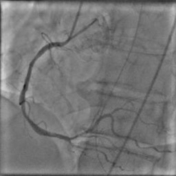
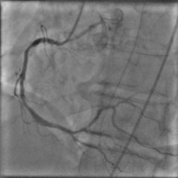
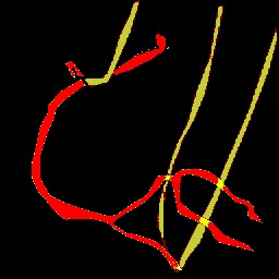
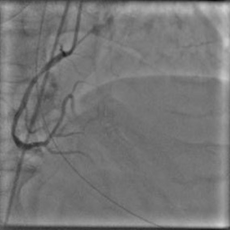
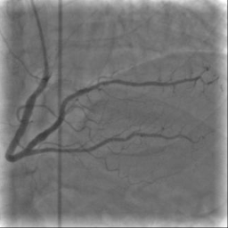
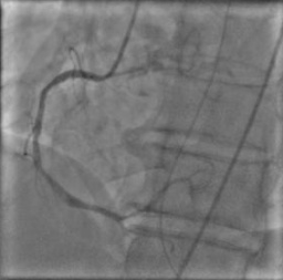
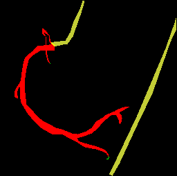
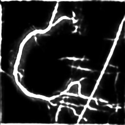
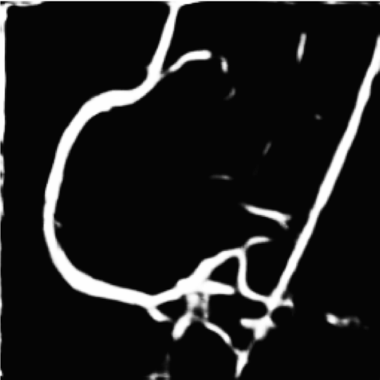
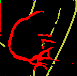
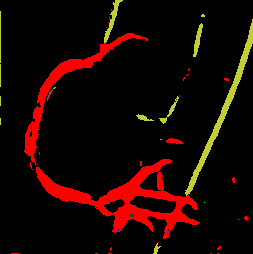
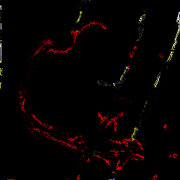
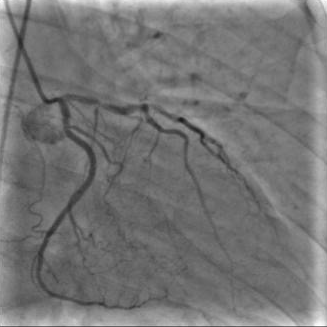
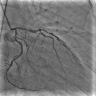
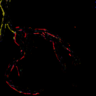
X-ray angiography is the most used imaging modality to visualise blood vessels for interventional purposes such as stenting of stenosed vessels or for diagnostic purposes such as assessment of myocardial perfusion or stenosis grading. To minimise ionising radiation exposure of the patient and medical personnel during image acquisition, low power X-Rays are used resulting in noisy and low contrast images. In the context of diagnosis, the main object of interest is the vascular tree, its branchings and variations in thickness. It is therefore necessary to accurately highlight the vessels in consecutive frames to reduce the noise and improve contrast. In addition, in interventional procedures, identifying interventional instruments (catheter, wires) is also needed in order to better plan and control their positioning. Efficiently discriminating between instruments and vessels as well as other anatomical structures that may have similar appearance is crucial during the interventions. Figure 1(a-c) shows an example of an angiogram sequence. Note large non-rigid motion between frames as well as the ambiguity between vessels and the catheter. Figure 1(e) shows a frame from a different sequence of the same patient but taken at different scan and angle and (f) shows a different patient. There is a significant difference in vessel as well as catheter locations in all three sequences, which we consider as independent examples. Figure 1(d) shows the ground truth segmentation of the first frame.

CNN based methods provide state of the art segmentation results but need to be trained with a large number of examples with ground truth segmentations that are typically obtained by manual annotation by experts. The presence of noise and low contrast make this task particularly challenging and methods with handcrafted features lead to inaccurate object detection. Neural networks were demonstrated to achieve outstanding performance in vision tasks when trained from well annotated and large datasets. This is in contrast to unsupervised training where limited success has been achieved so far. We make a step towards that direction by developing an approach to automatically generate annotated examples by exploiting the knowledge of the application context and the data.
In this paper we present a multistage architecture based on Convolutional Neural Networks for semantic segmentation of instruments and vessel tree. We propose a method to automatically generate label proposals that are successively refined in a multistage process. This data is then used to train a CNN in a way that exploits spatial and temporal continuity. This results in a network capable of generating accurate segmentations. In summary, our main contributions are:
-
•
We introduce a new approach for weakly supervised training of segmentation network applied to video angiograms. We propose a method that exploits low-level segmentations and optical flow to generate annotations for multi-class video segmentation.
-
•
We demonstrate that a convolutional network can be trained from automatically segmented sequences with noise, and can improve the quality of these unsupervised segmentations.
-
•
We improve the performance of the state of the art segmentation network U-Net [Ronneberger et al.(2015)Ronneberger, Fischer, and Brox] by 20%, processed with 90fps.
2 Related work
Segmentation.
Image segmentation has been a well researched topic, with new techniques arising frequently. Pixel-wise segmentation strives to create pixel masks (labels) that correspond to objects of interest in the image. One of the most common variational approaches from [Friedman and Russel(2013)] estimates the background using an incremental version of EM and then subsequently subtracts it from the images. Many variational methods rely on the calculation of a Hessian matrix to acquire a vesselness index, i.e. the probability of a given pixel belonging to a vessel. In [Frangi et al.(1998)Frangi, Niessen, Vincken, and Viergever],[B. Felfelian et al.(2016)B. Felfelian, Karimi, Soroushmehr, Samavi, Nallamothu, and Najarian] the authors construct a Hessian filter to enhance and segment coronary arteries, based on the Hessian matrix and the eigenvalues of the image. In [Truc et al.(2009)Truc, Khan, ans S. Lee, and Kim], a decimation free directional filter bank is used to decompose, analyse and then average the images to produce a final vesselness metric that is less sensitive to noise. In [Fazlali et al.(2015)Fazlali, Karimi, Soroushmehr, Sinha, Samavi, Nallamothu, and Najarian] the Hessian filter is enhanced with a pipeline consisting of a guided filter, Canny edge detection and block searching in order to determine the vessels of the angiography X-Rays; [Petkov et al.(2014)Petkov, Carrillo, P.Radeva, and C.Gatta] further extends this approach to address its diaphragm misclassification. An interesting variational method comes from [H.Ma et al.(2017)H.Ma, Hoongendoorn, Regar, Niessen, and van Walsum], which split the images into three layers; the breathing layer, the quasi-static layer and the vessel layer based on the movement of each of the layers. The layers are then processed using morphological operators and principal component analysis to segment the vessels. A Polygonal Path Image was introduced in [Bismuth et al.(2012)Bismuth, Vaillant, Talbot, and Najman] to unify local and global curvature structures with controllable smoothness to highlight guide wires.
Due to the data hungry nature of CNN approaches and the lack of annotated fluoroscopy data, the related work has been focusing on modalities like Magnetic Resonance Imaging (MRI), Computer Tomography (CT) or Positron Emission Tomography (PET). One of the most popular and effective architectures for medical image segmentation has been U-Net as introduced in [Ronneberger et al.(2015)Ronneberger, Fischer, and Brox] or 3D-Unet [Çiçek et al.(2016)Çiçek, Abdulkadir, Lienkamp, Brox, and Ronneberger], where a symmetric convolutional network is segmenting and reconstructing images. A residual neural network type architecture with two streams, a low resolution and a high resolution one, was investigated in [Kamnitsas et al.(2016)Kamnitsas, Ferrante, Parisot, Ledig, Nori, Criminisi, Rueckert, and Glocker] to segment brain lesions from MRI volumes. In [Rajchl et al.(2016)Rajchl, Lee, Oktay, Kamnitsas, Passerat-Palmbach, Bai, Demodaram, Rutherford, Hajnal, Kainz, and Rueckert], an extension of the well known GrabCut [C.Rother et al.(2004)C.Rother, Kolmogorov, and Blake] was introduced for pixel wise segmentation initialised by a bounding box of a of neo-natal brain lesion.
Tracking and registration.
Tracking of blood vessels in angiography is very challenging since vessels appear and disappear as contrast agent is injected and vessel shapes change because of the heart and lung motion. Due to changes in appearance, template trackers (e.g. [Comaniciu et al.(2003)Comaniciu, Ramesh, and Meer]) are less robust. Model based methods such as [Nwogu and Lorigo(2007)] regard blood vessels as active curves and deform the curves in the current frame to match the curves in the next frame. They are sensitive to noise and large change of the vessel shape. This problems led to classification-based tracking where pixels are classified in every frame as belonging to objects or background instead of transforming the appearance of objects from frame to frame. Such a tracker is robust against appearance changes and intermittent object presence. Classification-based trackers can be static e.g. [Avidan(2004)] where training is performed prior to tracking or adaptive, e.g. [Collins et al.(2005)Collins, Liu, and Leordeanu] where a classifier is built during tracking. Since our goal is to create a real-time pipeline, our preference is with static-type tracking using a convolutional neural network which can be very fast in inference mode while it can generalise well so that shape variability is accounted for.
A multi-resolution registration algorithm was proposed in [Yang et al.(2007)Yang, Wang, Tang, Zhou, Liu, and Chen] where the mask image is decomposed to coarse and fine sub-image blocks iteratively and each block is rigidly registered to the live image.
An iterative refinement algorithm was introduced in [Wang and Zhang(2009)] for registration in DSA. Nonrigid motion is iteratively modelled using thin-plate spline (TPS) calculated from a set of corresponding interest points. The iterative nature of such registration algorithms leads to high computational time, which makes them difficult to use in real-time clinical applications. In [Rivest-Henault et al.(2012)Rivest-Henault, Sundar, and
Cheriet] the authors use a prior CT Scan to perform a 3d to 2d registration of the coronary artery. The process however is not real time.
3 Weakly supervised segmentation
In this section we present our approach for generating segmented sequences that are then used to train an automatic segmentation method. There is a large number of available unannotated X-ray angiography sequences. These sequences are captured after a catheter is introduced and contrast agent injected. In the first few frames, only the catheter is visible, with vessels appearing once contrast agent is injected and propagates through the vessels. The goal is to classify pixels into three labels i.e. background, vessels with contrast agent, and a catheter.
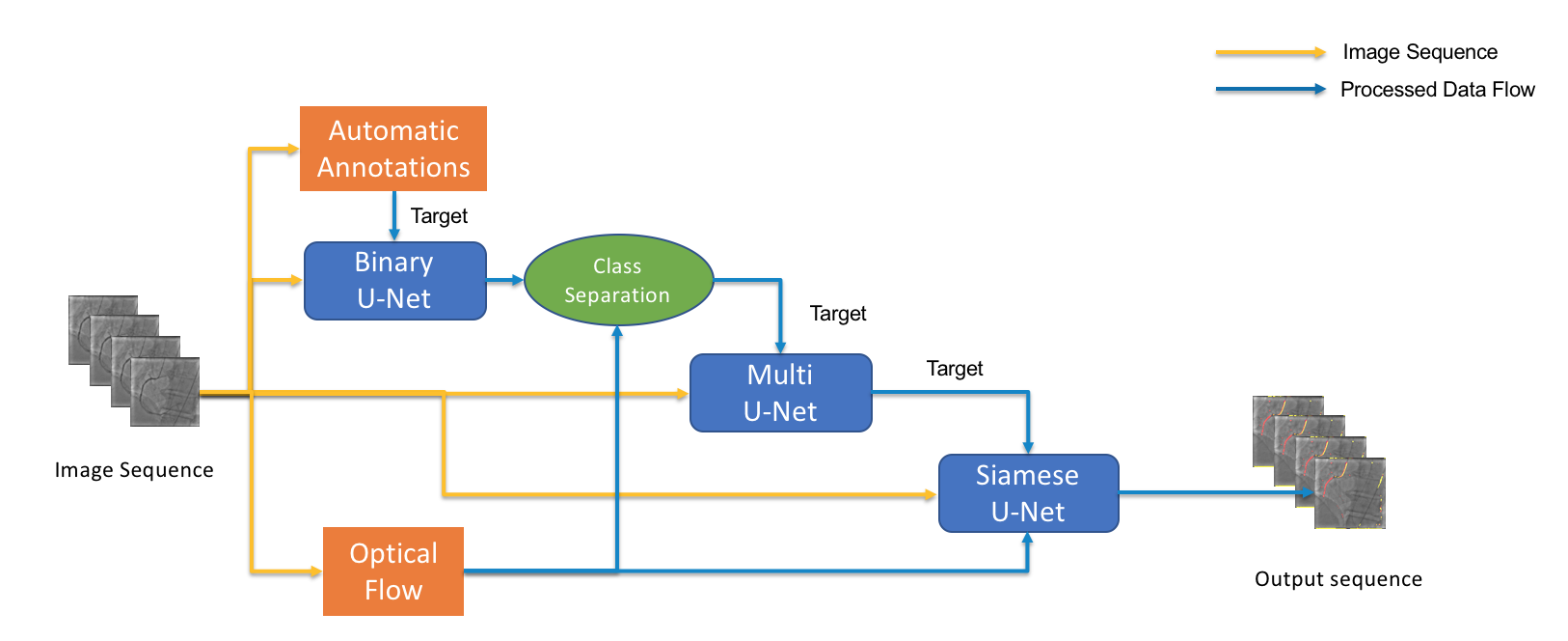
The proposed pipeline for annotating data and training Siamese U-Net is illustrated in Figure 2. We first employ a simple low-level morphological operator to generate binary masks for training a binary U-Net that discriminates between background and other labels. Such trained U-Net outputs segmentation masks with significantly reduced noise compared to the low level processing. Motion maps, generated by coarse-to-fine optical flow [Pathak et al.(2017)Pathak, Girshick, Dollár, Darrell, and Hariharan], provide additional information for more accurate segmentation and discrimination between catheter and contrast agent. The binary segmentation masks are then converted into three class labels using initial frames and their motion maps. This data is then used to train a multi class U-Net, which further improves multi-class segmentation. Finally, a Siamese U-Net is trained using image sequences, motion maps and refined multi-class segmentation masks. The approach trains a multi-class segmentation convolutional network with an unsupervised process, otherwise weakly supervised if a small amount of manually annotated data is used to pre-train or fine-tune the network in addition to the large volume of data that is automatically labelled. In the following we discuss each of the components in more details.
3.1 U-Net segmentation
Our segmentation pipeline relies on U-Net approach [Ronneberger et al.(2015)Ronneberger, Fischer, and Brox] at various processing stages, we therefore briefly explain its architecture here. U-Net consists of two streams; a convolutional (downstream) and a de-convolutional (upstream) path. The convolutional path is based upon traditional convolutional neural network layers including spatial convolutions, max pooling and activation functions (ReLU). The outputs of consecutive layers are down-sampled with a stride of 2 while increasing the number of features at each level by 2. There are 4 levels with 3 convolutional layers each, that generate a compressed representation of 1024 features. This is followed by a de-convolutional stream which up-converts the feature maps while concatenating them with the corresponding ones from the convolutional stream using skip connections to preserve details. ReLU layers are used after each deconvolution. This architecture was designed to learn a segmentation process of still images and training is typically performed with binary cross-entropy loss between original images and their segmentation masks.
3.2 Multi-class separation
Background and motion maps are used to infer the three label segmentations masks from a video sequence.
Background segmentation
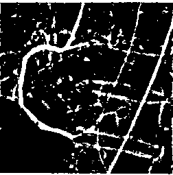
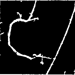
is performed for every input frame. Since interventional tools and vessels with contrast agent appear darker than the surrounding pixels, we employ the black top-hat morphological operator [Gonzalez and Woods(2018)], defined as the difference between the closing of the image and the original one to produce approximate binary masks. The operator is used with a structuring element. We eliminate small artefacts produced by the morphological operation using connected components analysis. The generated data still contains some noise as well as irregular vessel and catheter boundaries. Moreover there are some significant parts of vessels missing in such segmentations. We observed that a U-Net trained with such data can significantly reduce the occurrence of noise while successfully adding the missing parts. Figure 4 shows an example of such segmentation. We therefore improve the binary segmentation by using U-Net pretrained with the initial binary masks.
Catheter transfer.
The binary masks do not distinguish between vessels and catheters. According to standard medical practice, the angiography recording is started before introducing the contrast agent, since the latter is absorbed within a few seconds. Thus the first few frames, typically the second, as well as their binary segmentations can be used to discriminate between background and the catheter. In order to transfer the catheter label from the initial frames to those with contrast agent and visible blood vessels we use dense optical flow vectors efficiently calculated with coarse to fine approach [Pathak et al.(2017)Pathak, Girshick, Dollár, Darrell, and Hariharan]. Motion maps are then used to transform the binary mask of frame with the catheter only, onto frame that contains both catheter and vascular tree. We denote the result of the non-rigid warping . The segmentation labels that appear in both, and , correspond to the catheter while the binary labels in that find no correspondence in are classified as contrast agent (blood vessel). Thus, we generate multi-class labels using the binary masks and the optical flow. Similarly to the processing of binary masks we use this data to train a multi-class U-Net and apply it to improve the segmentation results. The multi-class U-Net is based upon the earlier binary block with the substitution of the last convolution layer with a layer. In addition we use categorical cross entropy (cf. Equation 1) as the loss function as opposed to the binary cross entropy from the earlier version. The input to the multi-class U-Net are the raw frames and the optical flow class separated masks as target labels.
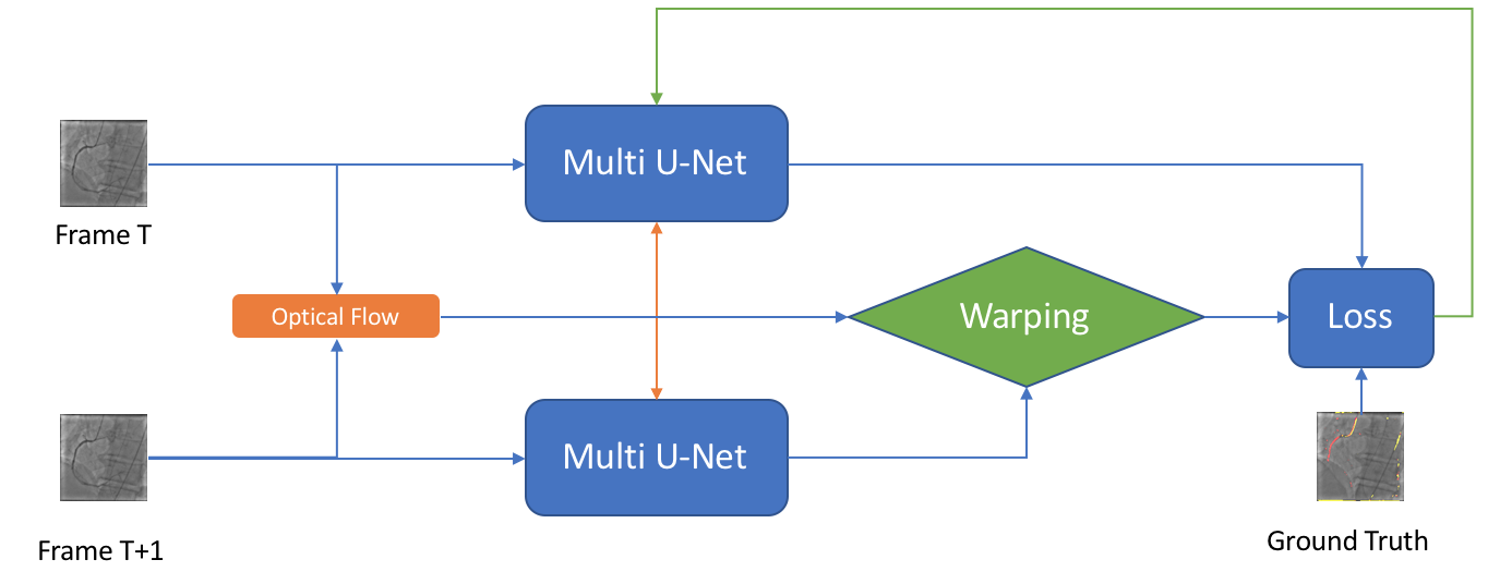
3.3 Siamese U-Net
U-Net as proposed in [Ronneberger et al.(2015)Ronneberger, Fischer, and Brox] and discussed in Section 3.1 is designed to segment static images. We extend this approach to make use of the temporal information captured by dense motion maps. Inspired by the success of Siamese architecture in vision related tasks such as matching two images, we train a Siamese network that analyses pairs of frames from different time instances and enforces temporal consistency between their segmentations.
The architecture is illustrated in Figure 3 and consist of two U-Nets that share their weights. At each training iteration the Siamese U-Net takes two frames, and , producing segmentations and . The dense motion map generated by optical flow between the two frames is used to transform into . In case of errorless optical flow and spatial segmentation, and should be identical. The multi-class cross entropy loss is
| (1) |
where are automatically generated segmentation labels as discussed in Section 3.2 and index corresponds to one of the three classes in the segmentation and ground truth masks i.e. background, catheter, and vessels.
4 Experimental results
In this section we evaluate our proposed approach and discuss results. We note that we have evaluated the pipeline both in its unsupervised capacity as well as the weakly supervised by injecting a small amount of manually annotated images for finetuning.
Dataset
The dataset consists of anonymised fluoroscopy X-Rays of 26 different patients. The images were acquired during stent placement using a General Electric Innova 2000 system and stored according to standard medical protocol in DICOM format. In total the dataset includes 36000 frames corresponding to 365 distinct video sequences with an average of 98 frames each. Different sequences of the same patient were taken at different angles and stages of the procedure therefore they differ significantly as shown in Figure 1(c)(e)(f). Each frame is rescaled from to due to memory constraints.
We randomly sampled 3000 pairs of frames not used in the test set, and used them to automatically generate annotations for training as discussed in Section 4.1 and Section 4.2. Furthermore, two sequences of 85 frames each, were manually annotated to serve as our test set for quantitative evaluation. Finally, we randomly selected 85 training frames for which we performed manual annotation using MIT’s CSAIL LabelMe tools [Russel and Torralba()]. These images were then augmented 10-fold by applying random rotations within range , and used in data augmentation experiments.
4.1 Qualitative evaluation
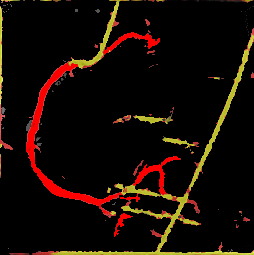
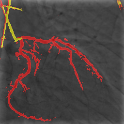
Using the approach discussed in section 3.2 we automatically generated annotations for 3000 randomly selected frames from sequences different than those with manual ground truth. In order to highlight the difference between the manual and automatic segmentation we present both in figure 4. The ground truth mask includes three labels i.e. background (black), vessel tree (red), and catheter (yellow). Black top-hat based segmentation mask contains significant noise, boundary irregularities and large parts of vessels as well as catheter are missing. The masks filtered with connected components seem to reduce the amount of noise but also further remove some small valid segments. This is crucial when comparing quantitative results of this simple segmentation method with and without connected component filter in Section 4.2.


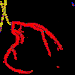
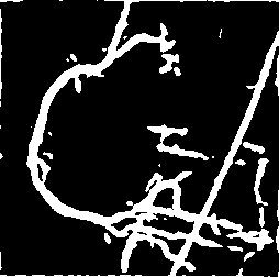
3000 pairs of frames with their optical flow from the automatically generated dataset were used to train the binary U-Net and the Siamese U-Net models presented in Sections 3.1 and 3.3. As illustrated in Figure 4(f) the Siamese U-Net is more accurate than the baseline U-Net. U-Net fine-tuned with manually annotated and augmented data provides masks with less noise and smooth object boundaries, although the same segments are successfully extracted in both, unsupervised Siamese U-Net and the fine-tuned one.
Figure 5 compares multi-class segmentation obtained with optical flow, multi-class U-Net and Siamese U-Net. All methods were trained with automatically generated annotations without fine-tuning. Both methods provide visually comparable segmentations therefore in the following section we perform a quantitative analysis of the segmentation accuracy.
Some of the errors appearing in the segmentation (Figure 5(g),5(n)) can be attributed to the use of simple interpolation layers instead of transposed convolutions. Despite the use of skip connections between encoding and decoding sections of the U-Net, the use of simple up sampling layers introduces loss of details and thickening of the vessels by a few pixels. We believe that use of transposed convolutions would improve results slightly. This will be the subject of future study.
4.2 Quantitative evaluation
In this section we present quantitative results for our proposed segmentation approach. The comparison is done using the dice index defined between segmentation masks and
| (2) |
where stands for the true positive number of labeled pixels, is the false positive number and are false negative pixels. Dice index, however, is best suited for binary segmentation, therefore, to address this shortcoming we report per class dice index in addition to an overall score related to the binary segmentation by the proposed methods. In the multi-class case we define the binary mask as the union of the catheter and contrast agent masks. Table 1 shows the results for various stages of the binary segmentation pipeline noting the variant of the approach. Interestingly, according to the dice index, the black top-hat segmentation produces more accurate masks than the connected components filter. As discussed in Figure 4(d) the connected components filter also removes some true positive segments in the mask. Unsupervised binary U-Net significantly improves the results. The proposed Siamese U-Net provides the best results with dice score of 0.98. Note that the multi-class labels are converted here to binary masks of background and the remaining two labels considered as one.
| Architecture | Dice |
|---|---|
| Black top-hat + CC | 0.77 |
| Black top-hat | 0.85 |
| U-Net | 0.92 |
| U-Net + CC | 0.89 |
| Siamese U-Net | 0.95 |
| Siamese U-Net + CC | 0.98 |
We further evaluate our models in multi-class segmentation task and report the dice scores for each label in table 2. Again, our proposed Siamese U-Net consistently improves upon the state of the art U-Net from [Ronneberger et al.(2015)Ronneberger, Fischer, and Brox]. We test two models trained with data augmented with different transformations i.e. small random rotation & translation in Augm_1 vs. rotation only in Augm_2. The latter seems to be more suited for the type of transformations that the input sequences typically include. Our best performing Siamese U-Net outperforms the original approach from [Ronneberger et al.(2015)Ronneberger, Fischer, and Brox] by more than 20%.
| Architecture | Catheter | Vessels |
|---|---|---|
| U-Net | 0.49 | 0.30 |
| U-Net + CC | 0.44 | 0.36 |
| Siamese U-Net + CC | 0.51 | 0.50 |
| Siamese U-Net + Augm_1 | 0.61 | 0.49 |
| Siamese U-Net + Augm_2 | 0.69 | 0.54 |
5 Efficiency
The proposed method was implemented in Python and the Keras framework within Tensorflow and tested with a single NVidia Titan-X GPU. In inference mode, the processing speed of U-Net trained in Siamese setup is 90 frames per second, which is suitable for real time segmentation of angiography sequences. Training time ranges from 20 min up to 8 hours when all 3000 automatically generated annotations are used for training.
6 Conclusions
We have introduced an approach for weakly-supervised training of a multi-class segmentation network in medical application of angiogram sequences. Our pipeline exploits automatic low level binary segmentations as well as motion vectors from optical flow to generate data annotations for multi-class segmentations thus avoiding tedious manual segmentation of many training examples. We show that automatic annotations that include some noise can still be used to train a CNN approach and lead to improved results. We also demonstrated how to enforce the temporal consistency between segmented frames within a siamese training architecture. Multi-class U-Net trained in siamese settings significantly improves the accuracy of segmentations compared to the original U-Net approach. Our network achieves processing speed of 90 frames per second, far beyond the speed capabilities of most of the existing interventional systems which makes it suitable for a real time applications. More experiments with different type of medical data using similar training setup will further validate this approach.
Acknowledgements
The authors would like to thank Dr Tsagarakis, the chair of the scientific and ethical boards of the Evangelismos General Hospital, Athens, Greece for providing anonymized angiography sequences. This work was supported by EPSRC grant EP/N007743/1.
References
- [Avidan(2004)] S. Avidan. Support vector tracking. TPAMI, 2004.
- [B. Felfelian et al.(2016)B. Felfelian, Karimi, Soroushmehr, Samavi, Nallamothu, and Najarian] H.R. Fazlali B. Felfelian, N. Karimi, SMR Soroushmehr, S. Samavi, B. Nallamothu, and K. Najarian. Vessel segmentation in low contrast x-ray angiogram images. ICIP, 2016.
- [Bismuth et al.(2012)Bismuth, Vaillant, Talbot, and Najman] V. Bismuth, R. Vaillant, H. Talbot, and L. Najman. Curvilinear structure enhancement with the polygonal path image - application to guide-wire segmentation in x-ray fluoroscopy. MICCAI, 2012.
- [Çiçek et al.(2016)Çiçek, Abdulkadir, Lienkamp, Brox, and Ronneberger] Ö. Çiçek, A. Abdulkadir, S. S. Lienkamp, T. Brox, and O. Ronneberger. 3d u-net: Learning dense volumetric segmentation from sparse annotation. arXiv:1606.06650 [cs.CV], 2016.
- [Collins et al.(2005)Collins, Liu, and Leordeanu] R. Collins, Y. Liu, and M. Leordeanu. Online selection of discriminative tracking features. TPAMI, 2005.
- [Comaniciu et al.(2003)Comaniciu, Ramesh, and Meer] D. Comaniciu, V. Ramesh, and P. Meer. Kernel-based object tracking. TPAMI, 2003.
- [C.Rother et al.(2004)C.Rother, Kolmogorov, and Blake] C.Rother, V. Kolmogorov, and A. Blake. Grubcut: interactive foreground extraction using iterated graph cuts. ACM TOG, 2004.
- [Fazlali et al.(2015)Fazlali, Karimi, Soroushmehr, Sinha, Samavi, Nallamothu, and Najarian] H.R. Fazlali, N. Karimi, S.M.R. Soroushmehr, S. Sinha, S. Samavi, B. Nallamothu, and K. Najarian. Vessel region detection in coronary x-ray angiograms. ICIP, 2015.
- [Frangi et al.(1998)Frangi, Niessen, Vincken, and Viergever] A.F. Frangi, W.J. Niessen, K.L. Vincken, and M.A. Viergever. Multiscale vessel enhancement filtering. MICCAI, 1998.
- [Friedman and Russel(2013)] N. Friedman and S. Russel. Image segmentation in video sequences: A probabilistic approach. arXiv:1302.1539 [cs.CV], 2013.
- [Gonzalez and Woods(2018)] R. C. Gonzalez and R. E. Woods. Digital Image Processing. Pearson, 4th edition, 2018.
- [H.Ma et al.(2017)H.Ma, Hoongendoorn, Regar, Niessen, and van Walsum] H.Ma, A. Hoongendoorn, E. Regar, W.J. Niessen, and T. van Walsum. Automatic online layer separation for vessel enhancement in x-ray angiograms for percutaneous coronary interventions. MIA, 2017.
- [Kamnitsas et al.(2016)Kamnitsas, Ferrante, Parisot, Ledig, Nori, Criminisi, Rueckert, and Glocker] K. Kamnitsas, E. Ferrante, S. Parisot, C. Ledig, A. Nori, A. Criminisi, D. Rueckert, and B. Glocker. Deepmedic for brain tumor segmentation. BRATS Challenge, 2016.
- [Nwogu and Lorigo(2007)] I. Nwogu and L. Lorigo. Fast temporal tracking and 3d reconstruction of a single coronary vessel. ICIP, 2007.
- [Pathak et al.(2017)Pathak, Girshick, Dollár, Darrell, and Hariharan] D. Pathak, R. Girshick, P. Dollár, T. Darrell, and B. Hariharan. Learning feautres by watching objects move. CVPR, 2017.
- [Petkov et al.(2014)Petkov, Carrillo, P.Radeva, and C.Gatta] S. Petkov, X. Carrillo, P.Radeva, and C.Gatta. Diaphragm border detection in coronary x-ray angiographies: New method and applications. CMIG, 2014.
- [Rajchl et al.(2016)Rajchl, Lee, Oktay, Kamnitsas, Passerat-Palmbach, Bai, Demodaram, Rutherford, Hajnal, Kainz, and Rueckert] M. Rajchl, M. C.H. Lee, O. Oktay, K. Kamnitsas, J. Passerat-Palmbach, W. Bai, M. Demodaram, M. A. Rutherford, J. V. Hajnal, B. Kainz, and D. Rueckert. Deepcut: Object segmentation from bounding box annotations using convolutional neural networks. arXiv:1605.07866 [cs.CV], 2016.
- [Rivest-Henault et al.(2012)Rivest-Henault, Sundar, and Cheriet] D. Rivest-Henault, H. Sundar, and M. Cheriet. Nonrigid 2d/3d registration of coronary artery models with live fluoroscopy for guidance of cardiac interventions. TMI, 2012.
- [Ronneberger et al.(2015)Ronneberger, Fischer, and Brox] O. Ronneberger, P. Fischer, and T. Brox. U-net: Convolutional networks for biomedical image segmentation. MICCAI, 2015.
- [Russel and Torralba()] B. Russel and A. Torralba. Labelme - open annotation tool. URL http://labelme.csail.mit.edu/Release3.0/.
- [Truc et al.(2009)Truc, Khan, ans S. Lee, and Kim] P.T. Truc, M.A. Khan, Y.K. Lee ans S. Lee, and T.S. Kim. Vessel enhancement filter using directional filter bank. CVIU, 2009.
- [Wang and Zhang(2009)] J. Wang and JQ Zhang. An iterative refinement dsa image registration algorithm using structural image quality measure. In IIH-MSP, 2009.
- [Yang et al.(2007)Yang, Wang, Tang, Zhou, Liu, and Chen] J. Yang, Y. Wang, S. Tang, S. Zhou, Y. Liu, and W. Chen. Multiresolution elastic registration of x-ray angiography images using thin-plate spline. TNS, 2007.