Urea-Mediated Anomalous Diffusion in Supported Lipid Bilayers
Abstract
Diffusion in biological membranes is seldom simply Brownian motion; instead, the rate of diffusion is dependent on the timescale of observation and so is often described as anomalous. In order to help better understand this phenomenon, model systems are needed where the anomalous subdiffusion of the lipid bilayer can be tuned and quantified. We recently demonstrated one such model by controlling the excluded area fraction in supported lipid bilayers (SLBs) through the incorporation of lipids derivatised with polyethylene glycol. Here we extend this work, using urea to induce anomalous subdiffusion in SLBs. By tuning incubation time and urea concentration, we produce DCPC bilayers that exhibit anomalous behaviour on the same scale observed in biological membranes.
Key Words
Anomalous, diffusion, lipid bilayers, membranes, urea.
Introduction
Diffusion is a vital process that underpins many cellular functions, including protein organisation [1], signalling [2, 3], and cell survival [4]. In living systems diffusion rarely follows the Brownian motion predicted by a simple random walk model but instead exhibits ‘anomalous’ subdiffusion, whereby the rate of diffusion is dependent on the timescale of observation [5]. Anomalous subdiffusion has been observed in 3D in the cytosol [6] and in 2D in plasma membranes [7, 8, 9]. The underlying mechanism for anomalous subdiffusion in membranes is thought to involve molecular crowding [10], with contributions from slower-moving obstacles [11, 12], pinning sites, and compartmentalisation [10, 8, 13]; reviewed comprehensively elsewhere [14]. The notion that the cell membrane is a homogenous entity in which lipids and proteins are free to diffuse unhindered, as per the ‘fluid mosaic model’ [15], has in recent years been re-evaluated to accommodate increased levels of complexity [10].
Anomalous diffusion can be modelled by a power law:
| (1) |
where the conventional diffusion coefficient D is replaced by an anomalous transport coefficient , whose dimensions change for different degrees of anomalous behaviour.The anomalous coefficient defines whether the diffusion is normal (), sub-diffusive () or super-diffusive (). The units of vary with the degree of anomalous behaviour, which presents a challenge of interpretation. However, by de-dimensionalising the observation time [5] with a ‘jump time’ ,
| (2) |
the length-scale associated with the 2D anomalous behaviour can be defined ().
Artificial bilayers have been critical in furthering our understanding of anomalous diffusion [16, 17, 18, 19, 20, 21]. In supported lipid bilayers (SLBs), phase separation [17], protein binding [18], and defect formation [22] have been used to generate anomalous diffusion. Simulations have also played a vital role[5, 23, 24, 25, 26, 27, 28, 29, 30], in particular those linking the role of mobile and immobile obstacles within the bilayer to the phenomenon [11, 12]. Simulations have also provided the means to better interpret single particle tracking (SPT) data [31], as well as methods for discriminating between distinct classes of anomalous diffusion [32].
In order to elucidate the specific molecular mechanisms giving rise to anomalous subdiffusion in vivo, there is a need for experimental models which are able to exhibit readily tuneable anomalous subdiffusion of a biologically relevant magnitude [14]. Recently we used SPT to sample anomalous behaviour over four orders of magnitude of time by forming SLBs containing varying mole fractions of lipids functionalised with polyethylene glycol (PEG), thereby controlling nanoscale obstacle formation [22]. Here, we make use of urea as a chaotropic agent, with reported ability to alter the physical properties of lipid bilayers [33, 34, 35, 36]. Urea is present at high concentrations in the tissues of deep-sea elasmobranchs (sharks, skates, rays) [37] and is also part of the Natural Moisturising Factor in skin [38], where it is thought to offer cell membranes protection from osmotic shock due to highly saline or dehydrating conditions by stabilising the lamellar liquid phase. Here we use single-molecule total internal reflection fluorescence (smTIRF) and perform SPT to evaluate urea as a means to induce anomalous diffusion in pre-formed SLBs.
Materials and Methods
Materials
1,2-dicapryl-sn-glycero-3-phosphocholine (DCPC) was purchased from Avanti Polar Lipids (Alabaster, AL). Texas Red 1,2-dihexadecanoyl-sn-glycero-3-phosphoethanolamine triethylammonium salt (TR-DHPE) and 1,2-dipalmitoyl-sn-glycero-3-phosphoethanolamine-N-[methoxy(polyethylene glycol) - 5000] ammonium salt (PEG(5K)-DPPE) was purchased from Lipoid (Ludwigshafen, Germany). Unless stated, all other chemicals were purchased from Sigma-Aldrich. All aqueous solutions were prepared using doubly deionized 18.2 M cm MilliQ water.
Supported Lipid Bilayers
SLBs were prepared on glass coverslips by fusion of small unilamellar vesicles (SUVs) [39] made from 1.77 mM DCPC doped with 1.0 mol% PEG(5K)-DPPE and mol% TR-DHPE. The addition of PEG-functionalised DPPE (below the mol% required to induce anomalous diffusion [22]) helps improve bilayer fluidity by raising the bilayer, thereby reducing interactions between the lipids in the lower leaflet and underlying glass [40]. Texas Red-labelled DHPE was also included in order to assess the diffusive properties of the bilayer using smTIRF.
Lipid mixtures were first dried with nitrogen and placed under vacuum overnight. The dried lipids were hydrated with water and vortexed before tip sonication (Vibracell VCX130PB with CV188 tip, Sonics & Materials, Newtown, CA) for 15 minutes at 25% amplitude. The resulting clear vesicle suspension was centrifuged (3 minutes; 14000 g) before the supernatant was retained and any titanium residue (from the sonicator probe) was discarded. SUV preparations were stored at 4∘C for up to 48 hours.
Glass coverslips were rigorously cleaned using stepwise bath sonication with DECON-90, MilliQ water, and propan-2-ol for 20 minutes each. Immediately before use, the glass was dried under nitrogen and cleaned with oxygen-plasma treatment for 3 minutes (Diener Electronic, Femto). A well was created on each coverslip using vacuum grease (Dow Corning). The coverslip was heated to 37∘C before 50 L of SUV stock were diluted 1:1 in buffer (250 mM NaCl, 10 mM EDTA, 20 mM Tris pH 7.0) and added to the chamber immediately. DCPC SLBs were produced by fusion of the SUVs onto the glass coverslip. The vesicles were incubated for 30 minutes before the membranes were washed thoroughly with degassed MilliQ water followed by buffer.
Urea was added (or removed) by buffer exchange via pipetting; all but 50 L of fluid above the SLB was replaced with 200 l of the new buffer (containing 0.2, 0.5, or 1M urea), a minimum of 5 times. Bilayers were imaged 15 seconds after buffer exchange.
Total Internal Reflection Fluorescence Microscopy
532 nm continuous-wave laser light was focussed at the back aperture of an objective lens (60 TIRF oil-immersion NA 1.49, Nikon, 1.4 kW cm-2) such that total internal reflection occurred at the coverslip/sample interface. The excited TR-DHPE fluorescence was transmitted through 545 nm dichroic and 550 nm longpass filters before being imaged with an electron-multiplying CCD camera (Andor iXon). The inverted microscope objective was heated to maintain 37∘C at the sample throughout imaging; above the transition temperature for this lipid to ensure the bilayer was in the liquid phase. Bilayers were imaged at an exposure time of 20 ms for 5000 frames.
Single Particle Tracking
Results
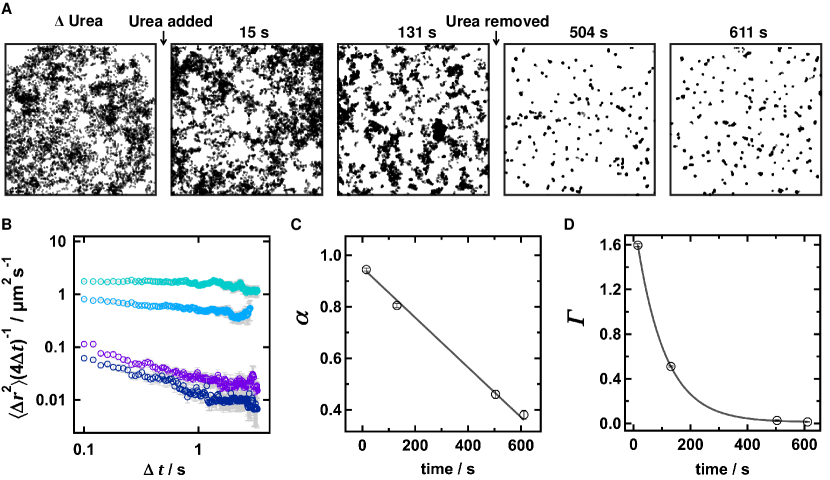
Diffusion of TR-DHPE in the DCPC SLB was fast (6 m2 s-1) and normal ( = 1.01 0.01) in the absence of urea (Fig. 1A&B). In the presence of 1M urea, the diffusion became slower and more anomalous over time (Fig. 1C). decreased roughly linearly to 0.38 and the transport coefficient () showed an approximately exponential decrease to 0.02 m2 s-α (Fig1D) over a 10 minute period. Although values cannot be directly compared (because they depend on , which is also changing), a linear change in would be expected to cause an overall exponential change in , as we report.
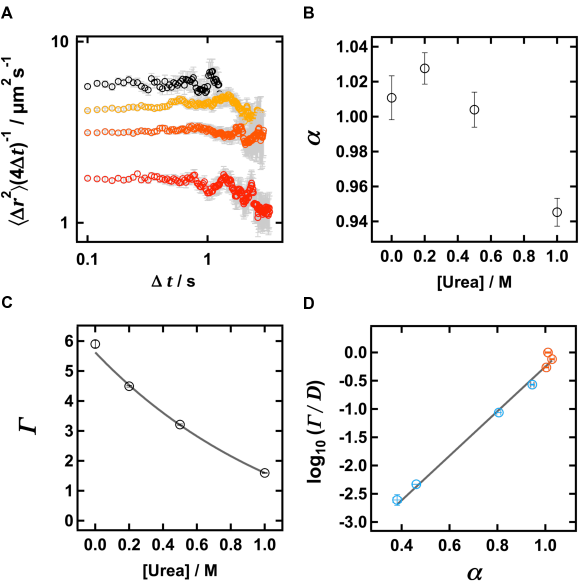
Increasing the urea concentration of the buffer surrounding the SLB incrementally from 0 to 1 M, with a fixed short incubation time (15 s), resulted in increasingly slower diffusion (Fig. 2A). The behaviour is largely normal at this short interval, with only a modest decrease of (to 0.94) at the highest concentration tested (Fig2B). An exponential decrease in with increasing urea concentration was observed (Fig. 2C). From the linear relationship between log10(/) and (Fig. 2D) the characteristic length-scale () associated with the system was calculated to be 45.1 nm, with a jump time () of 86.1 s.
Discussion
We observe that urea causes diffusion in DCPC SLBs to become irreversibly slower and more anomalous in a time and concentration-dependent manner. Given our previous experiments reporting defect-mediated anomalous diffusion using PEG-doping of SLBs [22], it is appealing to suggest that a similar mechanism must operate for urea. For this case, urea would associate with the bilayer, where its chaotropic nature would act to induce the removal of bilayer patches from the glass coverslip surface, producing defects visible as excluded areas of the surface corresponding to those observed in Fig 1A. However, there is little evidence that urea acts directly to solubilise or otherwise permeabilise lipid bilayers [34], and this hypothesis would rely on urea acting at the glass-lipid interface.
An alternative explanation for our results would be the action of urea to alter lipid phase behaviour, inducing phase co-existance phases[33]. Unfortunately, the evidence supports a mode of action whereby urea stabilizes the liquid disordered phase [33, 34], suppressing phase separation, rather than encourage it. In our experiments, we observe a decrease in the area fraction of mobile lipids, which is the opposite trend.
A final hypothesis would be the action of urea not on the bilayer, but on the PEG-DHPE. A chaotropic effect on the PEG might act to increase the area fraction occupied by the PEG, which would then again drive the formation of defects in the membrane [22].
The effect that urea has on diffusion appears not only irreversible, but appears to progress even once urea is removed from the bulk solution. The half-life for this process at 1M urea was short (69 s) and was finished after approximately 500 s. We speculate that either our (1000-fold dilution) washing procedure must be ineffective, or there is a more long-lived, direct, interaction between urea and the bilayer. Given the low partition coefficient for urea in lipid bilayers [43] and the evidence from studies of multilamellar phases that it remains primarily in the aqueous layers between bilayers [34], it is difficult to rationalize this as a possible mechanism.
Further work is needed to distinguish between these different possible mechanisms either by viewing the defects directly (e.g. by atomic force microscopy) or by restoring the defects by addition of fresh SUVs.
Conclusion
We have presented preliminary findings demonstrating a novel approach to controlling anomalous subdiffusion in SLBs on a scale relevant to biological systems [13, 16] by incorporating urea into the aqueous medium surrounding a supported lipid bilayer. Although this work involved the use of DCPC, it would be interesting to extend the method to other, more biologically-relevant lipid compositions. As a complementary method to the inclusion of PEG-lipids, we see potential for this approach for producing a simple membrane model with defined anomaleity,
Authors’ Contributions
EEW performed the experiments, HLEC and MRC performed the analysis, MIW secured the funding; all authors wrote and reviewed the manuscript.
Competing Interests
The authors declare no competing interests.
Funding
We thank the European Research Council for providing funding for this work (ERC-2012-StG-106913, CoSMiC).
References
References
- [1] Erin D. Sheets, Greta M. Lee, Rudolf Simson and Ken Jacobson “Transient confinement of a glycosylphosphatidylinositol-anchored protein in the plasma membrane” In Biochemistry 36.41, 1997, pp. 12449–12458 DOI: 10.1021/bi9710939
- [2] Daniel Choquet and Antoine Triller “The role of receptor diffusion in the organization of the postsynaptic membrane” In Nature Reviews Neuroscience 4.4, 2003, pp. 251–265 DOI: 10.1038/nrn1077
- [3] Boris N. Kholodenko “Cell-signalling dynamics in time and space” In Nature Reviews Molecular Cell Biology 7.3, 2006, pp. 165–176 DOI: 10.1038/nrm1838
- [4] Umber Cheema et al. “Oxygen diffusion through collagen scaffolds at defined densities: implications for cell survival in tissuemodels” In Journal of tissue engineering and regenerative medicine 4.7, 2011, pp. 524–531 DOI: 10.1002/term
- [5] M.. Saxton “Anomalous diffusion due to obstacles: a Monte Carlo study” In Biophysical Journal 66.2, 1994, pp. 394–401 DOI: 10.1016/S0006-3495(94)80789-1
- [6] Benjamin M. Regner et al. “Anomalous diffusion of single particles in cytoplasm” In Biophysical Journal 104.8, 2013, pp. 1652–1660 DOI: 10.1016/j.bpj.2013.01.049
- [7] Felix Höfling and Thomas Franosch “Anomalous transport in the crowded world of biological cells” In Reports on Progress in Physics 76.4, 2013 arXiv:1301.6990
- [8] Takahiro Fujiwara et al. “Phospholipids undergo hop diffusion in compartmentalized cell membrane” In Journal of Cell Biology 157.6, 2002, pp. 1071–1081 DOI: 10.1083/jcb.200202050
- [9] Yonatan Golan and Eilon Sherman “Resolving mixed mechanisms of protein subdiffusion at the T cell plasma membrane” In Nature Communications 8.May Nature Publishing Group, 2017, pp. 1–15 DOI: 10.1038/ncomms15851
- [10] Akihiro Kusumi et al. “Paradigm Shift of the Plasma Membrane Concept from the Two-Dimensional Continuum Fluid to the Partitioned Fluid: High-Speed Single-Molecule Tracking of Membrane Molecules” In Annual Review of Biophysics and Biomolecular Structure 34.1, 2005, pp. 351–378 DOI: 10.1146/annurev.biophys.34.040204.144637
- [11] M.. Saxton “Lateral diffusion in an archipelago. The effect of mobile obstacles” In Biophysical Journal 52.6 Elsevier, 1987, pp. 989–997 DOI: 10.1016/S0006-3495(87)83291-5
- [12] Hugues Berry and Hugues Chaté “Anomalous diffusion due to hindering by mobile obstacles undergoing Brownian motion or Orstein-Ulhenbeck processes” In Physical Review E - Statistical, Nonlinear, and Soft Matter Physics 89.2, 2014, pp. 1–9 DOI: 10.1103/PhysRevE.89.022708
- [13] Kotono Murase et al. “Ultrafine membrane compartments for molecular diffusion as revealed by single molecule techniques” In Biophysical Journal 86.6, 2004, pp. 4075–4093 DOI: 10.1529/biophysj.103.035717
- [14] Michael J. Saxton “Wanted: A positive control for anomalous subdiffusion” In Biophysical Journal 103.12 Biophysical Society, 2012, pp. 2411–2422 DOI: 10.1016/j.bpj.2012.10.038
- [15] S.. Singer and Garth L. Nicholson “The fluid mosaic model of the structure of cell membranes” In Science 175, 1972, pp. 720–731
- [16] G J Schütz, H Schindler and T Schmidt “Single-molecule microscopy on model membranes reveals anomalous diffusion.” In Biophysical journal 73.2, 1997, pp. 1073–1080 DOI: 10.1016/S0006-3495(97)78139-6
- [17] Timothy V. Ratto and Marjorie L. Longo “Anomalous subdiffusion in heterogeneous lipid bilayers” In Langmuir 19.5, 2003, pp. 1788–1793 DOI: 10.1021/la0261803
- [18] Margaret R. Horton, Felix Höfling, Joachim O. Rädler and Thomas Franosch “Development of anomalous diffusion among crowding proteins” In Soft Matter 6.12, 2010, pp. 2648 DOI: 10.1039/b924149c
- [19] Katelyn M. Spillane et al. “High-speed single-particle tracking of gm1 in model membranes reveals anomalous diffusion due to interleaflet coupling and molecular pinning” In Nano Letters 14.9, 2014, pp. 5390–5397 DOI: 10.1021/nl502536u
- [20] Hsiao Mei Wu, Ying Hsiu Lin, Tzu Chi Yen and Chia Lung Hsieh “Nanoscopic substructures of raft-mimetic liquid-ordered membrane domains revealed by high-speed single-particle tracking” In Scientific Reports 6.January Nature Publishing Group, 2016, pp. 1–10 DOI: 10.1038/srep20542
- [21] Markus Rose, Nehad Hirmiz, Jose M. Moran-Mirabal and Cécile Fradin “Lipid diffusion in supported lipid bilayers: A comparison between line-scanning fluorescence correlation spectroscopy and single-particle tracking” In Membranes 5.4, 2015, pp. 702–721 DOI: 10.3390/membranes5040702
- [22] H L E Coker et al. “Controlling Anomalous Diffusion in Lipid Membranes” In arXiv:1709.04698 [physics.bio-ph] arXiv: arxiv:1709.04698%20[physics.bio-ph]
- [23] M.. Saxton “Lateral diffusion in an archipelago. Distance dependence of the diffusion coefficient” In Biophysical Journal 56.3 Elsevier, 1989, pp. 615–622 DOI: 10.1016/S0006-3495(89)82708-0
- [24] M.J. Saxton “Anomalous diffusion in fluorescence photobleaching recovery : a Monte Carlo study” In Biophys.J. 81.October, 2001, pp. 2226–2240
- [25] Sławomir Stachura and Gerald R. Kneller “Anomalous lateral diffusion in lipid bilayers observed by molecular dynamics simulations with atomistic and coarse-grained force fields” In Molecular Simulation 40.1-3, 2014, pp. 245–250 DOI: 10.1080/08927022.2013.840902
- [26] Yousof Mardoukhi, Jae-Hyung Jeon and Ralf Metzler “Geometry controlled anomalous diffusion in random fractal geometries: looking beyond the infinite cluster” In Physical Chemistry Chemical Physics 17.44 Royal Society of Chemistry, 2015, pp. 30134–30147 DOI: 10.1039/C5CP03548A
- [27] Heidi Koldsø et al. “Membrane Compartmentalization Reduces the Mobility of Lipids and Proteins within a Model Plasma Membrane” In J. Phys. Chem. B 120.34, 2016, pp. 8873–8881 DOI: 10.1021/acs.jpcb.6b05846
- [28] Jae Hyung Jeon et al. “Protein crowding in lipid bilayers gives rise to non-Gaussian anomalous lateral diffusion of phospholipids and proteins” In Physical Review X 6.2, 2016, pp. 1–17 DOI: 10.1103/PhysRevX.6.021006
- [29] Evangelos Bakalis, Siegfried Höfinger, Alessandro Venturini and Francesco Zerbetto “Crossover of two power laws in the anomalous diffusion of a two lipid membrane” In Journal of Chemical Physics 142.215102, 2015
- [30] Matti Javanainen et al. “Anomalous and normal diffusion of proteins and lipids in crowded lipid membranes” In Faraday Discuss. 161, 2013, pp. 397–417 DOI: 10.1039/C2FD20085F
- [31] Eldad Kepten et al. “Guidelines for the fitting of anomalous diffusion mean square displacement graphs from single particle tracking experiments” In PLoS ONE 10.2, 2015, pp. 1–10 DOI: 10.1371/journal.pone.0117722
- [32] Ralf Metzler, Jae-Hyung Jeon, Andrey G. Cherstvy and Eli Barkai “Anomalous diffusion models and their properties: non-stationarity, non-ergodicity, and ageing at the centenary of single particle tracking” In Phys. Chem. Chem. Phys. 16.44 Royal Society of Chemistry, 2014, pp. 24128–24164 DOI: 10.1039/C4CP03465A
- [33] A. Nowacka et al. “Small polar molecules like glycerol and urea can preserve the fluidity of lipid bilayers under dry conditions” In Soft Matter 8.5, 2012, pp. 1482–1491 DOI: 10.1039/C1SM06273E
- [34] Fátima O. Costa-Balogh, Håkan Wennerström, Lars Wadsö and Emma Sparr “How small polar molecules protect membrane systems against osmotic stress: The urea-water-phospholipid system” In Journal of Physical Chemistry B 110.47, 2006, pp. 23845–23852 DOI: 10.1021/jp0632440
- [35] Z W Yu et al. “Study of the effect of urea on the phase transition of phospholipid by synchrotron X-ray diffraction” In High Energy Physics and Nuclear Physics-Chinese Edition 25, 2001, pp. 75–80
- [36] Philip L. Yeagle and Arindam Sen “Hydration and the Lamellar to Hexagonal II Phase Transition of Phosphatidylethanolamine” In Biochemistry 25.23, 1986, pp. 7518–7522 DOI: 10.1021/bi00371a039
- [37] Homer W Smith “The Composition of the Body Fluids of Elasmobranchs” In J. Biol. Chem. 81, 1929, pp. 407–419
- [38] A.. Rawlings and C.. Harding “Moisturization and skin barrier function” In Dermatologic Therapy 17, 2004, pp. 43–48
- [39] A A Brian and H M McConnell “Allogeneic stimulation of cytotoxic T cells by supported planar membranes.” In Proceedings of the National Academy of Sciences of the United States of America 81.19, 1984, pp. 6159–63 DOI: 10.1073/pnas.81.19.6159
- [40] Fernando Albertorio et al. “Fluid and air-stable lipopolymer membranes for biosensor applications” In Langmuir 21.16, 2005, pp. 7476–7482 DOI: 10.1021/la050871s
- [41] Jean Yves Tinevez et al. “TrackMate: An open and extensible platform for single-particle tracking” In Methods 115 The Author(s), 2017, pp. 80–90 DOI: 10.1016/j.ymeth.2016.09.016
- [42] Caroline A. Schneider, Wayne S. Rasband and Kevin W. Eliceiri “NIH Image to ImageJ: 25 years of image analysis” In Nature Methods 9.7 Nature Publishing Group, 2012, pp. 671–675 DOI: 10.1038/nmeth.2089
- [43] J.M. Diamond and Y. Katz “Interpretation of nonelectrolyte partition coefficients between dimyristoyl lecithin and water” In The Journal of Membrane Biology 17.1, 1974, pp. 121–154 DOI: 10.1007/BF01870176