Hyperfine level structure in nitrogen-vacancy centers near the ground-state level anticrossing
Abstract
Energy levels of nitrogen-vacancy centers in diamond were investigated using optically detected magnetic-resonance spectroscopy near the electronic ground-state level anticrossing (GSLAC) at an axial magnetic field around 102.4 mT in diamond samples with a nitrogen concentration of 1 ppm and 200 ppm. By applying radiowaves in the frequency ranges from 0 to 40 MHz and from 5.6 to 5.9 GHz, we observed transitions that involve energy levels mixed by the hyperfine interaction. We developed a theoretical model that describes the level mixing, transition energies, and transition strengths between the ground-state sublevels, including the coupling to the nuclear spin of the NV center's 14N and 13C atoms. The calculations were combined with the experimental results by fitting the ODMR spectral lines based on a theoretical model, which yielded information about the polarization of nuclear spins. This study is important for the optimization of experimental conditions in GSLAC-based applications, e.g., microwave-free magnetometry and microwave-free nuclear-magnetic-resonance probes.
pacs:
76.30.Mi,76.70.Hb,75.10.DgI Introduction
Nitrogen-vacancy (NV) color centers in diamond crystals currently are used in a broad range of applications. They serve as qubits Popkin (2016) or quantum-memory elements Heshami et al. (2016) for quantum computers, or probes for various physical quantities like magnetic field Zheng et al. (2017); Wickenbrock et al. (2016), electric field Dolde et al. (2011); Michl et al. (2019), strain Tamarat et al. (2008); Manson et al. (2006), rotation Ledbetter et al. (2012); Wood et al. (2018); Ajoy and Cappellaro (2012) or temperature Clevenson et al. (2015). They can also be used to detect the properties of electronic and nuclear spins on the surface or in the interior of a diamond crystal Mamin et al. (2013); Staudacher et al. (2013); Loretz et al. (2014); Müller et al. (2014), such as substitutional nitrogen (P1) centers Wood et al. (2016); Wickenbrock et al. (2016); Hall et al. (2016), atoms Wood et al. (2016), and cross-relaxation with other point defects in the diamond lattice Wang et al. (2014). The presence and properties of other spin centers can be ascertained by measuring longitudinal () or transverse () relaxation times of the polarization of the NV centers’ ground-state electron spins.
For these applications it is crucially important to know in detail the energy level structure of the NV center, including its hyperfine structure, which arises from the interaction of the electron spin with the nuclear spin of the 14N atom which is a part of the NV center. One electronic magnetic sublevel split by the hyperfine interaction interacts with another electronic magnetic sublevel split by the hyperfine interaction. Near the magnetic field values at which magnetic sublevels cross or have avoided crossings (e.g., GSLAC), this interaction leads to strong hyperfine level mixing and alters the transition probabilities that involve these mixed levels.
The interaction of NV centers with nearby atoms and their nuclear spin polarization has been studied using electron spin resonance at low magnetic fields and near the excited state level anti-crossing at 512 G Dreau et al. (2012). It has been shown by studying optically detected magnetic resonance (ODMR) Negyedi et al. (2017) signals that nuclei adjacent to the vacancy can be polarized using only optical methods at specific values of the magnetic field near the GSLAC. The hyperfine manifold and level anticrossings of the NV center with the nuclear spin of and has been studied in the presence of a magnetic field of several tens of gauss transverse to the NV axis Clevenson et al. (2016). All-optical methods have been used to study the hyperfine structure induced by the interaction of NV centres with their nitrogen atoms for the case of and Broadway et al. (2016), whereas the dynamic nuclear polarization of as a function of magnetic field was modelled up to the GSLAC Ivady et al. (2015).
In this study, we used the ODMR method to investigate the ground state electron spin transitions and to study the hyperfine level structure of NV-center ensembles in the vicinity of the GSLAC. We calculated the level structure of these electron-spin states and the microwave-field-induced transition strengths between these levels. Then we used a parameter-optimization procedure to fit the experimentally measured curves with the results of the theoretical calculation. This fitting procedure yielded information about the degree of nuclear polarization of the spin in the vicinity of the GSLAC. The theoretical model gave the relative intensities of the transitions and, by adding coupling to , made it possible to describe additional transitions in the measured signals. The model applied a Monte Carlo approach to include the interaction with nuclei (which make up 1.1% of the carbon nuclei) for those lattice positions out to a distance of 5 Å that have significant coupling.

II Hyperfine level anticrossing in NV centers in diamond
The NV center is composed of a substitutional nitrogen atom and an adjacent vacancy. It exists in different charged states NV0 and NV-. In this work we focus on the energy levels of the NV- and refrain from writing out the charge state. The NV center has an electron spin in the ground state. There are both triplet and singlet excited states as shown in Fig. 1(a).
In the absence of an external magnetic field the NV center has a splitting between the ground-state magnetic sublevels of the electron spin and due to the spin-spin interaction Doherty et al. (2011). This zero-field splitting corresponds to 2.87 GHz in the 3A2 ground state and about 1.41 GHz in the 3E excited state. If an external magnetic field is applied along the NV axis, the magnetic sublevels of the electron spin acquire additional energy equal to
| (1) |
where GHz/T is the electron gyromagnetic ratio, is the magnetic quantum number of the electron spin, and is the magnetic field strength.
In addition to the electronic states, interactions with nearby nuclear spins must be considered. In all cases, the nucleus of the nitrogen atom associated with the NV center interacts with the NV electron spin. The vast majority (99.6%) of these nitrogen nuclei are whose nuclear spin is . Furthermore, although nuclei have zero nuclear spin, some (%) of the nearby carbon nuclei are with nuclear spin . There are interactions between the NV center and nearby P1 centers but we will not consider this interaction here.
In this approximation the Hamiltonian for the NV center in its ground state can be written as Wood et al. (2016):
| (2) |
where describes the ground state of the NV center with electron spin , gyromagnetic ratio , and zero-field splitting GHz; describes the nucleus with spin , electric quadrupole interaction parameter MHz, and gyromagnetic ratio MHz/T; describes nuclei with nuclear spin and gyromagnetic ratio MHz/T in the external magnetic field; describes the hyperfine interaction of the NV center with the 14N nucleus via the diagonal hyperfine interaction tensor ; and describes the interaction of the nucleus and the NV center. The last term requires special consideration, since the strength of the interaction depends on the distance between the NV center and the nucleus or nuclei and their relative orientation. In this section we will describe the calculation of the energy levels and interaction strengths without the or terms. In Sec. IV.4 we will discuss the effect of adding in the interactions with 13C.
The matrix is a diagonal hyperfine-interaction tensor between the electron spin of the NV center and nuclear spin of the 14N nucleus that belongs to the NV center,
| (3) |
where the hyperfine-interaction parameters are MHz, MHz. The values of the constants are taken from Wood et al. (2016) and references therein.
A crossing between and occurs when the Zeeman splitting compensates the zero-field splitting at a magnetic field value of 102.4 mT [see Fig. 1(b)]. Owing to the hyperfine interaction each of the electron-spin substates are split into hyperfine components. Some of the hyperfine components exhibit avoided crossings [see Fig. 1(c)].
The contribution of the sublevel and its hyperfine components can be plausibly ignored when calculating the eigenvalues and eigenvectors of the and hyperfine components near the GSLAC since the sublevel is separated from the other two by an energy corresponding to 5740 MHz. After calculating the eigenvalues and eigenvectors of the and submanifold, we added the states , , and [see (5)], which belong the the manifold, unmixed in this approximation, to have a full set of levels. Thus, we used a “truncated” Hamiltonian that includes only the electron spin sublevels with and and the nuclear spin sublevels with , to obtain approximate analytical solutions for the energy levels and wave functions of the hyperfine states. The energies of these components are
| (4a) | ||||
| (4b) | ||||
| (4c) | ||||
| (4d) | ||||
| (4e) | ||||
| (4f) | ||||
| (4g) | ||||
| (4h) | ||||
| (4i) | ||||
The wave functions can be written in the uncoupled basis as follows:
| (5a) | ||||
| (5b) | ||||
| (5c) | ||||
| (5d) | ||||
| (5e) | ||||
| (5f) | ||||
| (5g) | ||||
| (5h) | ||||
| (5i) | ||||
where
| (6a) | ||||
| (6b) | ||||
and
| (7) |
In the magnetic field some hyperfine levels become mixed. Although the magnetic quantum numbers and cease to be good quantum numbers as a result of this mixing, their sum still is preserved [see Eq. (5) (c)–(f)]. Only states with equal interact [see the interactions denoted in Fig. 1 (c)]. In particular, the states and remain unmixed even in the strong magnetic field that corresponds to the GSLAC. The other four states form two pairs of mixed states. One pair consists of states and , the other of states and . This information about the state mixing will be important when we analyze which transitions are allowed and which are forbidden when the magnetic field value is close to mT.
Magnetic-dipole transitions between various states can be driven with an applied radio-frequency magnetic field. The selection rules for these transitions are and MacQuarrie et al. (2013); Sannigrahi (1982); Lee et al. (2017).
Having calculated the energy levels and wave functions in (4) and (5), respectively, we now want to describe transitions between different states. We will consider only transitions that change the electron spin state in (2). In order to describe electron spin transitions we must add to the Hamiltonian a term to describe the interaction with the microwave (MW) field. This term can be constructed starting from the raising and lowering operators:
| (8) |
where are the spin operators for . However, we need a matrix to describe the raising and lowering of the electron spin in a system that contains also a nuclear spin . This operator can be obtained by taking the outer product with the three-dimensional identity matrix and folding in the matrix of wave functions whose columns are the ground-state eigenvectors . Then the interaction term can be written as
| (9) |
where is the Rabi frequency of the microwave radiation at the given transition frequency. We thus obtain a block-diagonal matrix of magnetic dipole transition elements . From these transition matrix elements the transition probabilities between levels and can be obtained:
| (10) |
To obtain actual transition intensities we multiply the transition probabilities by a term that takes into account the actual populations of each level, which depends on the polarization of . We describe this polarization using the concept of “spin temperature” Budker et al. (2004), which is defined as
| (11) |
Now the observed transition strengths will be the product of the transition probability and a term that depends on the populations of the levels involved in the transition. We define a matrix whose dimensions match the matrix of transition probabilities in (10), and whose elements de Lima Bernardo (2017), where is the nuclear spin of the initial state in the transition and is the nuclear spin of the final state. Now we multiply the terms in (10) and to obtain the transition intensities:
| (12) |
We can then construct the calculated ODMR spectrum in which the integral under the ODMR peak that corresponds to the energy difference between levels and is proportional to transition strength .
III Experimental set-up
We measured ODMR signals from ensembles of NV centers in two different samples. One sample was produced by chemical vapor deposition with a nitrogen concentration around 1 ppm (low-density sample). The other sample was a dense high-pressure, high-temperature (HPHT) crystal with a relatively high concentration of nitrogen of around 200 ppm (high-density sample). The measurements with the low-density sample were performed at the Johannes Gutenberg-University in Mainz, whereas the measurements with the high-density sample were performed at the Laser Centre of the University of Latvia in Riga. The NV centers were irradiated with green 532 nm light from a Nd:YAG laser (Coherent Verdi) and optically polarized to the state while the luminescence from the state was monitored [see the transition diagram in Fig. 1(a)]. Following the ODMR method, a microwave field was applied to induce transitions between the ground-state sublevels. The NV centers’ electrons were continuously pumped to the state. When a MW field is on resonance with a transition from an hyperfine component to an hyperfine component, the fluorescence intensity decreases.

Figure 2(a) shows the experimental setup used for the high-density sample in Riga. The magnetic field was produced by a custom-built magnet initially designed for electron paramagnetic resonance (EPR) experiments. It consists of two 19 cm diameter iron poles with a length of 13 cm each, separated by a 5.5 cm air gap. This magnet could provide a highly homogeneous field (0.0002 mT over the sensing volume, estimated by a simulation software COMSOL modeling). The diamond sample under investigation was held in place using a nonmagnetic holder (custom-made by STANDA), which provided three axes of rotation to align the NV axis with the applied magnetic field. Light with a wavelength of 532 nm (Coherent Verdi Nd:YAG) was delivered to the sample via an optical fiber with a core diameter of 400 micrometers (numerical aperture of 0:39). The same fiber was used to collect red fluorescence light, which was separated from the residual green reflections by a dichroic mirror and a long-pass filter (Thorlabs DMLP567R and FEL0600) and focused onto an amplified photodiode (Thorlabs PDA36A-EC). The signals were recorded and averaged on a digital oscilloscope (Agilent DSO5014A or Yokogawa DL6154) or a DAQ card (Measurement Computing USB-1408FS).
Figure 2(b) shows the experimental setup used for the low-density sample in Mainz. A custom-made electromagnet was used with 200 turns wound on a water-cooled copper mount. The electromagnet produced a field of 2.9 mT/A and could achieve magnetic fields up to 103.5 mT. The diamond could be rotated around the z-axis (NV axis). Moreover, the electromagnet could be moved with a computer-controlled 3D translation stage (Thorlabs PT3-Z8) and a rotation stage (Thorlabs NR360S, y-axis). In this way, all degrees of freedom for centering the diamond in the magnet and aligning the NV axis to the magnetic field were available. An accousto-optic modulator (AOM) in combination with a photodiode and a proportional-integral-derivative (PID) controller served to stabilize the laser intensity.
In both setups, the microwave field was generated and amplified using two sets of devices depending on the required frequency range. In Riga, for low frequencies a TTi TG5011 generator (0.001 mHz to 50 MHz) and for high frequencies a function generator (SRS SG386) with a power amplifier (Minicircuits ZVE-3W-83+) provided up to +30dBm. In Mainz, an SRS SG386 was used as a function generator over the entire range, in conjunction with power amplifiers. At high frequencies, an RFLU PA0706GDRF amplifier (Lambda) was used. It was replaced at low frequencies with a (Minicircuits ZHL-32A+) amplifier.
IV Experimental results and analysis
IV.1 ODMR signals for the transition
Figure 3 depicts ODMR signals for transitions in the frequency range 5.6–5.9 GHz, where the MW field is resonant with transitions from the mixed and hyperfine levels, to the hyperfine levels [see Fig. 1 (b)]. Experimentally measured signals are depicted together with curves obtained from a model calculation with some parameters obtained from a fitting procedure as explained below. Figures 3 (a)–(c) depict magnetic sublevels at a given magnetic field and indicate the allowed microwave transitions as arrows. The wave functions – are given in Eq. (5). The middle row [Fig. 3 (d)–(f)] shows the experimental signals for the low-density sample, and the bottom row [Fig. 3 (g)–(i)] shows the signals for the high-density sample.

We used a parameter-optimization procedure based on a test to determine the contribution of each transition in Fig. 3 (a)–(c) to the overall lineshapes in Fig. 3 (d)–(i). The reduced value is defined as , where are the number of points, the are the measured data points, the are the results of the model, and the are the mean square errors on the data points, which we set to unity here. To illustrate the procedure, let us consider Fig. 3 (d). This signal was recorded at a magnetic field value far away from the GSLAC. As a result, the mixing of the sublevels is insignificant, and a contribution to the signal is expected only from the following three transitions: (blue), (purple), and (green). In these transitions the nuclear spin projection does not change. We assumed that each transition has a Lorentzian lineshape centered at its respective transition frequency, which follows from the differences in level energies in Eq. (4). The transition strengths for these three transitions are equal, which is indicated by the fact that all three arrows have the same width. Nevertheless, the relative contributions (peak amplitudes) of each transition may differ because of differences in the populations of the three ground states involved: , , and , corresponding to the nuclear spin polarization of .
Next we attempted to find the spin temperature and magnetic field value that minimized the reduced value for the hypothesis that our theoretical model with these parameters described the measured data. Far away ( mT) from the GSLAC the magnetic field value in the fit was allowed to vary over a small range since the position of the ODMR peak depends not only on , but also on the nuclear spin polarization, which affects the contributions of transitions from the different nuclear spin components and thus can shift the ODMR peak position. Aside from the immediate vicinity of the GSLAC, the plot of ODMR peak position versus magnet coil current showed that the polarization does not change very much with magnetic field over the range of 101.0 mT to 103.5 mT. Indeed, the fitted peak positions at each point produced a straight line as a function of current in the magnet coil that could be extrapolated through the region near the GSLAC point. In this way, a calibration curve for as a function of the magnet current was obtained, which was used to obtain the magnetic field value in the region near the GSLAC, where only the spin temperature was allowed to vary.
For each possible set of parameters and , we calculated the corresponding populations of the eigenstates in (5). We obtained the transition strengths from the calculated eigenvalues of these states. The amplitude of each transition peak is calculated according to (12), the states are assumed to be “empty”. Then we used the SciPy optimize function Jones et al. (2001) to determine the widths of the Lorentzians corresponding to each of the peaks, each one of which corresponds to a component of the hyperfine transition. This step was important because the width of the Lorentzians for our sample is around 1 MHz, and the peaks of nearby hyperfine components partially overlap. We assumed that all hyperfine components at a particular field strength had the same width. At the GSLAC the width of the Lorentzians increases due to increased relaxation rates arising from increased interaction between the hyperfine levels Wood et al. (2016).
We calculated the reduced value using this set of parameters. We repeated the procedure for the next set of parameters and stored those that yielded the smallest reduced value. The peak amplitudes thus obtained are shown as the length of the colored bars in Fig. 3 (d). The color of each bar corresponds to the color of the arrow that represents the corresponding transition in Fig. 3 (a). We proceeded in a similar fashion for all subfigures. Near the GSLAC, there are more possible transitions that must be considered as a result of hyperfine level mixing [which follows from Eqn. (5)–(7)]. The number of possible transitions and their relative transition strengths are indicated by the number of arrows in Fig. 3 (b) and their widths.

IV.2 ODMR signals for the transition
We also measured ODMR signals for the transition within mT of the GSLAC, which corresponds to microwave frequencies below 40 MHz, and some results are shown in Fig. 4. Experimentally measured signals are plotted together with signals from a model calculation using parameters that were obtained in a similar way as in Sec. IV.1. The top row (a)–(c) shows the magnetic-sublevel structure in a particular magnetic field with the allowed transitions depicted by arrows whose width indicates the relative transition strength. The middle row (d)–(f) shows the measured signals for the low-density sample, and the bottom row (g)–(i) shows the corresponding signals for the high-density sample. Again, above and below the GSLAC, the signals consist of three components, which correspond to the allowed transitions between hyperfine levels that are weakly mixed in the magnetic field, but at the GSLAC, there is strong mixing and more transitions must be taken into account, as indicated by the number of arrows in Fig. 4 (b). Above and below the GSLAC, the agreement between measured and calculated curves is quite good. However, right near the GSLAC there are some discrepancies in the amplitudes of the peaks. These discrepancies are particularly significant in the high-density sample, for which the model essentially fails at the GSLAC. Possible reasons for the discrepancies might be inhomogeneities in the microwave power, in the diamond crystal lattice or in the magnetic field, or interactions with other nearby spins, such as P1 centers or 13C nuclei. Interactions with nearby spins and unknown defects might be one more reason for the failure of the model at the GSLAC for the high-density sample. We consider the effect of 13C nuclei in Sec. IV.4.

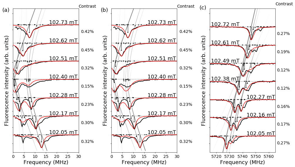
Figures 5(a) and (b) show in more detail ODMR signals measured near the GSLAC for the transition, and Fig. 5(c) shows signals measured for the transition in the low-density sample. The black curve shows the experimentally measured signals, while the red curve represents the result of the theoretical model calculation with parameters obtained by the same fitting procedure as described in connection with Figs. 3 and 4. The percentages to the right of each frame show the actual ODMR contrast measured at that magnetic field value as given in Fig. 7. The curves plotted here are normalized, although the curve with a contrast of 0.45% has in reality twice the amplitude as the curve with a contrast of 0.23%. Again, the nuclear spin populations and peak widths within mT of the GSLAC were taken from the values obtained for the high-frequency case at the same magnetic field value. Everywhere else the parameter optimization procedure was used as described in Sec. IV.1. The experimental data in Fig. 5(a) and Fig. 5(b) are identical, but for the calculated curves, the NV axis and the magnetic field vector were assumed to be parallel in the former case, whereas in the latter, an angle degrees between the NV axis and the magnetic field direction was assumed. This angle was found to give the best overall agreement between the experimentally measured values and the curves obtained from the model calculations with parameter fitting. In fact, it is difficult to align the magnetic field perfectly with the NV axis in the experiment. The signals in Fig. 3 are relatively insensitive to small misalignment angles, which is why they were not plotted.

Similar curves are presented for the high-density sample in Figure 6. In this case, the angle used in Fig. 6(b) was , which corresponds to a transverse magnetic field of 0.18 mT.
In all these experimentally measured signals, the overall peak intensities were normalized separately for each magnetic field value, because at the GSLAC, the contrast of the signals decreased strongly, as shown in Fig. 5. The decrease in contrast near the GSLAC is caused by energy level mixing, which redistributes the population between the and the levels. As a consequence, the time is drastically reduced, and so there are fewer NV centers in the ground state available for MW transitions. The vertical bars depict calculated ODMR peak positions and relative intensities. It can be seen that below and above the GSLAC the measured and calculated signals agree rather well. However, right at the GSLAC the agreement is not as good, and in particular, the model fails for the high-density sample at low frequencies in a range of mT of the GSLAC. The high-density sample may present additional defects that are not accounted for in the model or additional interactions among defects.

IV.3 Nuclear spin polarization
The peak amplitudes from the fit in Fig. 4(d)–(f), Fig. 5, and Fig. 3 (d)–(f) contain information about the relative populations of the ground-state hyperfine levels and thus the nuclear spin polarization He et al. (1993); Fuchs et al. (2008); Jacques et al. (2009); Smeltzer et al. (2009); Gali (2009); Smeltzer et al. (2011); Dreau et al. (2012); Fischer et al. (2013); Ivady et al. (2015). Nuclear spin polarization arises from an interplay of optical pumping and sublevel mixing. For example, Fig. 1 (c) shows how the sublevel is mixed with the sublevel near the GSLAC, where they are nearly degenerate. Optical pumping in this situation tends to move population from the sublevel to the sublevel via the sublevel. In a similar way, because of the mixing between sublevel and the sublevel, population moves from the sublevel to the sublevel via the sublevel. However, the sublevel is not mixed with any sublevel, and so the population accumulates in this state.
Besides spin temperature, another way to characterize the nuclear spin polarization is based on multipole expansion Auzinsh et al. (2010), in which the rank-zero multipole moment (monopole ) corresponds to population, the rank-one moment (dipole moment ), to orientation, and the second rank moment (quadrupole moment ), to alignment. In absence of processes that create coherences between different spin components in our system, only longitudinal (along the magnetic field direction) and spin polarization components are created.
Based on the component intensities, orientation would be calculated as
| (13) |
where corresponds to the integral under the calculated curve that makes up all transitions from level (5), and we assume that the hyperfine levels are “empty”.
Alignment can be calculated in a similar way as
| (14) |
Figure 8 shows the degree of spin polarization as a function of magnetic field obtained for the low-density sample. The high-density sample is not used, since the model fails near the GSLAC. The populations of the nuclear spin components were obtained from the peak amplitudes obtained by the parameter fitting procedure described in Sec. IV.1, and then the orientation and alignment were calculated using Eqs. (13) and (14), respectively. The data for this procedure are shown in Fig. 3, supplemented by similar measurements at many more magnetic-field values. The population of each nuclear spin component is proportional to the integral under the corresponding calculated curve divided by the transition strength. The region at the center shaded in gray in Fig. 8 indicates the magnetic field values for which an additional peak poorly described by the model [see, for example, Fig. 3(e)] complicates the optimization procedure, and makes it difficult to guarantee that the polarization value obtained from this procedure fully describes the true polarization. The polarization values extracted by our method are plotted in this region as well, but they should be taken with caution. Both figures show nuclear spin polarization near the GSLAC with a minimum at the GSLAC position. In principle, the polarization should vanish far away from the GSLAC where there is no hyperfine level mixing Ivady et al. (2015). However, we did not observe this behavior over the measured range of magnetic field values.

IV.4 Influence of 13C Nuclei
We now consider the interaction of our system with a collection of nearby nuclei labeled by the index . The interaction between the NV center and these nuclei is described by the HamiltonianNizovtsev et al. (2010):
| (15) |
where labels the nuclear spin of the -th nucleus and is the Hamiltonian corresponding to the -th nucleus. The tensor has the same form as in eq. (3). For the case of a nucleus in one of the three lattice positions next to the vacancy, , MHz and MHz Gali (2009). We follow a procedure similar to the one outlined by Nizovtsev et al. Nizovtsev et al. (2010) to rotate this tensor from the principal coordinate axes of the carbon nucleus to the coordinate system of the NV center with the -axis parallel to the [111] crystal direction. The rotation takes the form of , where is the rotation matrix about the -axis by an angle that rotates the -axis in the frame of the carbon nucleus into the frame of the [111] crystal direction.
The principal values of the tensor that corresponds to other lattice positions besides the nearest neighbors were taken from density functional theory (DFT) calculations performed by Gali et al. Gali et al. (2008). The lattice positions can be classified according to families of lattice points that have the same tensor values Smeltzer et al. (2011) (see Fig. 4 and Table 1 in that publication, which also gives the multiplicities of each family.) The value of of the angle between the -axis of the carbon nucleus’s principal axis and the NV axis were taken from the results of another DFT calculation performed by Nizovtsev et al. A. P. Nizovtsev et al. (2014). We took into account the nearest neighbors of the vacancy site as well as families A through H, which corresponds to 39 lattice sites. Other lattice sites have significantly less influence.
Next we use a Monte Carlo method to average scenarios where 13C atoms are located in different lattice sites. For each iteration, we loop through the 39 lattice sites, each of which has a 1.1% probability of hosting a 13C atom. Those that contain a 13C atom are added to the Hamiltonian in (15) with the values of the hyperfine tensor as described in the previous paragraph. The number of energy levels of the system follows from the eigenvalues and eigenvectors of Eq. (2), and it depends on the number of atoms as . The spectrum generated depends on the locations of the 13C atoms, which can differ from iteration to iteration. Thus, several hundred iterations are averaged to take into account that each NV center contributing to our signal has a different arrangement of 13C atoms in its vicinity.
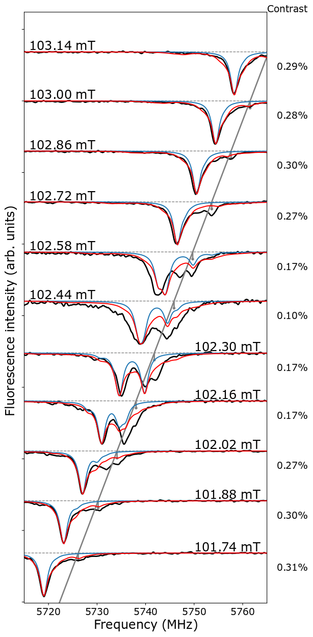
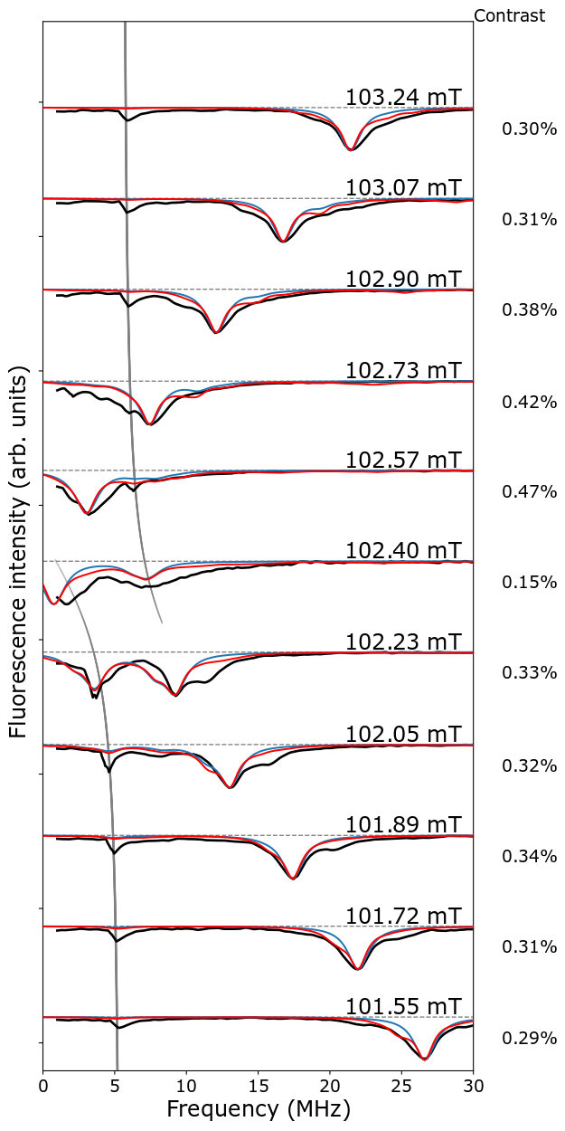
The results of the model calculations with 13C are shown in Fig. 9 for the transition and in Fig. 10 for the transition. In both figures, the blue curves show the calculations without the 13C interaction, whereas the red curves include the interaction with nearby 13C nuclei. One can see that the inclusion of the 13C interaction slightly improves the agreement with the calculations. Only the low-density sample was used for this comparison, since the model was more successful for this sample. Nevertheless, discrepancies remain, which might require the inclusion of more terms in the Hamiltonian, such as nearby P1 centers, which is outside the scope of this study.
The addition of 13C nuclei in the Hamiltonian [see Eq. (2)] allows for additional transitions to appear in the calculated signals. These new transitions are most apparent in describing the shoulder to the right of the main peak in the top three and bottom two panels of Fig. 10, as well as to the left of the main peak in the top two panels. Without the inclusion of nuclei in the model, these shoulders cannot be described. A similar effect can be observed in Fig. 9, but not as dramatically. The influence of nuclei allows otherwise forbidden transitions because the majority of them are off-axis, which effectively changes the angular momentum selection rules Wang et al. (2013). The gray curve in Fig. 9 corresponds to the transition, which generally appears in the experimentally measured signals, but is not so pronounced in either of the theoretical models, although the model that includes hints at it far away from the GSLAC position.
In some cases there remain significant discrepancies between the model and the experiment, even far from the GSLAC position. For example, the gray curve in Fig. 10 tracks the nominal transition. This transition corresponds to a strong peak in the experimentally measured signals; this peak appears also in the calculation, but is so weak that it cannot be distinguished at the magnification shown in the figure.
We conclude that the influence of reveals important aspects of the underlying physics and influences the strengths of the transitions that can take place, but fails to explain other significant aspects of the measured signals. Perhaps more detailed calculations that take into account the spin dynamics could reproduce the ODMR spectra more accurately.
We also note that there is a peak that appears in a number of the figures just to the right of the main peaks that cannot be explained by the model. Interestingly, this peak tracks the transition as is shown by the gray line in Fig. 9. This transition should not occur according to Eq. (10). However, the measurements suggest that some interaction (possibly off-axis as well) causes this transition to occur. Thus, a search for this interaction should also be considered in future theoretical work.


V Conclusions
In this work we have studied microwave-induced transitions between the hyperfine components of the ground-state sublevels of the NV center in diamond using the ODMR technique in two diamond samples with a nitrogen concentration of 1 ppm and 200 ppm. We have developed a straightforward theoretical model to describe these ODMR signals. The model describes the ODMR signals for magnetic field values in the vicinity of the GSLAC as well as away from it. Including the effects of nearby nuclei significantly improves the agreement between the model and the experimentally measured signals. The theoretical model allows to track and transitions enabled by the hyperfine level mixing. Within 0.5 mT of the GSLAC, the experimentally measured ODMR spectrum becomes rather complicated with some features that we have not been able to describe fully, although the general features are reproduced at least in the case of the low-density sample. In the case of the high-densitiy sample, the model fails at low frequency in the range of mT around the GSLAC position. Further investigation will be needed to find the interactions responsible for all of these features. Nevertheless, the model can be used to estimate the nuclear spin polarization of , which we have demonstrated in this study. We have performed ODMR measurements on the transition as well as on the transition. The latter transition is somewhat simpler, since the level is not involved in the hyperfine level mixing and anticrossing and thus serves as a useful cross-check to test the adequacy of the theoretical model. The ODMR technique has proven to be a useful tool for investigating how the hyperfine interaction influences the energy-level structure near the GSLAC and, with further improvements to the theoretical model, could shed more light on additional interactions and the process of nuclear spin polarization near the GSLAC. The results of this work will be used in the ongoing efforts to model and optimize NV-diamond based microwave-free sensors, in particular, magnetometers Zheng et al. (2017); Wickenbrock et al. (2016).
VI Acknowledgements
We thank Daniels Krimans for helpful contributions to the theoretical approach. The Riga group gratefully acknowledges the financial support from the M-ERA.NET project “Metrology at the Nanoscale with Diamonds” (MyND), from the Laserlab-Europe Project (EU-H2020 654148), and from the Base/Performance Funding Project Nr. AAP2016/B013, ZD2010/AZ27. A. Berzins acknowledges support from the PostDoc Latvia Project Nr. 1.1.1.2/VIAA/1/16/024 ”Two-way research of thin-films and NV centres in diamond crystal”.
The Mainz group acknowledges support by the German Federal Ministry of Education and Research (BMBF) within the Quantumtechnologien program (FKZ 13N14439) and the DFG through the DIP program (FO 703/2-1). H. Zheng acknowledges support from the GRK Symmetry Breaking (DFG/GRK 1581) program. This work was also supported by the EU FET OPEN Flagship Project ASTERIQS (action 820394) and the Cluster of Excellence Precision Physics, Fundamental Interactions, and Structure of Matter (PRISMA+ EXC2118/1) funded by the German Research Foundation (DFG) within the German Excellence Strategy (Project ID 39083149).
References
- Popkin (2016) G. Popkin, Science 354, 1090 (2016).
- Heshami et al. (2016) K. Heshami, D. G. England, P. C. Humphreys, P. J. Bustard, V. M. Acosta, J. Nunn, and B. J. Sussman, Journal of Modern Optics 63, 2005 (2016).
- Zheng et al. (2017) H. Zheng, G. Chatzidrosos, A. Wickenbrock, L. Bougas, R. Lazda, A. Berzins, F. H. Gahbauer, M. Auzinsh, R. Ferber, and D. Budker, in Society of Photo-Optical Instrumentation Engineers (SPIE) Conference Series (2017), vol. 10119 of Society of Photo-Optical Instrumentation Engineers (SPIE) Conference Series, p. 101190X, eprint 1701.06838.
- Wickenbrock et al. (2016) A. Wickenbrock, H. Zheng, L. Bougas, N. Leefer, S. Afach, A. Jarmola, V. M. Acosta, and D. Budker, Applied Physics Letters 109, 053505 (2016).
- Dolde et al. (2011) F. Dolde, H. Fedder, M. W. Doherty, T. Nöbauer, F. Rempp, G. Balasubramanian, T. Wolf, F. Reinhard, L. C. L. Hollenberg, F. Jelezko, et al., Nature Physics 7, 459 (2011).
- Michl et al. (2019) J. Michl, J. Steiner, A. Denisenko, A. Buelau, A. Zimmermann, K. Nakamura, H. Sumiya, S. Onoda, P. Neumann, J. Isoya, et al., ArXiv e-prints (2019), eprint 1901.01614v1.
- Tamarat et al. (2008) P. Tamarat, N. B. Manson, J. P. Harrison, R. L. McMurtrie, A. Nizovtsev, C. Santori, R. G. Beausoleil, P. Neumann, T. Gaebel, F. Jelezko, et al., New J. Phys. 10, 045004 (2008).
- Manson et al. (2006) N. B. Manson, J. P. Harrison, and M. J. Sellars, Phys. Rev. B 74, 104303 (2006).
- Ledbetter et al. (2012) M. P. Ledbetter, K. Jensen, R. Fischer, A. Jarmola, and D. Budker, Phys. Rev. A 86, 052116 (2012).
- Wood et al. (2018) A. A. Wood, E. Lilette, Y. Y. Fein, N. Tomek, L. P. McGuinness, L. C. L. Hollenberg, R. E. Scholten, and A. M. Martin, Science Advances 4, no. 5, eaar7691 (2018).
- Ajoy and Cappellaro (2012) A. Ajoy and P. Cappellaro, Phys. Rev. A 86, 062104 (2012).
- Clevenson et al. (2015) H. Clevenson, M. E. Trusheim, C. Teale, T. Schröder, D. Braje, and D. Englund, Nature Physics 11, 393 (2015).
- Mamin et al. (2013) H. J. Mamin, M. Kim, M. H. Sherwood, C. T. Rettner, K. Ohno, D. D. Awschalom, and D. Rugar, Science 339, 558 (2013).
- Staudacher et al. (2013) T. Staudacher, F. Shi, S. Pezzagna, J.Meijer, J. Du, C. A. Meriles, F. Reinhard, and J. Wrachtrup, Science 339, 561 (2013).
- Loretz et al. (2014) M. Loretz, S. Pezzagna, J. Meijer, and C. L. Degen, Appl. Phys. Lett. 104, 033102 (2014).
- Müller et al. (2014) C. Müller, X. Kong, J.-M. Cai, K. Melentijević, A. Stacey, M. Markham, D. Twitchen, J. Isoya, S. Pezzagna, J. Meijer, et al., Nat. Commun. 5, 4703 (2014).
- Wood et al. (2016) J. D. A. Wood, D. A. Broadway, L. T. Hall, A. Stacey, D. A. Simpson, J.-P. Tetienne, and L. C. L. Hollenberg, Phys. Rev. B 94, 155402 (2016).
- Hall et al. (2016) L. Hall, P. Kehayias, D. A. Simpson, A. Jarmola, A. Stacey, D. Budker, and L. C. L. Hollenberg, Nat. Commun. 7, 10211 (2016).
- Wang et al. (2014) H.-J. Wang, C. S. Shin, S. J. Seltzer, C. E. Avalos, A. Pines, and V. S. Bajaj, Nat. Commun. 5, 4135 (2014).
- Dreau et al. (2012) A. Dreau, J.-R. Maze, M. Lesik, J.-F. Roch, and V. Jacques, Phys. Rev. B 85, 134107 (2012).
- Negyedi et al. (2017) M. Negyedi, J. Palotás, B. Gyüre, S. Dzsaber, S. Kollarics, P. Rohringer, T. Pichler, and F. Simon, Rev. Sci. Instrum. 88, 013902 (2017).
- Clevenson et al. (2016) H. Clevenson, E. H. Chen, F. Dolde, C. Teale, D. Englund, and D. Braje, Phys. Rev. A 94, 021401(R) (2016).
- Broadway et al. (2016) D. A. Broadway, J. D. A. Wood, L. T. Hall, A. Stacey, M. Markham, D. A. Simpson, J.-P. Tetienne, and L. C. L. Hollenberg, Phys. Rev. Appl. 6, 064001 (2016).
- Ivady et al. (2015) V. Ivady, K. Szasz, A. L. Falk, P. V. Klimov, D. J. Christle, E. Janzen, I. A. Abrikosov, D. D. Awschalom, and A. Gali, Phys. Rev. B 92, 115206 (2015).
- Doherty et al. (2011) M. W. Doherty, N. B. Manson, P. Delaney, and L. C. L. Hollenberg, New J. Phys. 13, 025019 (2011).
- MacQuarrie et al. (2013) E. R. MacQuarrie, T. A. Gosavi, N. R. Jungwirth, S. A. Bhave, and G. D. Fuchs, Physical Review Letters 111, 227602 (2013).
- Sannigrahi (1982) A. B. Sannigrahi, Journal of Chemical Education 59, 819 (1982).
- Lee et al. (2017) D. Lee, K. W. Lee, J. V. Cady, P. Ovartchaiyapong, and A. C. Bleszynski Jayich, Journal of Optics 19, 033001 (2017).
- Budker et al. (2004) D. Budker, D. F. Kimball, and D. P. DeMille, Atomic Physics, An Exploration Through Problems and Solutions (Oxford University Press, 2004).
- de Lima Bernardo (2017) B. de Lima Bernardo, Physics Letters, Section A: General, Atomic and Solid State Physics (2017), ISSN 03759601, eprint 1606.00956.
- Jones et al. (2001) E. Jones, T. Oliphant, P. Peterson, et al., SciPy: Open source scientific tools for Python, http://www.scipy.org/ (2001).
- He et al. (1993) X.-F. He, N. B. Manson, and P. T. H. Fisk, Phys. Rev. B 47, 8809 (1993).
- Fuchs et al. (2008) G. D. Fuchs, V. V. Dobrovitski, R. Hanson, A. Batra, C. D. Weis, T. Schenkel, and D. D. Awschalom, Phys. Rev. Lett. 101, 117601 (2008).
- Jacques et al. (2009) V. Jacques, P. Neumann, J. Beck, M. Markham, D. Twitchen, J. Meijer, F. Kaiser, G. Balasubramanian, F. Jelezko, and J. Wrachtrup, Phys. Rev. Lett. 102, 057403 (2009).
- Smeltzer et al. (2009) B. Smeltzer, J. McIntyre, and L. Childress, Phys. Rev. A 80, 050302(R) (2009).
- Gali (2009) A. Gali, Phys. Rev. B 80, 241204(R) (2009).
- Smeltzer et al. (2011) B. Smeltzer, L. Childress, and A. Gali, New J. Phys. 13, 025021 (2011).
- Fischer et al. (2013) R. Fischer, A. Jarmola, P. Kehayias, and D. Budker, Phys. Rev. B 87, 125207 (2013).
- Auzinsh et al. (2010) M. Auzinsh, D. Budker, and S. Rochester, Optically Polarized Atoms, Understanding light-atom interactions (Oxford University Press, 2010).
- Nizovtsev et al. (2010) A. P. Nizovtsev, S. Y. Kilin, V. A. Pushkarchuk, A. L. Pushkarchuk, and S. A. Kuten’, Optics and Spectroscopy 108, 230 (2010), URL http://link.springer.com/10.1134/S0030400X10020128.
- Gali et al. (2008) A. Gali, M. Fyta, and E. Kaxiras, Phys. Rev. B 77, 155206 (2008).
- A. P. Nizovtsev et al. (2014) A. L. P. A. P. Nizovtsev, S. Ya. Kilin, V. A. Pushkarchuk, and F. Jelezko, New Journal of Physics 16, 083014 (2014).
- Wang et al. (2013) H.-J. Wang, C. S. Shin, C. E. Avalos, S. J. Seltzer, D. Budker, A. Pines, and V. S. Bajaj, Nat. Commun. 4, 1940 (2013).