Nuclear resonant scattering from 193Ir as a probe of the electronic and magnetic properties of iridates
Abstract
The high brilliance of the modern synchrotron radiation sources facilitates experiments with high energy x-rays. In this Letter we report on Nuclear Resonance Scattering at the 73 keV nuclear level in 193Ir. The transitions between the hyperfine split levels show an exceptionally large E2/M1 multi- polarity mixing ratio combined with an increased sensitivity to certain changes in the hyperfine field direction compared to non-mixing transitions. The method opens a new way for probing local magnetic and electronic properties of correlated materials containing iridium and provides novel insights into their anisotropic magnetism. In particular, unexpected out-of-plane components of magnetic hyperfine fields and non-zero electric field gradients in Sr2IrO4 have been detected and attributed to the presence of strong spin-orbit interaction. Due to the high, 62 natural abundance of the 193Ir isotope, no isotopic enrichment of the samples is required, qualifying the method for a broad range of applications.
There is burgeoning interest in understanding the physical properties of systems which are simultaneously subject to strong spin-orbit coupling (SOC) and electron correlations, as exemplified by recent studies which have revealed a range of novel electronic and magnetic phases displayed by various 4d and 5d transition metal oxides (TMOs) Kim et al. (2014); He et al. (2015); Crawford et al. (1994); Kim et al. (2009); Boseggia et al. (2013); Kim et al. (2008); Moon et al. (2008); Donnerer et al. (2016); Jackeli and Khaliullin (2009); Witczak-Krempa et al. (2014); Mattheiss (1976); Kim et al. (2012, 2016); de la Torre et al. (2015).
At one level, SOC introduces another competing energy scale, producing unexpected electronic states.This is the case for the so-called spin-orbit Mott insulator in iridate perovksites which would otherwise be expected to be metallic in the absence of SOC. At another, more profound level, the SOC fully entangles spin and orbital degrees of freedom such that the magnetic interactions acquire an anisotropic, bond-directional nature – the Kitaev interaction – augmenting the conventional isotropic Heisenberg term which dominates 3d systems Jackeli and Khaliullin (2009). The resulting Kitaev-Heisenberg model is proving to be extremely rich displaying a plethora of topological quantum phases including spin-liquids, superconductivity, etc., the exploration of which is in its infancy Witczak-Krempa et al. (2014); Schaffer et al. (2016). Further impetus for studying 4d and 5d TMOs stems from the rich possibilities offered by nano-structuring these materials, finding potential applications as biosensors, spintronic devices, catalysts, etc Lin et al. (2014); Qiu et al. (2015); Hirsch (1999); Fujiwara et al. (2013).
The iridate perovskites forming the Ruddlesden-Popper series of compounds Srn+1IrnO3n+1 play a central role in the evolution of the field of systems combining SOC and electron correlations. Sr2IrO4 (n=1) was the first example of the new class of spin orbit Mott insulators which has attracted considerable interest due to the similarities of its magnetism, and to a certain extent its electronic structure, to La2CuO4, the parent compound of high-temperature superconductors. Indeed potassium doped onto the surface of Sr2IrO4 has been shown to induce a d-wave gap similar to that displayed by superconducting cuprates, although definitive proof of superconductivity in the iridate perovskites has not yet been produced. Sr2I3O7 (n=2) is a marginal spin-orbit Mott insulator, in the sense that it can be transformed to unusual confined metallic phase (conducting in the ab plane only) for pressures above 55 GPa, although the details of key properties such as the magnetism of the high-pressure phase are unknown Crawford et al. (1994); Kim et al. (2009); Boseggia et al. (2013); Moon et al. (2008); Yan et al. (2015); Kim et al. (2016); Wang and Senthil (2011).
Indeed, revealing the nature of the electronic and magnetic correlations in iridates presents certain challenges which need to be overcome. These include the fact that the physics depends on a hierarchy of competing energy scales, requiring the characterisation of electronic and magnetic correlations over large ranges of energy and length scales. Second, single crystals of novel materials are often initially very small (in some cases no larger than 10 m), meaning that methods with high sensitivity have to be developed. X-ray resonant scattering, both elastic (REXS) and inelastic (RIXS), from the Ir 4d electrons has proven to be especially useful, particularly so as neutron techniques are less suitable due to the low sensitivity of the technique and the high neutron absorption cross section of Ir.
In this Letter we establish synchrotron based nuclear resonance scattering (NRS) on 193Ir at 73 keV as a complementary probe to REXS and RIXS for probing the electronic properties and magnetism of iridates. The main advantages of NRS are its exquisite sensitivity to the magnitude and direction of the electric and magnetic hyperfine fields, rendering it uniquely capable of revealing subtle changes to crystallographic and magnetic structures Röhlsberger (2004); Gerdau and DeWaard (1999). Moreover, the high photon energy of the 193Ir resonance Wagner and Zahn (1970) opens the possibility of studying iridates under extreme conditions of pressure, such as the insulator to metal transition displayed by Sr3Ir2O7 Donnerer et al. (2016); Ding et al. (2016).
Conventional Mössbauer spectroscopy on Ir has been performed several decades ago Wagner and Zahn (1970); Perlow et al. (1969), but did not become widespread, because the preparation of radioactive sources was notoriously difficult. NRS, on the other hand, does not require a radioactive source. Moreover, the narrow collimation and small beam size accessible at modern synchrotron radiation sources favor NRS studies of nanostructures Röhlsberger et al. (2001); Stankov et al. (2007) and small samples at extremely high pressures and temperatures Röhlsberger (2004); Gerdau and DeWaard (1999); Potapkin et al. (2013).
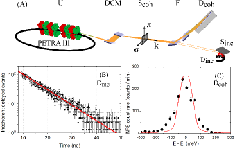
Natural Ir occurs in two stable isotopes, 191Ir and 193Ir. The 73 keV transition in 193Ir with nuclear spins of 3/2 and 1/2 of ground and excited state, respectively, is most favorable for NRS studies due to the high, 62% natural abundance of the 193Ir isotope and a comparatively long natural lifetime of 8 ns. The experiments were conducted at the Dynamics Beamline P01 at PETRA III (DESY, Hamburg) web site .
The storage ring was operated in 40-bunch top-up mode providing a
stable 100 mA ring current. The experimental setup
(Fig. 1 (A)) included a double-crystal Si(3 1 1) monochromator
which reduced the energy bandwidth of the 73 keV photons to about
8(1) eV. In order to reduce the energy bandwidth around the nuclear resonance
in 193Ir further, the energy bandwidth of photons transmitted
by the sample was filtered via Bragg reflections from two specially
designed Si crystals (F, Fig. 1 (A)). The first crystal
with an asymmetric (4 4 0) reflection collimates the beam for
matching the acceptance of the subsequent (6 4 2) reflection which
reduces the energy bandwidth to about 150 meV Sup . Tuning
of the photon energy to that of the nuclear resonance and
measurement of the lifetime of the excited state was performed by
monitoring delayed nuclear fluorescence from an Ir metal foil by a
large area avalanche photo diode (APD) detector (Dinc,
Fig. 1 (A)). The nuclear forward scattering (NFS) was
detected by a fast detector array consisting of 16 APDs (Dcoh,
Fig. 1 (A)) Baron et al. (2006); Sergueev et al. (2007); Sup . The fast
detector array and very high bunch purity in the PETRA
storage ring enabled the counting of delayed photons as
early as 3 ns after the excitation pulse with a time resolution of
about 0.6 ns Sup .
The nuclear resonance was found 3.211 keV below the K- edge of Ir at 76.111 keV, and its energy was determined to 72.90(8) keV. This value is in good agreement with the frequently reported literature value of 73.0(5) keV Achterberg et al. (2006), though it is lower than the more precise value 73.045(5) keV obtained in Ref. Kishimoto et al. (2005) from the measurement of internal conversion. The reason for the latter is unclear as in both measurements the maximum of the derivative of the edge absorption curve was used as a reference. Furthermore, the reference value of the edge used in Kishimoto et al. (2005) is 76.101 keV, which increases the disagreement. Fitting the time spectrum of delayed nuclear fluorescence with an exponential decay function (Fig. 1 (B)), we determined the natural lifetime to be 8.4(2) ns, in accordance with the lifetime value of 8.8(2) ns reported in Ref. Achterberg et al. (2006) and slightly lower than the one reported in Kishimoto et al. (2005) of 8.78 ns (no error given).
The corresponding resonance linewidth is 78(2) neV. Using the NFS setup, we measured an instrumental function of the filtering optics (Fig. 1 (C)). Its width of 158(8) meV (FWHM) is close to that of 112 meV (FWHM) predicted by dynamical theory (Fig. 1 (C), red line). The broadening can be related to the imperfections in the bulk silicon utilized for the crystals.
In order to demonstrate the feasibility of the technique we
performed NFS measurements on elemental Ir and on IrO2. Both
materials have been studied earlier by conventional Mössbauer
spectroscopy Wagner (1983); these are used here as references for
validation of data treatment routines in the time domain.
While elemental Ir shows a single resonance line, Ir in IrO2
exhibits an Ir4+ state with a pure electric hyperfine interaction Wagner (1983).
NRS time spectra of 100 m thick foil of elemental Ir have been
acquired in half an hour (Fig. 2 (A)) at signal
countrates of about 7 s-1. We observed a shift of beating
minima to later times with increasing temperature due to the
decrease of Lamb-Mössbauer factor (Fig. 2 (A),
lower graphs). Since the temporal beating pattern can be fully
described as dynamical beats Hannon and Trammell (1999), hyperfine interactions
can be ruled out, in accordance with the cubic lattice and
paramagnetism of the elemental Ir Kandiner (2013). Fitting the
temperature dependence of the Lamb-Mössbauer factor with the
Debye model Gütlich et al. (2011), we determined a Debye temperature of
309(30)K. This value is in good agreement with the literature
value of 335(13) K Steiner et al. (1969).
NFS time spectra of the IrO2 powder sample are shown in Fig.
2 (B). Fitting of the experimental data (Fig. 2 (B), upper
graph) was performed with the CONUSS software Sturhahn (2000, 2000).
To take the high mixing ratio of the E2/M1
multipole radiation into account Sturhahn and Gerdau (1994); Wagner (1983) CONUSS had to
be extended. Special cases of the NRS theory for ferro-magnetic and
anti-ferromagnetic arrangements considering high mixing ratios are
rolled out in detail in the Supplemental Material Sup . Where suitable, a
comparison to the simple M1 case in 57Fe is also given.
Fitting the data resulted in a quadrupole splitting of 2.76(2) mm/s (8.96(7)) was obtained ( is the elementary change, is the quadrupole moment, is the electric field gradient (EFG) along the quantization axis). This value is in excellent agreement with the value of 2.71(6) mm/s reported in Ref. Atzmony et al. (1967). We obtained an axially symmetric EFG with =1.71(1)1018 V/cm2 which is two orders of magnitude higher than in the isostructural 4d-RuO2 reported in Ref. Bessas et al. (2014). The EFG in IrO2 is therefore mostly determined by valence 5d-electrons because of: (i) three times lower shielding of the Ir nucleus from the valence electrons than from the surrounding ions Raghavan et al. (1976); Wagner (1983) and (ii) more elongated 5d-orbitals in IrO2 providing a potentially higher EFG Gütlich et al. (2011). In order to measure the isomer shift of Ir4+ in IrO2, we introduced an Ir metal foil as a single line reference absorber and acquired a NFS time spectrum of the combined setup (Fig. 2 (B), lower graph). From the evaluation of this dataset we obtained an isomer shift of - 0.89(5) mm/s in IrO2 relative to Ir metal, which is in good agreement with the value of - 0.93(1) mm/s reported in Ref. Shenoy and Wagner (1984).

To develop the method for studies of magnetic materials, we measured NFS from the ferromagnetic alloy Fe0.98Ir0.02 in an external magnetic field of 0.53(5) T. Dilute alloys of Fe1-xIrx () show nearly pure magnetic hyperfine interactions Mössbauer et al. (1971); Salomon and Shirley (1974), and the hyperfine fields in these alloys are the highest for all known compounds with d-elements Rao (1975). The large hyperfine fields lead to very fast oscillations in the temporal beat patterns of NFS and therefore provide the best benchmark of time resolution of the setup. The NFS time spectrum of a 1.6 mm thick sample of Fe0.98Ir0.02 exhibits extremely fast oscillations with a period of ns (Fig. 2 (C)).
Notably, despite the aforementioned high E2/M1 mixing ratio the Fe0.98Ir0.02 NFS spectrum shows a very regular beating pattern, significant for an (almost pure) two transition line spectrum. At a first glance this is surprising as even the pure M1 case (e.g. for 57Fe) shows a more complicated spectrum. The reason for this is that due to the E2/M1 mixing parameter, which value is close the square root of 1/3, M1 and E2 transition amplitudes in the mixed M1/E2 case can cancel each other for specific transitions in 193Ir (for more details see Supplemental Material Sup , part C). We refine the value of the hyperfine field to 133(1) T, which is in good agreement with the value of 140(2) T reported for Fe0.973Ir0.027 in Ref. Wagner and Zahn (1970).
Having validated the NRS technique by studying relevant reference samples, we applied it for exploring magnetism and electronic properties of two iridates from the series of Srn+1IrnO3n+1 Ruddlesden-Popper phases, i.e. SrIrO3 and Sr2IrO4.
SrIrO3 is a paramagnetic metal with electronic structure determined by both strong spin-orbit interaction and large electronic correlation at temperatures higher than 50 K Moon et al. (2008); Zhao et al. (2008). Due to its impact on the electronic anisotropy and EFG resulting from it, the spin-orbit interaction in SrIrO3 can be studied by probing the quadrupole splitting of the 193Ir nuclear levels Ingalls (1964). For a SrIrO3 powder sample we obtained a quadrupole splitting of 1.24(5) mm/s (4.0(2)) at 15 K (Fig. 3 (A), upper graph), in very good agreement with the value of 1.26 mm/s measured at 4 K in Ref. Shenoy and Wagner (1984). Magnetic hyperfine interactions can be ruled out, in accordance with paramagnetism in this compound in the temperature range under scope Zhao et al. (2008). The quadrupole splitting decreases with temperature and reaches a value of 1.08(5) mm/s (3.5(2)) at 108 K (Fig. 3 (A), lower graph), which can be related to the presence of a gap in the electronic ground state. To the best of our knowledge, no change of Ir coordination symmetry is reported for the temperature range investigated. Therefore the temperature dependent change in quadrupole splitting can be exclusively addressed to the thermal population of electronic levels, supporting the evidence of semimetal-like electronic band structure Nie et al. (2015) in SrIrO3 and showing the decisive impact of the large distortions in IrO6 octahedra onto the electronic structure in this compound.
It is the unique strength of the NRS technique to provide local information on the magnitude and orientation of magnetic fields and electric field gradients at the Ir sites Sup . This allowed us to gain new insights into the magnetic order of the Sr2IrO4 perovskite. Sr2IrO4 crystals have a form of platelets with lateral size of 2x3 mm2 and thickness of about 30-70 m; the whole incident beam was accepted by the sample. The Sr2IrO4 sample was aligned with the () plane perpendicular to the incident beam (inset Fig. 3 (B,C)) and five crystals have been stacked in the same orientation along the beam in order to increase the NFS signal Sup . EDX and magnetisation measurements suggest slight oxygen deficiency in the Sr2IrO4 sample Sup .
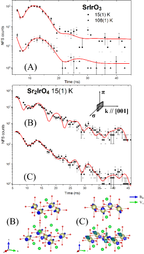
The temporal beat pattern in the time spectrum of Sr2IrO4 is
determined by both magnetic and electric hyperfine interactions
(Fig. 3 (B,C)). We obtained a hyperfine field of 24.2(2) T
which is in a very good agreement with the value of 24 T reported
by Mössbauer spectroscopy in Ref. Wagner (1983). Whilst NRS at
193Ir shows high sensitivity to the orientation of hyperfine
fields Sup , the model with in-plane hyperfine fields fails
to explain the measured time spectrum (Fig. 3 (B)).
Taking into account oxygen deficiency Sup and associated
distortion of tetragonal symmetry Ye et al. (2013), the local symmetry
of Ir in Sr2IrO4 permits the existence of magnetic components
along the c-axis Crawford et al. (1994); Ye et al. (2013). Introducing a 30o tilting
angle of hyperfine fields to the a-b plane into the model fit
provides a very good statistical quality of the fit to the measured
time spectrum (Fig. 3 (C)), supporting the existence
of out-of-plane components of the magnetic field at the Ir sites
which was not observed before. One has to note that the direction of
hyperfine field and magnetic moment do not need to coincide
Schünemann and Winkler (2000). Especially, the effect is expected in the
presence of significant orbital field contribution and interaction
with the lattice as reported in Ref. Gretarsson et al. (2016). An enhanced
electronegativity of Ir also favors high covalency of Ir-O bonds,
reducing the Fermi contact field and increasing the orbital field
contribution to the hyperfine field Oosterhuis and Lang (1969); Henning (1967); Herlitschke et al. (2014).
In accordance with this statement, a strong anisotropy in electronic
-factors in Sr2IrO4 was observed by ESR in Ref. Bogdanov et al. (2015).
Considering the electric field at the Ir nuclei in Sr2IrO4, we
observe an axially symmetric EFG with a magnitude of
V/cm2 in the [] direction. The
presence of an EFG is reasonable in view of distortion of the
IrO6 octahedra Crawford et al. (1994), oxygen deficiency, and the evidence
of a non-zero EFG in the isostructural Sr2RuO4
Ishida et al. (1997). The non-zero EFG in Sr2IrO4 and the
out-of-plane components of the magnetic hyperfine field found here
might be addressed to the non-zero angular momentum
of the outer electrons, arising from the reduced symmetry of the IrO6 octahedra;
temperature dependent measurements can provide further information on
the origin of this phenomenon.
In conclusion, we have established Nuclear Resonance Scattering at the 72.90(8) keV level in 193Ir as a new synchrotron-based technique for the studies of magnetism and electronic properties of iridates. A huge 133(1) T hyperfine field in dilute Fe0.98Ir0.02 alloy has been detected via NRS. Moreover, we found a thermally induced decrease of the electric field gradient across the Ir nuclei in SrIrO3 and observed a non-zero EFG and tilting of hyperfine fields from the basal plane in Sr2IrO4 that should stimulate further investigations to relate structural and electronic properties in the iridates. All samples contained 193Ir in its natural abundance; no preparation of radioactive sources is required and no line broadening due to the source is present. NRS at 193Ir is sensitive to dilute systems and spin structures, providing a valuable input for studies to relate magnetism and spin-orbit interactions in iridates, e.g. in strong magnetic fields Kim et al. (2009); Perlow et al. (1969), or under confinement in nanomaterials Hirsch (1999); Lin et al. (2014); Fujiwara et al. (2013) and heterostructures Nichols et al. (2016). The oxidation state of iridium and crystal fields at Ir ions can be tracked via measurements of isomer shift and quadrupole interactions at the Ir nucleus, respectively.
Acknowledgements.
We acknowledge support of the Helmholtz association via project oriented funds. The PETRA machine operation group is gratefully acknowledged for establishing a beam cleaning procedure and maintaining high bunch purity. Wolfgang Sturhahn is greatly acknowledged for extending the CONUSS software for calculations of mixed multipole radiation. The authors are thankful to Hlynur Gretarsson, Christian Donnerer, and Raphaël P. Hermann for fruitful discussions on the physics of iridates. We thank Manfred Spiwek and Frank-Uwe Dill for the preparation of the silicon crystals and setup at the beamline. Thomas F. Keller (DESY NanoLab) is acknowledged for EDX measurements on Sr2IrO4. Work at UCL was supported by the Engineering and Physical Sciences Research Council (grants EP/N027671/1 and EP/N034694/1).References
- Kim et al. (2014) Y. K. Kim, O. Krupin, J. D. Denlinger, A. Bostwick, E. Rotenberg, Q. Zhao, J. F. Mitchell, J. W. Allen, and B. J. Kim, “Fermi arcs in a doped pseudospin-1/2 Heisenberg antiferromagnet,” Science 345, 187–190 (2014).
- He et al. (2015) J. He, H. Hafiz, Th. R. Mion, T. Hogan, C. Dhital, X. Chen, Q. Lin, M. Hashimoto, D. H. Lu, Y. Zhang, R. S. Markiewicz, A. Bansil, S. D. Wilson, and Rui-Hua He, “Fermi Arcs vs. Fermi Pockets in Electron-doped Perovskite Iridates,” Sci. Rep. 5, 8533 (2015).
- Crawford et al. (1994) M. K. Crawford, M. A. Subramanian, R. L. Harlow, J. A. Fernandez-Baca, Z. R. Wang, and D. C. Johnston, “Structural and magnetic studies of Sr2IrO4,” Phys. Rev. B 49, 9198–9201 (1994).
- Kim et al. (2009) B. J. Kim, H. Ohsumi, T. Komesu, S. Sakai, T. Morita, H. Takagi, and T. Arima, “Phase-sensitive observation of a spin-orbital mott state in Sr2IrO4,” Science 323, 1329–1332 (2009).
- Boseggia et al. (2013) S. Boseggia, H.C. Walker, J. Vale, R. Springell, Z. Feng, R.S. Perry, M.M. Sala, H.M. Rønnow, S.P. Collins, and D.F. McMorrow, “Locking of iridium magnetic moments to the correlated rotation of oxygen octahedra in Sr2IrO4 revealed by x-ray resonant scattering,” J. Phys. Condens. Matter 25, 422202 (2013).
- Kim et al. (2008) B. J. Kim, Hosub Jin, S. J. Moon, J.-Y. Kim, B.-G. Park, C. S. Leem, Jaejun Yu, T. W. Noh, C. Kim, S.-J. Oh, J.-H. Park, V. Durairaj, G. Cao, and E. Rotenberg, “Novel Mott State Induced by Relativistic Spin-Orbit Coupling in Sr2IrO4,” Phys. Rev. Lett. 101, 076402 (2008).
- Moon et al. (2008) S. J. Moon, H. Jin, K. W. Kim, W. S. Choi, Y. S. Lee, J. Yu, G. Cao, A. Sumi, H. Funakubo, C. Bernhard, and T. W. Noh, “Dimensionality-Controlled Insulator-Metal Transition and Correlated Metallic State in Transition Metal Oxides Srn+1IrnO3n+1 (, 2, and ),” Phys. Rev. Lett. 101, 226402 (2008).
- Donnerer et al. (2016) C. Donnerer, Z. Feng, J. G. Vale, S. N. Andreev, I. V. Solovyev, E. C. Hunter, M. Hanfland, R. S. Perry, H. M. Rønnow, M. I. McMahon, V. V. Mazurenko, and D. F. McMorrow, “Pressure dependence of the structure and electronic properties of Sr3Ir2O7,” Phys. Rev. B 93, 174118 (2016).
- Jackeli and Khaliullin (2009) G. Jackeli and G. Khaliullin, “Mott insulators in the strong spin-orbit coupling limit: From Heisenberg to a quantum compass and Kitaev models,” Phys. Rev. Lett. 102, 017205 (2009).
- Witczak-Krempa et al. (2014) W. Witczak-Krempa, G. Chen, Y.B. Kim, and L. Balents, “Correlated Quantum Phenomena in the Strong Spin-Orbit Regime,” Annu. Rev. Condens. Matter Phys. 5, 57–82 (2014).
- Mattheiss (1976) L. F. Mattheiss, “Electronic structure of RuO2, OsO2, and IrO2,” Phys. Rev. B 13, 2433–2450 (1976).
- Kim et al. (2012) J. Kim, D. Casa, M. H. Upton, T. Gog, Y.-J. Kim, J. F. Mitchell, M. van Veenendaal, M. Daghofer, J. van den Brink, G. Khaliullin, and B. J. Kim, “Magnetic Excitation Spectra of Sr2IrO4 Probed by Resonant Inelastic X-Ray Scattering: Establishing Links to Cuprate Superconductors,” Phys. Rev. Lett. 108, 177003 (2012).
- Kim et al. (2016) Y. K. Kim, N. H. Sung, J. D. Denlinger, and B. J. Kim, “Observation of a d-wave gap in electron-doped Sr2IrO4,” Nature Phys. 12, 37–41 (2016).
- de la Torre et al. (2015) A. de la Torre, S. McKeown Walker, F. Y. Bruno, S. Riccó, Z. Wang, I. Gutierrez Lezama, G. Scheerer, G. Giriat, D. Jaccard, C. Berthod, T. K. Kim, M. Hoesch, E. C. Hunter, R. S. Perry, A. Tamai, and F. Baumberger, “Collapse of the mott gap and emergence of a nodal liquid in lightly doped Sr2IrO4,” Phys. Rev. Lett. 115, 176402 (2015).
- Schaffer et al. (2016) R. Schaffer, E. Kin-Ho Lee, B.-J. Yang, and Y. B. Kim, “Recent progress on correlated electron systems with strong spin–orbit coupling,” Reports on Progress in Physics 79, 094504 (2016).
- Lin et al. (2014) Z. C. Lin, C. Xie, Y. Osakada, Y. Cui, and B. Cui, “Iridium oxide nanotube electrodes for sensitive and prolonged intracellular measurement of action potentials,” Nat. Commun. 5, 3206 (2014).
- Qiu et al. (2015) Zh. Qiu, D. Hou, T. Kikkawa, K. Uchida, and E. Saitoh, “All-oxide spin Seebeck effects,” Appl. Phys. Express 8, 083001 (2015).
- Hirsch (1999) J. E. Hirsch, “Spin hall effect,” Phys. Rev. Lett. 83, 1834–1837 (1999).
- Fujiwara et al. (2013) K. Fujiwara, Y. Fukuma, H. Matsuno, J.and Idzuchi, Y. Niimi, Y. Otani, and H. Takagi, “5d iridium oxide as a material for spin-current detection,” Nat. Comm. 4, 2893 (2013).
- Yan et al. (2015) Y. J. Yan, M. Q. Ren, H. C. Xu, B. P. Xie, R. Tao, H. Y. Choi, N. Lee, Y. J. Choi, T. Zhang, and D. L. Feng, “Electron-doped Sr2IrO4: An analogue of hole-doped cuprate superconductors demonstrated by scanning tunneling microscopy,” Phys. Rev. X 5, 041018 (2015).
- Wang and Senthil (2011) F. Wang and T. Senthil, “Twisted Hubbard Model for Sr2IrO4: Magnetism and Possible High Temperature Superconductivity,” Phys. Rev. Lett. 106, 136402 (2011).
- Röhlsberger (2004) R. Röhlsberger, Nuclear condensed matter physics with synchrotron radiation: basic principles, methodology and applications, Springer Tracts in Modern Physics, Vol. 208 (Springer, Heidelberg, 2004).
- Gerdau and DeWaard (1999) E. Gerdau and H. DeWaard, eds., Nuclear resonant scattering of synchrotron radiation, Vol. 123-124 (Springer International Publishing, 1999).
- Wagner and Zahn (1970) F. Wagner and U. Zahn, “Mössbauer isomer shifts, hyperfine interactions, and magnetic hyperfine anomalies in compounds of iridium,” Z. Phys. 233, 1–20 (1970).
- Ding et al. (2016) Yang Ding, Liuxiang Yang, Cheng-Chien Chen, Heung-Sik Kim, Myung Joon Han, Wei Luo, Zhenxing Feng, Mary Upton, Diego Casa, Jungho Kim, Thomas Gog, Zhidan Zeng, Gang Cao, Ho-kwang Mao, and Michel van Veenendaal, “Pressure-induced confined metal from the mott insulator ,” Phys. Rev. Lett. 116, 216402 (2016).
- Perlow et al. (1969) G. J. Perlow, W. Henning, D. Olson, and G. L. Goodman, “Hyperfine anomaly in 193Ir by Mössbauer effect, and its application to determination of the orbital part of hyperfine fields,” Phys. Rev. Lett. 23, 680–682 (1969).
- Röhlsberger et al. (2001) R. Röhlsberger, J. Bansmann, V. Senz, K. L. Jonas, A. Bettac, O. Leupold, R. Rüffer, E. Burkel, and K. H. Meiwes-Broer, “Perpendicular Spin Orientation in Ultrasmall Fe Islands on W(110),” Phys. Rev. Lett. 86, 5597–5600 (2001).
- Stankov et al. (2007) S. Stankov, R. Röhlsberger, T. Slezak, M. Sladecek, B. Sepiol, G. Vogl, A. I. Chumakov, R. Rüffer, N. Spiridis, J. Lazewski, K. Parlinski, and J. Korecki, “Phonons in Iron: From the Bulk to an Epitaxial Monolayer,” Phys. Rev. Lett. 99, 185501 (2007).
- Potapkin et al. (2013) V. Potapkin, C. McCammon, K. Glazyrin, A. Kantor, I. Kupenko, C. Prescher, R. Sinmyo, G.V. Smirnov, A.I. Chumakov, and R. Rüffer, “Effect of iron oxidation state on the electrical conductivity of the Earth’s lower mantle,” Nat. Commun. 4, 1427 (2013).
- (30) Dynamics Beamline P01 web site, http://photon-science.desy.de/facilities .
- (31) See Supplemental Material at (URL will be inserted by publisher) for details on x-ray filter, APD detector array, beam cleaning in the PETRA ring, sensitivity of NRS to direction of hyperfine fields in iridates, and on alignment of the Sr2IrO4 single crystals which includes Refs. Sergueev et al. (2007); Gerdau and DeWaard (1999); Röhlsberger (2004); Keil and Ehrlichmann (2016); Klute et al. (2011); Nawrocky et al. (1993); Sturhahn (2000); Sturhahn and Gerdau (1994); Sung et al. (2016); Gretarsson et al. (2016).
- Baron et al. (2006) Alfred Q. R. Baron, Shunji Kishimoto, John Morse, and Jean-Marie Rigal, “Silicon avalanche photodiodes for direct detection of X-rays,” Journal of Synchrotron Radiation 13, 131–142 (2006).
- Sergueev et al. (2007) I. Sergueev, A. I. Chumakov, T. H. Deschaux Beaume-Dang, R. Rüffer, C. Strohm, and U. van Bürck, “Nuclear forward scattering for high energy Mössbauer transitions,” Phys. Rev. Lett. 99, 097601 (2007).
- Achterberg et al. (2006) E. Achterberg, O.A. Capurro, G.V. Marti, V.R. Vanin, and R.M. Castro, “Nuclear Data Sheets for A = 193,” Nuclear Data Sheets 107, 1–224 (2006).
- Kishimoto et al. (2005) S. Kishimoto, Y. Yoda, Y. Kobayashi, S. Kitao, R. Haruki, and M. Seto, “Evidence for nuclear excitation by electron transition on 193Ir and its probability,” Nucl. Phys. A 748, 3–11 (2005).
- Wagner (1983) F. E. Wagner, “Mössbauer spectroscopy with 191,193Ir,” Hyperfine Interact. 13, 149–173 (1983).
- Hannon and Trammell (1999) J.P. Hannon and G.T. Trammell, “Coherent -ray optics,” Hyperfine Interact. 123, 127–274 (1999).
- Kandiner (2013) H.J. Kandiner, Iridium, Gmelin Handbook of Inorganic and Organometallic Chemistry - 8th edition (Springer Berlin Heidelberg, 2013).
- Gütlich et al. (2011) Ph. Gütlich, E. Bill, and A. X. Trautwein, Mössbauer Spectroscopy and Transition Metal Chemistry Fundamentals and Applications (Springer, Heidelberg, 2011).
- Steiner et al. (1969) P. Steiner, E. Gerdau, W. Hautsch, and D. Steenken, “Determination of the mean life of some excited nuclear states by Mössbauer experiments,” Z. Phys. A 221, 281–290 (1969).
- Sturhahn (2000) W. Sturhahn, “CONUSS and PHOENIX: Evaluation of nuclear resonant scattering data,” Hyperfine Interact. 125, 149–172 (2000).
- Sturhahn and Gerdau (1994) W. Sturhahn and E. Gerdau, “Evaluation of timedifferential measurements of nuclearresonance scattering of xrays,” Phys. Rev. B 49, 9285–9294 (1994).
- Atzmony et al. (1967) U. Atzmony, E. R. Bauminger, D. Lebenbaum, A. Mustachi, S. Ofer, and J. H. Wernick, “Mössbauer effect in 193Ir in intermetallic compounds and salts of iridium,” Phys. Rev. 163, 314–323 (1967).
- Bessas et al. (2014) D. Bessas, D. G. Merkel, A. I. Chumakov, R. Rüffer, R. P. Hermann, I. Sergueev, A. Mahmoud, B. Klobes, M. A. McGuire, M. T. Sougrati, and L. Stievano, “Nuclear forward scattering of synchrotron radiation by 99Ru,” Phys. Rev. Lett. 113, 147601 (2014).
- Raghavan et al. (1976) P. Raghavan, E. N. Kaufmann, R. S. Raghavan, E. J. Ansaldo, and R. A. Naumann, “Sign and magnitude of the quadrupole interaction of 111Cd in noncubic metals: Universal correlation of ionic and electronic field gradients,” Phys. Rev. B 13, 2835–2847 (1976).
- Shenoy and Wagner (1984) G. K. Shenoy and F.E. Wagner, “Mössbauer-Effect Isomer Shifts,” in Mössbauer Spectroscopy Applied to Inorganic Chemistry, edited by G. J. Long (Springer US, Boston, MA, 1984) pp. 57–76.
- Mössbauer et al. (1971) R.L. Mössbauer, M. Lengsfeld, W. von Lieres, W. Potzel, P. Teschner, F.E. Wagner, and G. Kaindl, “Nuclear gamma resonance study of the IrFe and IrNi alloy systems,” Z. Naturforsch. A 26, 343 (1971).
- Salomon and Shirley (1974) D. Salomon and D. A. Shirley, “Quadrupole coupling at 193Ir nuclei in iron,” Phys. Rev. B 9, 29–31 (1974).
- Rao (1975) G.N. Rao, “Dilute-impurity hyperfine fields in Fe, Co, Ni, and Gd,” At. Data Nucl. Data Tables 15, 553 – 576 (1975).
- Zhao et al. (2008) J.G. Zhao, L.X. Yang, Y. Yu, F.Y. Li, R.C. Yu, Z. Fang, L.C. Chen, and C.Q. Jin, “High-pressure synthesis of orthorhombic sriro3 perovskite and its positive magnetoresistance,” J. of Appl. Phys. 103, 103706 (2008).
- Ingalls (1964) R. Ingalls, “Electric-Field Gradient Tensor in Ferrous Compounds,” Phys. Rev. 133, A787–A795 (1964).
- Nie et al. (2015) Y. F. Nie, P. D. C. King, C. H. Kim, M. Uchida, H. I. Wei, B. D. Faeth, J. P. Ruf, J. P. C. Ruff, L. Xie, X. Pan, C. J. Fennie, D. G. Schlom, and K. M. Shen, “Interplay of Spin-Orbit Interactions, Dimensionality, and Octahedral Rotations in Semimetallic SrIrO3,” Phys. Rev. Lett. 114, 016401 (2015).
- Ye et al. (2013) Feng Ye, S. Chi, B. C. Chakoumakos, J. A. Fernandez-Baca, T. Qi, and G. Cao, “Magnetic and crystal structures of Sr2IrO4: A neutron diffraction study,” Phys. Rev. B 87, 140406 (2013).
- Schünemann and Winkler (2000) V. Schünemann and H. Winkler, “Structure and dynamics of biomolecules studied by Mössbauer spectroscopy,” Rep. Prog. Phys. 63, 263 (2000).
- Gretarsson et al. (2016) H. Gretarsson, N. H. Sung, M. Höppner, B. J. Kim, B. Keimer, and M. Le Tacon, “Two-magnon raman scattering and pseudospin-lattice interactions in Sr2IrO4 and Sr3Ir2O7,” Phys. Rev. Lett. 116, 136401 (2016).
- Oosterhuis and Lang (1969) W. T. Oosterhuis and G. Lang, “Mössbauer effect in K3FeCN6,” Phys. Rev. 178, 439–456 (1969).
- Henning (1967) J.C.M. Henning, “Covalency and hyperfine structure of ions in crystal fields,” Phys. Lett. A 24, 40 – 42 (1967).
- Herlitschke et al. (2014) M. Herlitschke, A. L. Tchougreeff, A. V. Soudackov, B. Klobes, L. Stork, R. Dronskowski, and R. P. Hermann, “Magnetism and lattice dynamics of FeNCN compared to FeO,” New J. Chem. 38, 4670–4677 (2014).
- Bogdanov et al. (2015) N. A. Bogdanov, V. M. Katukuri, J. Romhányi, V. Yushankhai, V. Kataev, B. Büchner, J. van den Brink, and L. Hozoi, “Orbital reconstruction in nonpolar tetravalent transition-metal oxide layers,” Nat. Comm. 6, 73061–73069 (2015).
- Ishida et al. (1997) K. Ishida, Y. Kitaoka, K. Asayama, S. Ikeda, S. Nishizaki, Y. Maeno, K. Yoshida, and T. Fujita, “Anisotropic pairing in superconducting Sr2RuO4: Ru NMR and NQR studies,” Phys. Rev. B 56, R505–R508 (1997).
- Nichols et al. (2016) J. Nichols, X. Gao, S. Lee, T. L. Meyer, J. W. Freeland, V. Lauter, D. Yi, J. Liu, D. Haskel, and J. R. Petrie, “Emerging magnetism and anomalous Hall effect in iridate-manganite heterostructures,” Nat. Comm. 7, 12721 (2016).
- Keil and Ehrlichmann (2016) J. Keil and H. Ehrlichmann, “Bunch Purity Measurements at PETRA III,” (7th International Particle Accelerator Conference, Busan (Korea), 8 May13 May, 2016) pp. 3434–3436.
- Klute et al. (2011) J. Klute, K. Balewski, A. Delfs, H. T. Duhme, M. Ebert, Ru. Neumann, and F. Obier, “The PETRA III Multibunch Feedback System,” (10th European Workshop on Beam Diagnostics and Instrumentation for Particle Accelerators, Hamburg (Germany), 16 May18 May, 2011) pp. 494–496.
- Nawrocky et al. (1993) R.J. Nawrocky, U. Bergmann, and D.P. Siddons, “A bunch killer for the nsls x-ray electron storage ring,” in Proceedings of International Conference on Particle Accelerators, Vol. 3 (1993) pp. 2145–2147.
- Sung et al. (2016) N.H. Sung, H. Gretarsson, D. Proepper, J. Porras, M. Le Tacon, A. V. Boris, B. Keimer, and B. J. Kim, “Crystal growth and intrinsic magnetic behaviour of Sr2IrO4,” Philos. Mag. 96, 413–426 (2016).