Application of computational breast phantoms to evaluate reconstruction methods for fluorescence molecular tomography
Abstract
Fluorescence molecular tomography (FMT) has potential of providing high contrast images for breast tumor detection. Computational phantom provides a convenient way to a wide variety of fluorophore distribution configurations in patients and perform comprehensive evaluation of the imaging systems and methods for FMT. In this study, a digital breast phantom was used to compare the performance of a novel sparsity-based reconstruction method and Tikhonov regularization method for resolving tumors with different amount of separation. The results showed that the proposed sparse reconstruction method yielded better performance. This simulation-based approach with computational phantoms enabled an evaluation of the reconstruction methods for FMT for breast-cancer detection.
I Introduction
Fluorescence imaging is an emerging optical technique that provides high contrast image with the use of fluorophores. Previously, Indocyanine Green (ICG) has been reported for fluorescence image-guided surgery for breast cancer[1]. Tomography enables 3D visualization and estimation of fluorescence distribution. Recently, clinical studies have been conducted to study the feasibility of fluorescence molecular tomography (FMT) for breast tumor detection[2]. The advent of sparse reconstruction methods have shown potential of providing more accurate estimation of fluorescence distribution and intensity compared to conventional method in FMT[3]. However, studies are required to assess whether the sparse reconstruction methods indeed yield improved performance for FMT-based breast-cancer imaging. Computational phantom provides a comparatively easier way to conduct these studies by allowing modeling of different fluroscence distributions in breast-cancer populations as opposed to having to actually image patients. In this study, we conducted a computational human-phantom-based study to compare the performance of sparse reconstruction method and a traditional Tikhonov regularization method for resolving tumors in patient images.
II Theory
II-A Forward model
The forward model of FMT can be described with two equations representing light propagation from source to fluorophore and from fluorophore to detector, respectively. Let , and denote positions of source, fluorophore and detector respectively, , denote the Green’s function of excitation and emission light, denote fluorescence yield, denote source function, , denote the support of source and fluorophore positions, respectively, and , denote fluence of source and fluorophore, then the forward model is written as:
| (1) |
and
| (2) |
From equation (1) and (2) we can derive the following linear matrix equation for measurements and unknown fluorophore distribution:
| (3) |
where is the system matrix.
II-B Proposed sparsity-driven reconstruction method
The inverse problem of FMT is usually ill-posed and regularization is required in order to solve the fluorescence distribution. Tikhonov regularization method is widely used and it can be expressed as follows:
| (4) |
where is typically chosen as . is identity matrix.
Breast tumor tends to concentrate in a small region compared with the whole breast, and fluorophore is designed to target the tumor. Thus, the inverse problem of FMT for breast tumor can also be regarded as a sparse reconstruction problem. We propose a sparse-reconstruction approach similar to another approach we developed for transcranial small-animal imaging previously[4]. The basic idea is to minimize the following regularized problem to solve for :
| (5) |
Sparse reconstruction algorithm requires that measurement matrix should be nearly orthogonal. For this purpose, the coherence of G needs to be reduced, for which we use the truncated SVD. Fast iterative shrinkage-thresholding algorithm (FISTA) is then used to find sparse solution for equation (5). Finally maximum-likelihood expectation maximization algorithm is implemented for noise reduction.
III Computational studies with breast phantoms
Simulation-based experiments were conducted to study the ability of FMT for resolving breast tumor. A breast phantom generated from MR database was used in this experiment. The phantom was segmented into four different tissues: blood vessels, skin layer, fat, and fibroglandular tissues. Different optical properties were assigned to each type of tissue, as described in [5]. Two tumors of size 1.6 cm were inserted in the breast phantom at difference seperation and fluorophore was assumed to concentrate in the tumors. 40 laser sources and 40 detectors were positioned around the phantom. Monte Carlo method was implemented to calculate the forward model[6]. Two different methods, Tikhonov regularization method and the proposed sparse reconstruction method were used for reconstruction of FMT.
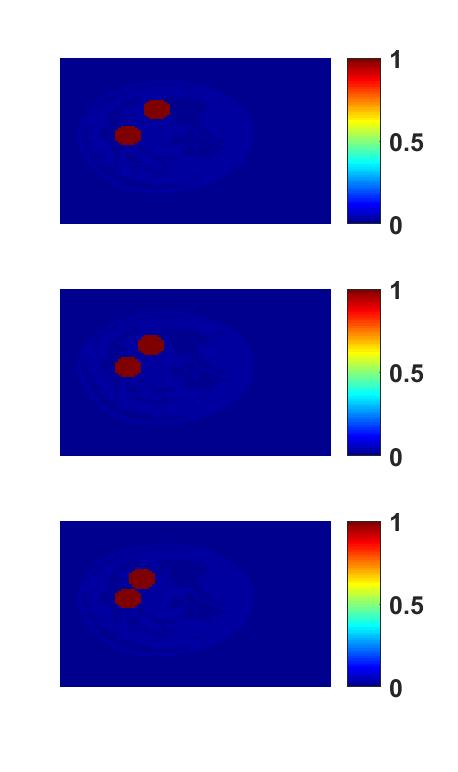
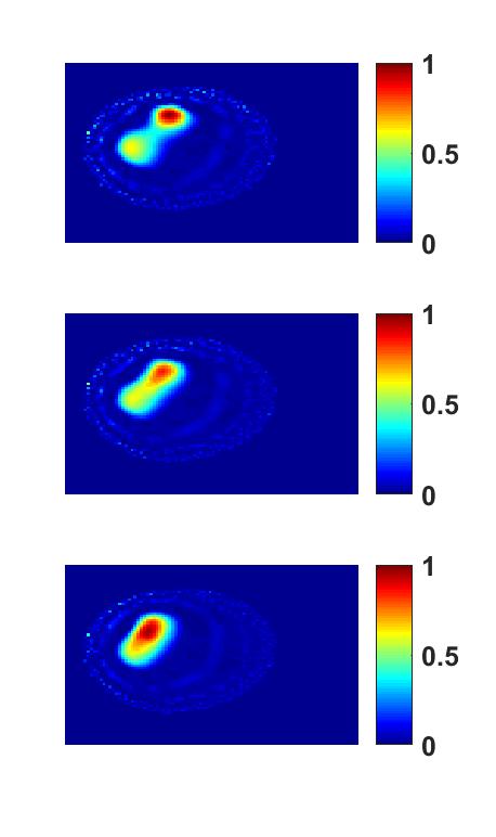
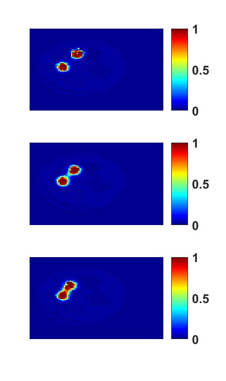
The result is shown in Figure (1).
IV Results and Conclusions
From figure (1), we notice that for larger separation between tumors (2.5cm), both Tikhonov method and sparse reconstruction method can differentiate the two tumors. When the separation between the tumors decreases, Tikhonov regularization method oversmooths the edge of the tumors, making it hard to distinguish them. However, the sparse reconstruction method is still able to resolve the two tumors. In addition, we also notice that compared to Tikhonov reconstruction method, sparse reconstruction provides more accurate estimation of fluorophore distribution and signal intensity.
In conclusion, this simulation-based approach with computational phantoms enables an evaluation of the reconstruction methods for FMT for breast-cancer detection. In future, we propose to conduct objective evaluation studies to further evaluate the performance of these reconstruction methods[7].
Acknowledgment
This work was supported by the NIH BRAIN Initiative Award R24 MH106083.
References
- [1] B. E. Schaafsma, J. S. D. Mieog, M. Hutteman, J. R. van der Vorst, P. J. Kuppen, C. W. Löwik, J. V. Frangioni, C. J. van de Velde, and A. L. Vahrmeijer, “The clinical use of indocyanine green as a near-infrared fluorescent contrast agent for image-guided oncologic surgery,” Journal of Surgical Oncology, vol. 104, no. 3, pp. 323–332, 2011.
- [2] A. Corlu, R. Choe, T. Durduran, M. A. Rosen, M. Schweiger, S. R. Arridge, M. D. Schnall, and A. G. Yodh, “Three-dimensional in vivo fluorescence diffuse optical tomography of breast cancer in humans,” Optics express, vol. 15, no. 11, pp. 6696–6716, 2007.
- [3] J. Dutta, S. Ahn, C. Li, S. R. Cherry, and R. M. Leahy, “Joint l1 and total variation regularization for fluorescence molecular tomography,” Physics in medicine and biology, vol. 57, no. 6, p. 1459, 2012.
- [4] Y. Zhu, A. K. Jha, J. K. Dreyer, H. N. D. Le, J. U. Kang, P. E. Roland, D. F. Wong, and A. Rahmim, “A three-step reconstruction method for fluorescence molecular tomography based on compressive sensing,” pp. 1 005 911–1 005 911–8, 2017.
- [5] Y. Lou, W. Zhou, T. P. Matthews, C. M. Appleton, and M. A. Anastasio, “Generation of anatomically realistic numerical phantoms for photoacoustic and ultrasonic breast imaging,” Journal of Biomedical Optics, vol. 22, no. 4, p. 041015, 2017.
- [6] Q. Fang and D. A. Boas, “Monte carlo simulation of photon migration in 3d turbid media accelerated by graphics processing units,” Opt. Express, vol. 17, no. 22, pp. 20 178–20 190, Oct 2009.
- [7] A. K. Jha, E. Clarkson, and M. A. Kupinski, “An ideal-observer framework to investigate signal detectability in diffuse optical imaging,” Biomedical optics express, vol. 4, no. 10, pp. 2107–2123, 2013.