Reliable and Practical Computational Prediction of Molecular Crystal Polymorphs
The ability to reliably predict the structures and stabilities of a molecular crystal and its (often numerous) polymorphs without any prior experimental information would be an invaluable tool for a number of fields, with specific and immediate applications in the design and formulation of pharmaceuticals Cruz-Cabeza et al. (2015). In this case, detailed knowledge of the polymorphic energy landscape for an active pharmaceutical ingredient yields profound insight regarding the existence and likelihood of late-appearing polymorphs Bučar et al. (2015), and hence has significant public health and economic implications. However, the computational prediction of the structures and stabilities of molecular crystal polymorphs is particularly challenging due to the high dimensionality of conformational and crystallographic space accompanied by the need for relative (free) energies to within kJ/mol per molecule. In this work, we combine the most successful crystal structure sampling strategy with the most accurate energy ranking strategy of the latest blind test of organic crystal structure prediction (CSP), organized by the Cambridge Crystallographic Data Centre (CCDC) Reilly et al. (2016); Gibney (2015). Our final energy ranking is based on first-principles density functional theory (DFT) calculations that include three key physical contributions: (i) a sophisticated treatment of Pauli exchange-repulsion and electron correlation effects with hybrid functionals, (ii) inclusion of many-body van der Waals dispersion interactions, and (iii) account of harmonic (and sometimes anharmonic) vibrational free energies. In doing so, this combined approach has an optimal success rate in producing the crystal structures corresponding to the five blind-test molecules and even predicts stable polymorphs that have not been observed to date. With this practical approach, we demonstrate the feasibility of obtaining reliable structures and stabilities for molecular crystals of pharmaceutical importance, paving the way towards an enhanced fundamental understanding of polymorphic energy landscapes and routine industrial application of molecular CSP methods.
Accurate and reliable CSP methods are able to furnish detailed knowledge of the energetic landscape corresponding to a given molecular crystal and its (often numerous) thermodynamically relevant polymorphs. With access to the structures and relative thermodynamical stabilities of such varied crystal-packing motifs, one can gain crucial insight into whether or not the existing structure of a pharmaceutical drug candidate is indeed the most thermodynamically stable solid form at ambient conditions. This in turn enables an informed and critical assessment of the potential risk associated with the assumed stable form disappearing at some point during the manufacturing process or consumable shelf life Bučar et al. (2015). In this case, a polymorph with similar stability but different (and often unwanted) properties could emerge as the dominant solid form—an event which can trigger a cascade of deleterious health-related, social, and financial repercussions. As such, the utilization of accurate and reliable computational CSP methods in conjunction with experimental polymorph screening efforts offers a comprehensive and sustainable solution to this grand challenge Price (2014).
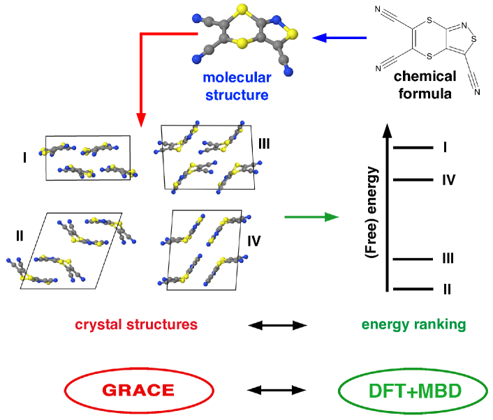
In general, the accuracy and reliability of a given CSP methodology depends on two distinct but equally important theoretical aspects: (i) sufficiently complete sampling of the conformational and crystallographic space spanned by a given molecular crystal and (ii) sufficiently accurate ranking of the numerous low-energy polymorphs according to their relative thermodynamic stabilities Reilly et al. (2016); Price (2014). In this regard, major advances have been made along both of these thrusts over the past few years, resulting in substantial progress in the field of molecular CSP Beran (2016); Yang et al. (2014); Nyman and Day (2015); Nyman et al. (2016); Schneider et al. (2016); Brandenburg and Grimme (2016); Price and Brandenburg (2017); Shtukenberg et al. (2017). By and large, the most important benchmarks for assessing the utility of a given molecular CSP approach are the regular blind tests organized by the CCDC Day et al. (2009); Bardwell et al. (2011); Reilly et al. (2016), wherein participants predict the structure of a given molecular crystal based solely on the two-dimensional (2D) chemical formula for the individual molecule(s) involved. Over the past few decades, the chemical diversity and complexity of the CCDC blind test has gradually increased and now includes small and rigid molecules as well as elaborate polymorphic systems involving large and flexible molecules, salts, and co-crystals. A general overview of the protocol employed in a typical molecular CSP methodology is illustrated in Fig. 1. Starting with the 2D chemical formula for each molecule, 3D molecular structures are first computed using standard geometry optimization techniques that are supplemented with additional sampling of all energetically relevant conformational isomers for flexible molecules. Next, a vast number of possible crystal-packing arrangements is generated by comprehensively sampling different intermolecular orientations, space groups, unit cell sizes, and molecular conformations. Finally, the generated crystal structures are ranked according to their relative (free) energies.
In this work, we demonstrate that an accurate, reliable, and computationally feasible protocol for the prediction of molecular crystal polymorphs can be obtained by combining the most successful crystal structure sampling strategy (Neumann et al.) with the most successful first-principles energy ranking strategy (Tkatchenko et al.) from the latest CCDC blind test Reilly et al. (2016); Gibney (2015). In this regard, the approach for generating crystal structures by Neumann et al. was able to correctly predict all experimentally observed structures (except for one) within the top 100 most stable structures, building on top of their major successes in previous blind tests Day et al. (2009); Bardwell et al. (2011); Neumann (2008). The fact that several experimental structures could only be found with this approach again highlights the complexity associated with sufficiently sampling wide swaths of crystallographic space. In this approach, initial molecular crystal structures are created with a Monte Carlo parallel tempering algorithm that employs a tailor-made force field within the Grace software package. Following this initial screening, a set of candidate crystal structures are then reoptimized in a hierarchical and statistically-controlled process using dispersion-inclusive DFT Neumann and Perrin (2005); Neumann (2008); Neumann et al. (2008); Reilly et al. (2016). Beyond the robust sampling of the essential regions of crystallographic space, these initial energy rankings can be substantially improved upon by employing state-of-the-art first-principles methodologies as detailed below.
To demonstrate this procedure, we start with the top 100 initial molecular crystal structures (for every system in the blind test) provided by Neumann et al. (see Supplementary Information of Ref. 3). Form E of system XXIII is the only experimental structure that was not present in this set of initial structures and is included for completeness. We note in passing that this form was in fact generated by Neumann et al., but was located just outside the energetic window considered for the structures. In total, this set includes structures (with unit cell sizes ranging from to atoms) and therefore provides a large-scale benchmark structural database under realistic CSP conditions.
Based on these initial molecular crystal structures, we have developed a robust hierarchical first-principles approach for energetically ranking all relevant polymorphs. This approach is directly applicable to pharmaceutically relevant systems and includes three important theoretical aspects that are commonly neglected in typical CSP protocols: (i) a sophisticated treatment of Pauli exchange-repulsion and electron correlation effects with hybrid functionals, (ii) inclusion of many-body dispersion interactions and dielectric screening effects, and (iii) an account of harmonic (and sometimes anharmonic) vibrational contributions to the free energy. In this regard, the hybrid PBE0 functional Adamo and Barone (1999) in conjunction with the many-body dispersion (MBD) model Tkatchenko et al. (2012); DiStasio Jr. et al. (2012); Ambrosetti et al. (2014); DiStasio Jr. et al. (2014); Blood-Forsythe et al. (2016) is able to predict absolute experimental lattice energies to within kcal/mol Reilly and Tkatchenko (2013); Otero-de-la Roza and Johnson (2012) and relative stabilities of several polymorphic systems to within kJ/mol Marom et al. (2013); Reilly and Tkatchenko (2013, 2015); Shtukenberg et al. (2017). Hence, the PBE0+MBD approach is used for all calculations of static lattice energies. Geometry and lattice optimizations, as well as vibrational free energies are computed with the PBE functional Perdew et al. (1996) in conjunction with the effective-pairwise Tkatchenko-Scheffler (TS) dispersion correction Tkatchenko and Scheffler (2009) (denoted as PBE+TS). A detailed description of the computational approaches employed in this work is available below in the Methods section.
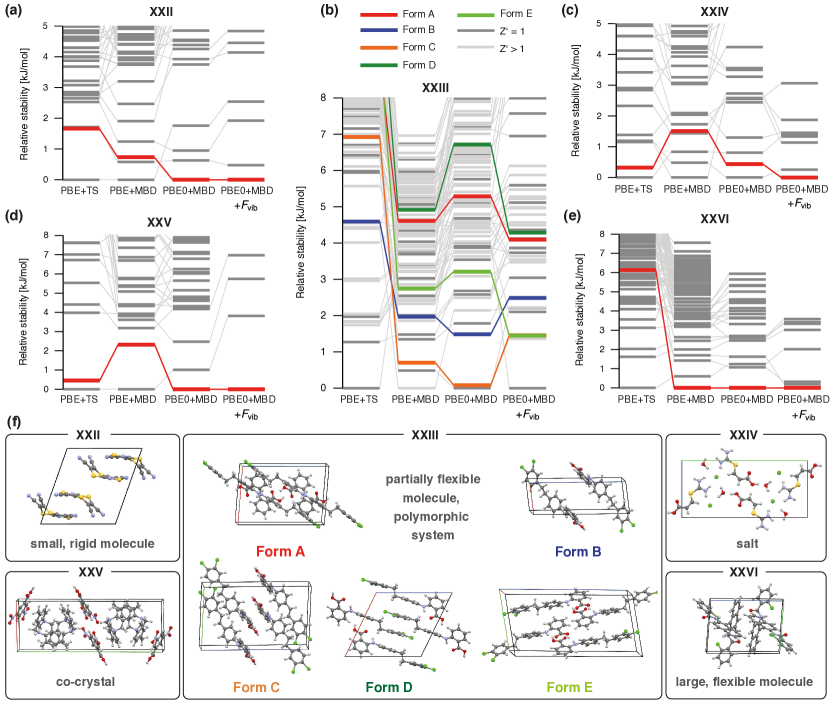
The stability rankings obtained for the five blind-test systems are shown in Fig. 2 (with all structures and energies (Tables S6-S10) available in the Supplementary Information (SI)). In this figure, we not only show the final stability rankings, but also several intermediary steps, in which one or more of the three aforementioned theoretical contributions are not accounted for in the rankings. The first ranking considers only static lattice energies computed at the PBE+TS level, while the second ranking accounts for beyond-pairwise many-body dispersion interactions (PBE+MBD). In the third ranking, we include a more sophisticated treatment of Pauli exchange-repulsion via PBE0+MBD. In doing so, the deleterious effects of self-interaction error (a DFT artifact in which an electron interacts with itself) are significantly ameliorated, which leads to a substantial improvement in the description of electrostatic and charge-transfer effects. In the final ranking, we supplement the PBE0+MBD energies with harmonic vibrational free energy contributions at the PBE+TS level (). This leads to a stability ranking based on Helmholtz free energies which accounts for thermal entropic effects.
We first concentrate our discussion on systems XXII, XXIV, XXV, and XXVI. For all of these systems, our final stability ranking at the PBE0+MBD+ level predicts the experimental structure as the most stable form—the ideal outcome of any CSP protocol. As seen from the intermediate stability rankings, all of the three previously mentioned theoretical effects are required to obtain this result. For example, Pauli exchange-repulsion (through the PBE0 functional) plays a crucial role for system XXII Curtis et al. (2016), while many-body dispersion effects are the most important factor for system XXVI. In addition, all structures with free energies that are within kJ/mol of the experimental structure are essentially minor variations of the latter (see SI), which demonstrates the robustness of our CSP approach in dealing with pharmaceutically-relevant systems like salts, co-crystals, and molecular crystals involving large and flexible molecules.
Now we focus our discussion on the most challenging system in the blind test (XXIII). This system involves a conformationally flexible molecule and has five experimentally confirmed polymorphs Reilly et al. (2016). The fact that this compound is also a former drug candidate Simons et al. (2009) makes it an ideal testing ground for CSP of pharmaceutically-relevant molecules. The low-energy barriers between different conformers in this molecule lead to a fairly complex polymorphic landscape with numerous crystal structures located within a very small energy window. As shown in Fig. 2, the PBE+TS method is again insufficient for quantitative energy ranking predictions and places all experimentally observed structures within the top 11 kJ/mol—an energy window containing 84 structures. Each refinement of the energetic rankings changes their relative stabilities, with all experimental structures observed within the top kJ/mol ( kcal/mol) in the final ranking with PBE0+MBD+. At this level, all experimental structures were found within an energy interval of kJ/mol, the expected energy range associated with co-existing polymorphs Cruz-Cabeza et al. (2015). We note here that our procedure finds one structure (Str. N70) that is kJ/mol more stable than all experimentally observed structures, a remarkable finding that is discussed in more detail below.
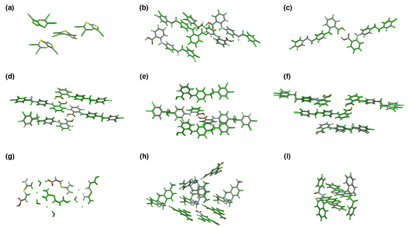
For all systems with only one known polymorph, the systematic and hierarchical energy ranking protocol presented herein correctly produced the experimental structure as the most stable forms (Rank 1). This represents a significant improvement over the Ranks (XXII), (XXIV), (XXV), and (XXVI) obtained by the unrefined results of Neumann et al., which again stresses the critical importance of an energy ranking protocol based on state-of-the-art first-principles based methodologies. For system XXIII, all experimental structures were found within the top 18 structures, with the two structures with Ranks (Form E) and (Form C). When only considering the structures, we find all three experimental structures among the top 10 structures, as compared to the top 26 in the initial ranking by Neumann et al.. Moreover, all of our predicted structures agree to within Å of the experimental structures as quantified by the root-mean-square deviation (RMSD) measure. These RMSD values are also within Å of the initial structures obtained by Neumann et al., which again stresses the need for such a comprehensive crystal structure sampling protocol (see Table S4 in the SI). Overlays of the predicted and experimental structures are provided in Extended Data Fig. 4.
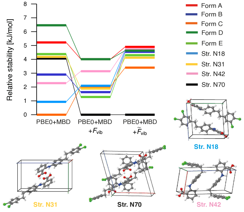
With the exceptions of systems XXII and XXIV, our computational protocol underestimates unit cell volumes by % on average, which is an expected discrepancy that originates from the fact that the geometry and lattice optimizations did not include temperature (thermal expansion) effects. In this regard, a majority of the thermal expansion in molecular crystals can be accounted for via the quasi-harmonic approximation (QHA) Allen and De Wette (1969); Hoja et al. (2017); Erba et al. (2016); Heit et al. (2016). Therefore, we have also computed the unit cell volumes at K for all of the experimental structures for XXIII (Forms A, B, C, D, E) as well as the first four structures (Str. N70, N31, N18, N42), which have yet to be experimentally observed. With this approach, we are now able to predict unit cell volumes to within % on average. As such, the QHA provides a simple but effective way of including thermal effects in molecular crystal structures using first-principles based methodologies. Stability rankings based on these thermally-expanded structures are shown in Fig. 3. The stabilities computed with the QHA can be interpreted as relative Gibbs free energies and the largest observed change stemming from the use of thermally-expanded structures amounts to kJ/mol at the PBE0+MBD+ level.
In addition to thermal expansion, the vibrational contributions to the free energy also contain anharmonic effects, which can be accounted for by utilizing Morse oscillators (see Methods). The corresponding free energy stability rankings with such an anharmonic treatment of the vibrational free energy are denoted by PBE0+MBD+ and shown in Fig. 3. At this level, all experimental structures are found within an energy window of only kJ/mol, which is well within the expected energy range for co-existing polymorphs. We note in passing that Brandenburg and Grimme have also studied the experimental structures of system XXIII utilizing a semi-empirical tight-binding approach within the QHA; however, their values lie within a much larger energy window of kJ/mol Brandenburg and Grimme (2016).
Quite interestingly, the unobserved polymorph of XXIII (Str. N70) is significantly more stable than any of the experimentally determined crystal structures, even after accounting for thermal expansion in the underlying crystal structures as well as anharmonic vibrational free energy contributions. In this regard, this polymorph is actually further stabilized by vibrational entropy and shares many structural features with form A. The most notable difference is the stacking pattern of the molecular sheets (see Figure S1). As such, we hypothesize that Form A might be kinetically favored over Str. N70 and this hitherto unobserved polymorph could potentially be crystallized by slowly melting Form A or introducing surfactants during the crystallization procedure (see SI for a detailed discussion). In addition, from a thermodynamic standpoint, Str. N18, N31, and N42 might also be observed experimentally, although Str. N42 involves a twisted molecular conformation which might not be easily accessible in solution. Experimental evidence Reilly et al. (2016) suggests that Form A should be the most stable structure at low temperatures and Form D the most stable structure at room temperature. Indeed, we observe that Form D is stabilized by thermal effects and predicted to be more stable than Form A at the PBE0+MBD+ level. In addition, inclusion of anharmonic vibrational free energies brings all of the experimentally determined structures closer together, i.e., all of the structures are now within kJ/mol.
In a broad context of crystal polymorphism, our findings suggest that late-appearing crystal forms Bučar et al. (2015) are ubiquitous for molecules of pharmaceutical interest, further reinforcing recent experimental and computational predictions for coumarin Shtukenberg et al. (2017), dalcetrapib Neumann et al. (2015), rotigotine Wolff et al. (2010); Bučar et al. (2015), and ritonavir Bauer et al. (2001). In the case of system XXIII, the stability of a new potential form N70 is substantially higher (by 3 kJ/mol) than that of all experimentally discovered forms. Systematic tests carried out in this work ensure the reliability of our CSP procedure to 1-2 kJ/mol, suggesting that the so far unobserved Str. N70 should be the thermodynamically stable form at ambient conditions. Obviously, experimental confirmation of this fact would be desirable and our suggestions for crystallization experiments should be useful in this endeavor. We also stress that further improvements of the presented CSP procedure are desirable and possible. For example, the efficiency could be further improved by using DFT+MBD energies for constructing tailor-made force fields during the crystal generation step of the CSP. In addition, one could improve the accuracy of free energy calculations by employing more advanced dynamical approaches, by using either path-integral molecular dynamics Rossi et al. (2016) or the vibrational self-consistent field approach Monserrat et al. (2013); Drummond et al. (2015); Engel et al. (2015).
In summary, we have introduced a robust and computationally feasible procedure that yields accurate and reliable predictions of the structures and stabilities of the thermodynamically relevant polymorphs associated with complex molecular crystals, including salts, co-crystals, and flexible large molecules of pharmaceutical interest. Our approach explicitly accounts for all relevant enthalpic and entropic effects, including sophisticated treatments of Pauli exchange-repulsion, many-body dispersion interactions, and vibrational free energies at finite temperatures, all of which are directly obtained from quantum-mechanical calculations. The approach presented herein takes us one step closer to obtaining an enhanced fundamental understanding of polymorphic energy landscapes and routinely employing computational molecular crystal structure prediction in conjunction with experimental polymorph screening. Such a joint theoretical-experimental procedure offers a comprehensive and sustainable solution to the grand challenges associated with molecular crystals polymorphs, whose very existence offers us the promise of novel and hitherto unexplored pharmaceutical agents on one hand, and quite devastating public health and economic repercussions on the other.
I Acknowledgements
J.H. and A.T. acknowledge support from the Deutsche Forschungsgemeinschaft under the program DFG-SPP 1807. H-Y.K., R.A.D., and R.C. acknowledge support from the Department of Energy (DOE) under Grant Nos. DE-SC0008626. R.A.D. also acknowledges partial support from start-up funding from Cornell University. This research used computational resources provided by the Argonne Leadership Computing Facility at Argonne National Laboratory (supported by the Office of Science of the U.S. Department of Energy under Contract No. DE- AC02-06CH11357), the National Energy Research Scientific Computing Center (supported by the Office of Science of the U.S. Department of Energy under Contract No. DE-AC02-05CH11231), the Terascale Infrastructure for Groundbreaking Research in Science and Engineering (TIGRESS) High Performance Computing Center and Visual itization Laboratory at Princeton University, the Fritz Haber Institute of the Max Planck Society (FHI-aims and Mercury), and the High Performance Computing facilities of the University of Luxembourg–see http://hpc.uni.lu.
II Author Contributions
J.H., M.A.N, and A.T. designed the research. M.A.N. provided the initial crystal structures. J.H., H-Y.K., and R.A.D. carried out the polymorph ranking calculations. J.H., M.A.N., R.C., R.A.D., and A.T. analyzed the data. J.H., R.A.D., and A.T. wrote the paper with contributions from all authors.
III Methods
For each system in the latest blind test, we utilized the top crystal structures from the submission of Neumann et al. (which are available in the Supplementary Information of Ref. 3) as initial structures for this study. For systems with two submitted lists, we used the list which also included structures, i.e., structures which have two molecules in the asymmetric unit. Form E of system XXIII was the only experimental structure not present in this set and was therefore added for completeness. All calculations were performed using the all-electron FHI-aims code Blum et al. (2009); Marek et al. (2014); Auckenthaler et al. (2011); Havu et al. (2009). Throughout this work, we utilized two different accuracy levels in FHI-aims, which are denoted as light and tight. For the light level, we use the light species default setting in FHI-aims for all numerical atom-centered basis functions and integration grids. The number of -points () in each direction was determined by the smallest integer satisfying Å, with being the unit cell length in a given direction. For the tight level, we use the tight species default settings in FHI-aims and the number of -points is determined by the criterion that Å. Many-body dispersion (MBD) interactions were evaluated at the MBD@rsSCS level with a reciprocal-space implementation that utilized the same -point mesh as the DFT calculations Tkatchenko et al. (2012); Ambrosetti et al. (2014). Convergence criteria of eV, electrons/Å3, eV/Å, and eV were used for the total energy, charge density, forces, and sum of eigenvalues, respectively.
First, we performed full lattice and geometry relaxations (without any symmetry constraints) using the PBE functional Perdew et al. (1996) in conjunction with the effective-pairwise Tkatchenko-Scheffler (TS) dispersion correction Tkatchenko and Scheffler (2009), ensuring that the smallest force component is less than eV/Å. Duplicate structures were identified using Mercury Macrae et al. (2008). Structures were considered similar if out of molecules within the crystals matched within % in terms of distances and within in terms of angles, and the corresponding root-mean-square deviation (RMSD20) is smaller than Å. Two similar structures were considered to be identical if their PBE+TS energy (light) agreed to within kJ/mol. Only the most stable structure among identical structures was retained throughout the protocol. These optimized structures were symmeterized using PLATON Spek (2009) and are provided in the SI. All structures were named according to their rank in the initial ranking by Neumann et al. In order to determine if an experimental structure was found, we used the same settings for the crystal similarity search as described above.
Next, relative energetic stabilities were computed based on these PBE+TS optimized structures by using PBE+TS and PBE+MBD Tkatchenko et al. (2012); Ambrosetti et al. (2014) with tight settings. In order to ensure the convergence of the relative energies, we have created a benchmark set consisting of small structures of system XXII and small structures of system XXIV. For these structures, PBE+MBD energies were computed using really tight settings for the integration grids and tier basis functions. When considering all possible relative energies between structures from the same system, the mean absolute deviation (MAD) for the tight settings amounts to only kJ/mol with a maximal deviation of kJ/mol. This illustrates the fact that tight settings provide converged relative energies. Relative stabilities of these benchmark systems are available in Table S1 of the SI.
Since PBE0 Adamo and Barone (1999) calculations with tight settings are not possible for all of the studied systems due to the massive computational cost and memory requirements, we approximate the PBE0+MBD energies by adding the difference between PBE0+MBD and PBE+MBD evaluated at the light level to the PBE+MBD energies calculated at the tight level. For the aforementioned benchmark set, this approximation has a MAD of only kJ/mol with a maximum deviation of kJ/mol, when compared to PBE0+MBD energies evaluated with tight settings (see Table S3 in the SI). In contrast, PBE0+MBD energies at the light level yield a MAD of kJ/mol with a maximum deviation of kJ/mol. Therefore, our approximation provides relative energies that are in very good agreement with tight PBE0+MBD energies. PBE0+MBD energies were computed for all structures of systems XXII, XXIII, and XXIV. For the remaining systems, PBE0+MBD calculations are available for (at least) the structures located within the top kJ/mol of the PBE+MBD rankings.
Vibrational free energies () were computed at the PBE+TS level with light settings by utilizing the phonopy code Togo and Tanaka (2015) and the finite-difference method within the harmonic approximation. The final stability rankings in Fig. 2 were always based on PBE0+MBD+ energies evaluated at temperatures correspond to the experimental crystal structure measurements. For the finite-difference calculations, we used displacements of Å and (when necessary) supercells that ensure cell lengths greater than Å in every direction. Furthermore, the vibrational free energy was evaluated in reciprocal space, where the number of -points () in each direction is determined by the smallest integer satisfying Å. All structures had no imaginary frequencies at the -point and the magnitude of the three acoustic modes was smaller than cm-1 in most cases and always smaller than cm-1. Vibrational free energies were calculated for (at least) all structures that are located within the top kJ/mol according to the PBE0+MBD ranking. For system XXIII, vibrational free energies were calculated for all structures and for all structures containing up to molecules per unit cell within the top kJ/mol of the PBE0+MBD ranking. One possibility to further improve this procedure is to include MBD interactions in the geometry optimizations and vibrational free energy calculations in addition to the static lattice energy rankings Blood-Forsythe et al. (2016). This would increase the computational cost by approximately 60% on average and will be discussed elsewhere.
For the quasi-harmonic approximation (QHA), we performed PBE+TS lattice and geometry optimizations of several structures from system XXIII using light settings with external hydrostatic pressures of , , , , and GPa. Then, harmonic vibrational free energies were computed for all of the obtained structures. Based on the light PBE+TS energies and harmonic vibrational free energies, the unit cell volume corresponding to K was determined via the Murnaghan equation of state Murnaghan (1944). Based on these thermally-expanded structures, the stability rankings were calculated as described above.
For all thermally-expanded structures of system XXIII, we computed the anharmonic vibrational contributions to the free energies by replacing the harmonic oscillators by Morse oscillators. This is done for all phonon modes at the -point of cells containing molecules, i.e., for Forms A, C, D, E, and Str. N70, this corresponds to the unit cell, while for Form B and Str. N18, N31, and N42, this corresponds to a supercell. The structures were displaced along all normal modes in both directions, corresponding to energy changes of and according to the harmonic approximation, where is the Boltzmann constant and K. The energies of all displaced structures were calculated with PBE+TS using light settings. To have a consistent sampling of the thermally-accessible energy window, we demanded that the largest observed energy change with respect to the optimized thermally-expanded structure always lies between and . Therefore, the displacement amplitudes of a few low-frequency modes had to be reduced in order to sample the desired energy window. Next, we fitted a Morse potential Morse (1929); Dahl and Springborg (1988), given by
| (1) |
to the obtained data points for each mode. In this expression, is the displacement amplitude, and the parameters , , and describe the well depth, the width of the potential, and the minimum of the potential, respectively. The energy of a vibrational mode in state can be calculates by
| (2) |
with
| (3) |
where is the reduced mass. The anharmonic vibrational free energy () at the -point was computed according to
| (4) |
with
| (5) |
where runs over phonon modes. This approach yields anharmonic vibrational free energies at the -point for cells including molecules. In order to account also for other -points, we rely on the harmonic approximation and calculate the total vibrational free energies according to:
| (6) |
where is the fully converged harmonic vibrational free energy and is the harmonic vibrational free energy evaluated at the point only for the cells described above.
References
- Cruz-Cabeza et al. (2015) A. J. Cruz-Cabeza, S. M. Reutzel-Edens, and J. Bernstein, Chem. Soc. Rev. 44, 8619 (2015).
- Bučar et al. (2015) D. K. Bučar, R. W. Lancaster, and J. Bernstein, Angew. Chem. Int. Ed. 54, 6972 (2015).
- Reilly et al. (2016) A. M. Reilly, R. I. Cooper, C. S. Adjiman, S. Bhattacharya, A. D. Boese, J. G. Brandenburg, P. J. Bygrave, R. Bylsma, J. E. Campbell, R. Car, D. H. Case, R. Chadha, J. C. Cole, K. Cosburn, H. M. Cuppen, F. Curtis, G. M. Day, R. A. DiStasio Jr., A. Dzyabchenko, B. P. van Eijck, D. M. Elking, J. A. van den Ende, J. C. Facelli, M. B. Ferraro, L. Fusti-Molnar, C.-A. Gatsiou, T. S. Gee, R. de Gelder, L. M. Ghiringhelli, H. Goto, S. Grimme, R. Guo, D. W. M. Hofmann, J. Hoja, R. K. Hylton, L. Iuzzolino, W. Jankiewicz, D. T. de Jong, J. Kendrick, N. J. J. de Klerk, H.-Y. Ko, L. N. Kuleshova, X. Li, S. Lohani, F. J. J. Leusen, A. M. Lund, J. Lv, Y. Ma, N. Marom, A. E. Masunov, P. McCabe, D. P. McMahon, H. Meekes, M. P. Metz, A. J. Misquitta, S. Mohamed, B. Monserrat, R. J. Needs, M. A. Neumann, J. Nyman, S. Obata, H. Oberhofer, A. R. Oganov, A. M. Orendt, G. I. Pagola, C. C. Pantelides, C. J. Pickard, R. Podeszwa, L. S. Price, S. L. Price, A. Pulido, M. G. Read, K. Reuter, E. Schneider, C. Schober, G. P. Shields, P. Singh, I. J. Sugden, K. Szalewicz, C. R. Taylor, A. Tkatchenko, M. E. Tuckerman, F. Vacarro, M. Vasileiadis, A. Vazquez-Mayagoitia, L. Vogt, Y. Wang, R. E. Watson, G. A. de Wijs, J. Yang, Q. Zhu, and C. R. Groom, Acta Crystallogr. Sect. B: Struct. Sci. 72, 439 (2016).
- Gibney (2015) E. Gibney, Nature 527, 20 (2015).
- Price (2014) S. L. Price, Chem. Soc. Rev. 43, 2098 (2014).
- Beran (2016) G. J. O. Beran, Chem. Rev. 116, 5567 (2016).
- Yang et al. (2014) J. Yang, W. Hu, D. Usvyat, D. Matthews, M. Schutz, and G. K.-L. Chan, Science 345, 640 (2014).
- Nyman and Day (2015) J. Nyman and G. M. Day, Cryst. Eng. Comm. 17, 5154 (2015).
- Nyman et al. (2016) J. Nyman, O. S. Pundyke, and G. M. Day, Phys. Chem. Chem. Phys. 18, 15828 (2016).
- Schneider et al. (2016) E. Schneider, L. Vogt, and M. E. Tuckerman, Acta Crystallogr. Sect. B Struct. Sci. Cryst. Eng. Mater. 72, 542 (2016).
- Brandenburg and Grimme (2016) J. G. Brandenburg and S. Grimme, Acta Crystallogr. Sect. B Struct. Sci. Cryst. Eng. Mater. 72, 502 (2016).
- Price and Brandenburg (2017) S. L. Price and J. G. Brandenburg, in Non-covalent Interactions in Quantum Chemistry and Physics, edited by A. O. de la Roza and G. A. DiLabio (Elsevier, 2017) pp. 333–363.
- Shtukenberg et al. (2017) A. G. Shtukenberg, Q. Zhu, D. J. Carter, L. Vogt, J. Hoja, E. Schneider, H. Song, B. Pokroy, I. Polishchuk, A. Tkatchenko, A. R. Oganov, A. L. Rohl, M. E. Tuckerman, and B. Kahr, Chem. Sci. 8, 4926 (2017).
- Day et al. (2009) G. M. Day, T. G. Cooper, A. J. Cruz-Cabeza, K. E. Hejczyk, H. L. Ammon, S. X. M. Boerrigter, J. S. Tan, R. G. Della Valle, E. Venuti, J. Jose, S. R. Gadre, G. R. Desiraju, T. S. Thakur, B. P. van Eijck, J. C. Facelli, V. E. Bazterra, M. B. Ferraro, D. W. M. Hofmann, M. A. Neumann, F. J. J. Leusen, J. Kendrick, S. L. Price, A. J. Misquitta, P. G. Karamertzanis, G. W. A. Welch, H. A. Scheraga, Y. A. Arnautova, M. U. Schmidt, J. van de Streek, A. K. Wolf, and B. Schweizer, Acta Crystallogr. Sect. B Struct. Sci. 65, 107 (2009).
- Bardwell et al. (2011) D. A. Bardwell, C. S. Adjiman, Y. A. Arnautova, E. Bartashevich, S. X. M. Boerrigter, D. E. Braun, A. J. Cruz-Cabeza, G. M. Day, R. G. Della Valle, G. R. Desiraju, B. P. van Eijck, J. C. Facelli, M. B. Ferraro, D. Grillo, M. Habgood, D. W. M. Hofmann, F. Hofmann, K. V. J. Jose, P. G. Karamertzanis, A. V. Kazantsev, J. Kendrick, L. N. Kuleshova, F. J. J. Leusen, A. V. Maleev, A. J. Misquitta, S. Mohamed, R. J. Needs, M. a. Neumann, D. Nikylov, A. M. Orendt, R. Pal, C. C. Pantelides, C. J. Pickard, L. S. Price, S. L. Price, H. A. Scheraga, J. van de Streek, T. S. Thakur, S. Tiwari, E. Venuti, and I. K. Zhitkov, Acta Crystallogr. Sect. B Struct. Sci. 67, 535 (2011).
- Neumann (2008) M. A. Neumann, J. Phys. Chem. B 112, 9810 (2008).
- Neumann and Perrin (2005) M. A. Neumann and M.-A. Perrin, J. Phys. Chem. B 109, 15531 (2005).
- Neumann et al. (2008) M. A. Neumann, F. J. J. Leusen, and J. Kendrick, Angew. Chem. Int. Ed. 47, 2427 (2008).
- Adamo and Barone (1999) C. Adamo and V. Barone, J. Chem. Phys. 110, 6158 (1999).
- Tkatchenko et al. (2012) A. Tkatchenko, R. A. DiStasio Jr., R. Car, and M. Scheffler, Phys. Rev. Lett. 108, 236402 (2012).
- DiStasio Jr. et al. (2012) R. A. DiStasio Jr., O. A. von Lilienfeld, and A. Tkatchenko, Proc. Natl. Acad. Sci. U.S.A. 109, 14791 (2012).
- Ambrosetti et al. (2014) A. Ambrosetti, A. M. Reilly, R. A. DiStasio Jr., and A. Tkatchenko, J. Chem. Phys. 140, 18A508 (2014).
- DiStasio Jr. et al. (2014) R. A. DiStasio Jr., V. V. Gobre, and A. Tkatchenko, J. Phys. Condens. Matter 26, 213202 (2014).
- Blood-Forsythe et al. (2016) M. A. Blood-Forsythe, T. Markovich, R. A. DiStasio Jr., R. Car, and A. Aspuru-Guzik, Chem. Sci. 7, 1712 (2016).
- Reilly and Tkatchenko (2013) A. M. Reilly and A. Tkatchenko, J. Chem. Phys. 139, 024705 (2013).
- Otero-de-la Roza and Johnson (2012) A. Otero-de-la Roza and E. R. Johnson, J. Chem. Phys. 137, 054103 (2012).
- Marom et al. (2013) N. Marom, R. A. DiStasio Jr., V. Atalla, S. Levchenko, A. M. Reilly, J. R. Chelikowsky, L. Leiserowitz, and A. Tkatchenko, Angew. Chem. Int. Ed. 52, 6629 (2013).
- Reilly and Tkatchenko (2015) A. M. Reilly and A. Tkatchenko, Chem. Sci. 6, 3289 (2015).
- Perdew et al. (1996) J. P. Perdew, K. Burke, and M. Ernzerhof, Phys. Rev. Lett. 77, 3865 (1996).
- Tkatchenko and Scheffler (2009) A. Tkatchenko and M. Scheffler, Phys. Rev. Lett. 102, 073005 (2009).
- Curtis et al. (2016) F. Curtis, X. Wang, and N. Marom, Acta Crystallogr. Sect. B Struct. Sci. Cryst. Eng. Mater. 72, 562 (2016).
- Simons et al. (2009) L. J. Simons, B. W. Caprathe, M. Callahan, J. M. Graham, T. Kimura, Y. Lai, H. LeVine, W. Lipinski, A. T. Sakkab, Y. Tasaki, L. C. Walker, T. Yasunaga, Y. Ye, N. Zhuang, and C. E. Augelli-Szafran, Bioorg. Med. Chem. Lett. 19, 654 (2009).
- Allen and De Wette (1969) R. E. Allen and F. W. De Wette, Phys. Rev. 179, 873 (1969).
- Hoja et al. (2017) J. Hoja, A. M. Reilly, and A. Tkatchenko, Wiley Interdiscip. Rev. Comput. Mol. Sci. 7, e1294 (2017).
- Erba et al. (2016) A. Erba, J. Maul, and B. Civalleri, Chem. Commun. 52, 1820 (2016).
- Heit et al. (2016) Y. N. Heit, K. D. Nanda, and G. J. O. Beran, Chem. Sci. 7, 246 (2016).
- Neumann et al. (2015) M. A. Neumann, J. van de Streek, F. P. A. Fabbiani, P. Hidber, and O. Grassmann, Nat. Commun. 6, 7793 (2015).
- Wolff et al. (2010) H.-M. Wolff, L. Quere, and J. Riedner, “Polymorphic form of rotigotine,” (2010), eP 2215072A2.
- Bauer et al. (2001) J. Bauer, S. Spanton, R. Henry, J. Quick, W. Dziki, W. Porter, and J. Morris, Pharm. Res. 18, 859 (2001).
- Rossi et al. (2016) M. Rossi, P. Gasparotto, and M. Ceriotti, Phys. Rev. Lett. 117, 115702 (2016).
- Monserrat et al. (2013) B. Monserrat, N. D. Drummond, and R. J. Needs, Phys. Rev. B 87, 144302 (2013).
- Drummond et al. (2015) N. D. Drummond, B. Monserrat, J. H. Lloyd-Williams, P. L. Ríos, C. J. Pickard, and R. J. Needs, Nat. Commun. 6, 7794 (2015).
- Engel et al. (2015) E. A. Engel, B. Monserrat, and R. J. Needs, Phys. Rev. X 5, 021033 (2015).
- Blum et al. (2009) V. Blum, R. Gehrke, F. Hanke, P. Havu, V. Havu, X. Ren, K. Reuter, and M. Scheffler, Comput. Phys. Commun. 180, 2175 (2009).
- Marek et al. (2014) A. Marek, V. Blum, R. Johanni, V. Havu, B. Lang, T. Auckenthaler, A. Heinecke, H.-J. Bungartz, and H. Lederer, J. Phys. Condens. Matter 26, 213201 (2014).
- Auckenthaler et al. (2011) T. Auckenthaler, V. Blum, H.-J. Bungartz, T. Huckle, R. Johanni, L. Krämer, B. Lang, H. Lederer, and P. Willems, Parallel Comput. 37, 783 (2011).
- Havu et al. (2009) V. Havu, V. Blum, P. Havu, and M. Scheffler, J. Comput. Phys. 228, 8367 (2009).
- Macrae et al. (2008) C. F. Macrae, I. J. Bruno, J. A. Chisholm, P. R. Edgington, P. McCabe, E. Pidcock, L. Rodriguez-Monge, R. Taylor, J. van de Streek, and P. A. Wood, J. Appl. Cryst. 41, 466 (2008).
- Spek (2009) A. L. Spek, Acta Crystallogr. Sect. D 65, 148 (2009).
- Togo and Tanaka (2015) A. Togo and I. Tanaka, Scr. Mater. 108, 1 (2015).
- Murnaghan (1944) F. D. Murnaghan, Proc. Natl. Acad. Sci. U.S.A. 30, 244 (1944).
- Morse (1929) P. M. Morse, Phys. Rev. 34, 57 (1929).
- Dahl and Springborg (1988) J. P. Dahl and M. Springborg, J. Chem. Phys. 88, 4535 (1988).