A computational perspective of the role of Thalamus in cognition
Abstract
Thalamus has traditionally been considered as only a relay source of cortical inputs, with hierarchically organized cortical circuits serially transforming thalamic signals to cognitively-relevant representations. Given the absence of local excitatory connections within the thalamus, the notion of thalamic ‘relay’ seemed like a reasonable description over the last several decades. Recent advances in experimental approaches and theory provide a broader perspective on the role of the thalamus in cognitively-relevant cortical computations, and suggest that only a subset of thalamic circuit motifs fit the relay description. Here, we discuss this perspective and highlight the potential role for the thalamus – and specifically mediodorsal (MD) nucleus – in dynamic selection of cortical representations through a combination of intrinsic thalamic computations and output signals that change cortical network functional parameters. We suggest that through the contextual modulation of cortical computation, thalamus and cortex jointly optimize the information/cost trade-off in an emergent fashion. We emphasize that coordinated experimental and theoretical efforts will provide a path to understanding the role of the thalamus in cognition, along with an understanding to augment cognitive capacity in health and disease.
Cortico-centric view of perceptual and cognitive processing
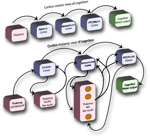
Until recently, cognition [in mammalian, bird and reptilian nervous system] has been viewed as a cortico-centric process, with thalamus considered to only play the mere role of a relay system. This classic view, much driven by the visual hierarchical model of the mammalian cortex (Felleman and Essen,, 1991), puts thalamus at the beginning of a feedforward hierarchy. The transmission of information from thalamus to early sensory cortex (V1 in the visual system for example), and the gradual increasing complex representations from V2 to MT/IT and eventually prefrontal cortex (PFC), constitute the core of the perceptual representation under the hierarchical model. A recent comparative study of biologically-plausible Convolutional Neural Networks (CNN) and the visual ventral stream, emphasizes on the feature enrichment through the network of hierarchically interconnected computational modules (layers in the neural network and areas in the ventral stream) (Yamins et al.,, 2014).
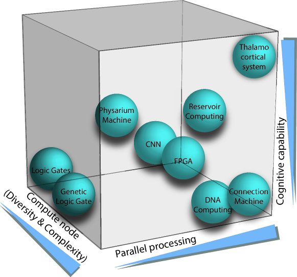
The strictly static feedforward model has since morphed to a dynamic hierarchical model due to the discoveries of the role of feedback from higher cortical areas to lower cortical areas (Heeger,, 2017). These dynamic hierarchies are even considered to be favorable for recursive optimizations, where the overall optimization can be achieved by breaking the problem into smaller ones to find the optimum for each of these smaller problems. Such recursive optimization does not need to be confined to just one cortical area and each of such distributed optimizations may even be solved differently. (Marblestone et al.,, 2016). This view of cortical computation is also paralleled with the growing use of recurrent neural networks (RNN) that can capture the dynamics of single neurons or neural population in a variety of tasks. As such, RNNs can mimic (a) context-dependent prefrontal response (Mante et al.,, 2013) or (b) can reproduce the temporal scaling of neural responses in medial frontal cortex and caudate nucleus (Wang et al.,, 2018). Although it is clear that higher level cortical feedback reaches all the way down to thalamus (Wimmer et al, other refs), the main attribute of perception/cognition remains cortico-centric under the umbrella of dynamic hierarchical models or RNN embodiment of cortical cognitive functions. Since the computation that is carried by the system should match the computing elements at the appropriate scale Dehghani, (2018) a mismatch between these presumed computational systems and the underlying circuitry becomes vividly apparent. First, associative cortex (and not just sensory cortex) receives thalamic input. Second, certain thalamic territories receive their primary (driving) input from the cortex, rather than sensory periphery, some of which are likely to be highly convergent on a single thalamic cell level, suggesting at least a cortical modulation of sensory input. Third, thalamic projections can be broad and diffuse, suggesting a modulatory, rather than a relay function. These points are indicative that thalamus may play a central part in cognitive processes. But what does including thalamus add that could not be achieved in cortical loops? One advantage that the thalamus could bring into the cortical equation is flexibility. For example, the same sensory cue may have different meanings according to a particular context a subject is in. A recent study Rikhye et al., (2018) shows that thalamus may be well suited to reorganize functional connectivity in frontal cortices in response to such contextual changes, allowing for a more flexible switching of rule to action mapping. We suggest that the unique cognitive capability of the thalamo-cortical system is tightly bound to parallel processing and contextual modulation that are enabled by the diversity of computing nodes (including thalamic and cortical structures) and complexity of the computing architecture (Fig. 1). We will start with a brief overview of the thalamic architecture, followed by experimental evidence and a computational perspective of the thalamic role in contextual cognitive computation.
Thalamic architecture: anatomical and functional features
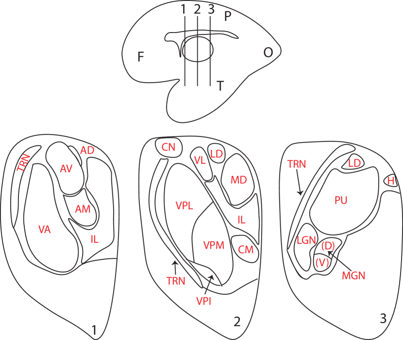
Traditionally, thalamic nuclei (see Fig. 2) are defined as collections of neurons that are segregated by gross features such as white matter tracts and various types of tissue staining procedures (Jones,, 1981). This gross anatomical classification has been equated with a functional one, where individual thalamic nuclei giving rise to a set of defined functions Jones, (1981, 1985). More recent fine anatomical studies challenge this notion, showing that within individual nuclei, single cell input/output connectivity patterns are quite variable.
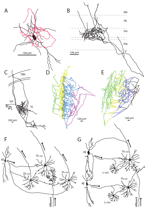
Thalamus lacks lateral excitatory connections and rather receives inputs from other subcortical structures and/or cortex. In fact, a major feature of forebrain expansion across evolution is the invasion of the thalamus by cortical inputs (Grant et al.,, 2012; Rouiller and Welker,, 2000). Most (90-95%) afferents to the relay nuclei are not from the sensory organs (Jones,, 1985; Sherman and Guillery,, 2013). Recent anatomical studies have shown a great diversity of cortical input type, strength and inferred degree of convergence, even within individual thalamic nuclei (Clasca et al.,, 2012) (see Fig. 3 for an example view of cell and network architecture diversity).
Excitatory inputs, mostly, arrive as feedbacks from layer 6 of the cortex as well as from brainstem reticular formation. In addition to the diversity of excitatory inputs, thalamic circuits receive a diverse set of inhibitory inputs (note that local GABAergic thalamic interneurons are mostly absent in non-LGN relay nuclei). The two major systems of inhibitory control are the thalamic reticular nucleus (TRN), a shell of inhibitory nucleus surrounding thalamic excitatory nuclei, and the extra-thalamic inhibitory system; a group of inhibitory projects across the fore-, mid- and hindbrain (see (Halassa and Acsády,, 2016) for a review on thalamic inhibition). Perhaps a major differentiating feature of these two systems (TRN and ETI) is temporal precision. One of the key characteristics of thalamus is lack of direct local loops. Only a very small group of inhibitory neurons with local connections exists in thalamus (Steriade and Llinás,, 1988). A mechanistic consequence of this architecture is the differential control of thalamic response gain and selectivity (Fig. 3 F and G), with the TRN controlling the first as observed in sensory systems (Pinault,, 2004), and ETI controlling the latter as observed in motor systems (Urbain and Deschênes,, 2007). For example, basal ganglia control of thalamic responses, a form of ETI control, would be implemented through thalamic disinhibition, which is not only dependent on ETI input, but also a special type of thalamic conductance that enables high frequency ‘bursting’ upon release from inhibition (Goldberg et al.,, 2013; Deniau and Chevalier,, 1985).
Overall, the variety of thalamic inputs (both excitatory and inhibitory), combined with intrinsic thalamic features such as excitability and morphology will determine the type of intrinsic computations that thalamus performs. This view appears to be consistent with recent observations of confidence encoding in both sensory (Komura et al.,, 2013) and motor systems (Ma and Jazayeri,, 2014; Jazayeri and Shadlen,, 2015).
Thalamic relay nuclei mostly project to the cortical middle layers in a topographic fashion. However, the majority of thalamic structures also project more diffusely to the cortical superficial layers, such as mediodorsal (MD), posteriodmedial complex (POm) and pulvinar for example (see Fig. 3 for an example of thalamic cell and circuit diversity). These diffuse projections seem poorly suited to relay information in a precise manner. Rather, they might have a modulatory role of cortical function. Further, a great degree of diversity can be observed at the level of thalamic axonal terminals within the cortex. While the idea of a thalamic relay was consolidated by observing that the main LGN neurons thought to be associated with form vision (M and P pathways) exhibit spatially compact cortical terminals, recent anatomical studies of individual neurons across the thalamus show a variety of terminal sizes and degree of spatial spread and intricate computational architecture (Fig. 3). This complexity of the architecture and diversity of the computing nodes are among the key factors that set apart the thalamo-cortical system from other conventional and unconventional computing engines (Fig. 1). Part of the complication in understanding how these anatomical types give rise to different functions is their potential for contacting different sets of excitatory and inhibitory cortical neurons.
Specifically, among thalamic nuclei, mediodorsal thalamus (MD) seems to have a connectivity pattern that is distinctively different from the classic sensory nuclei. Cortico-thalamic projections to MD originate from both layers V and VI of PFC (Goldman-Rakic and Porrino,, 1985; Giguere and Goldman-Rakic,, 1988; Mitchell,, 2015). But, in contrast to relay nuclei, cortical input to MD terminates both extraglomerularly and within the synaptic glomeruli, suggesting that cortex plays a different role in MD activity (in comparison to LGN for example) (Schwartz et al.,, 1991). Additionally, optogenetic and in vitro electrophysiology techniques have revealed that MD not only projects to the layer I but also has additional terminations in layer III (Cruikshank et al.,, 2012). MD projections to PFC synapse with both excitatory and inhibitory cells (Cruikshank et al.,, 2012) where the triggered triggered feedforward inhibition could play a variety of roles from regulating dendritic action potentials (Kim et al.,, 1995) to imposing a narrow temporal window within which the excitation can reach the target (Cruikshank et al.,, 2007). These elaborate input and output connectivities of the thalamocortical architecture point to the non-unitary computational role of thalamus. Among thalamic nuclei, specifically MD (and likely other MD-like nuclei) has the architecture necessary to be involved in modulatory computational roles rather than the well-known relay functions. In the next sections, we provide the experimental evidence and algorithmic designs pointing to this modulatory role. This idea of the thalamus controlling cortical state parameters is highlighted in Figs. 4, 5 and the next section.
Many facets of thalamic computation
It is commonly thought that processes like attention, decision making and working memory are implemented through distributed computations across multiple cortical regions (Corbetta,, 1998; Mesulam,, 1990; Scott et al.,, 2017). However, it is unclear how these computations are coordinated to drive relevant behavioral outputs. From an anatomical standpoint, the thalamus is strategically positioned to perform this function, but relatively little is known about its broad functional engagement in cognition. The thalamic cellular composition and network structure constrain how cortex receives and processes information. The thalamus is the major input to the cortex and interactions between the two structures are critical for sensation, action and cognition (Jones,, 1998; Sherman,, 2016; Nakajima and Halassa,, 2017). Despite recent studies showing that the mammalian thalamus contains several circuit motifs, each with specific input/output characteristics, the thalamus is traditionally viewed as a relay to or between cortical regions (Sherman and Guillery,, 2013). That the active role of thalamus in cognition is beyond relay stems from a) the fact that sensory input to thalamus is much limited in comparison to input from other structures, such as cortex and basal ganglia , b) the experimental evidence showing number of nuclei modulating cortical neural processing according to behavioral context and c) lesions of certain nuclei such as pulvinar and MD cause severe attention and memory deficits (Saalmann and Kastner,, 2015). Below we will discuss how the distinctive anatomical architecture and computational role of pulvinar and MD differ from relay nuclei such as LGN.
It is worth mentioning that this view of bona fide thalamic computations is quite distinct from the one in which thalamic responses reflect their inputs, with only linear changes in response size. This property of reflecting an input (with only slight modification of amplitude) was initially observed in the lateral geniculate nucleus (LGN), which receives inputs from the retina. LGN responses to specific sensory inputs (their receptive fields, RF) are very similar to those in the retina itself, arguing that there is little intrinsic computation happening in the LGN itself outside of gain control. Success in early vision studies (Hubel and Wiesel,, 1959, 1962) might have inadvertently given rise to the LGN relay function being generalized across the thalamus. The strictly feedforward thalamic role in cognition requires reconsideration (Halassa and Kastner,, 2017); only a few thalamic territories receive peripheral sensory inputs and project cortically in a localized manner, as the LGN does(Jones,, 1981; Raczkowski and Fitzpatrick,, 1990; Kakei et al.,, 2001; FitzGibbon et al.,, 2015; Sherman,, 2016).
The largest thalamic structures in mammals, the MD and pulvinar contain many neurons that receive convergent cortical inputs and project diffusely across multiple cortical layers and regions (Clasca et al.,, 2012; Rovo et al.,, 2012). For example, the primate pulvinar has both territories that receive topographical, non-convergent inputs from the striate cortex (Rovo et al.,, 2012) and others that receive convergent inputs from non-striate visual cortical (and frontal) areas (Mathers,, 1972). This same thalamic nucleus also receives inputs from the superior colliculus (Partlow et al.,, 1977), a subcortical region receiving retinal inputs. This suggests that the pulvinar contains multiple input ‘motifs’ solely based on the diversity of excitatory input. Such input diversity is not limited to the pulvinar, but is seen within many thalamic nuclei across the mammalian forebrain (Bickford,, 2016). Local inactivation of pulvinar neurons results in reduced neural activity in primary visual cortex (Purushothaman et al.,, 2012) suggesting a feedforward role. Recent studies, however, indicate that pulvinar may have additional roles. For example, pulvinar inactivation was shown to increase low-frequency cortical oscillations (Zhou et al.,, 2016). Given that such activity is often associated with inattention and sleep, this study suggested that pulvinar may keep cortical regions in an activated state that would allow responsiveness to top-down input from other areas to modulate ongoing activity according to attentional demands. A different study showed that during perceptual decision making pulvinar neurons encode choice confidence, rather than stimulus category (Komura et al.,, 2013). Together, these recent findings strongly argue for more pulvinar functions beyond relaying information.
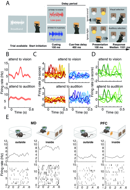
In the case of MD, direct sensory input is limited (Mitchell,, 2015) and the diffuse, distributed projections to cortex (Kuramoto et al.,, 2017) are poorly suited for information relay; this input/output connectivity suggests different functions. Recent studies (Bolkan et al.,, 2017; Schmitt et al.,, 2017; Rikhye et al.,, 2018) have begun to shed light on the type of computation that MD performs. Taking advantage of the genetic accessibility of the mouse for neuronal manipulations, these studies have revealed that MD coordinates task-relevant activity in the prefrontal cortex (PFC) in a manner analogous to a contextual signal regulating distinct attractor states within a computing reservoir. Specifically, in a task where animals had to keep a rule in mind over a brief delay period (Fig. 4A), PFC neurons show population-level persistent activity following the rule presentation, a sensory cue that instructs the animal to direct its attention to either vision or audition (Fig. 4B,C). MD neurons show responses that are devoid of categorical selectivity (Fig. 4D), yet are critical for selective PFC activity; optogenetic MD inhibition diminishes this activity, while MD activation augments it. The conclusion is that MD inputs enhance synaptic connectivity among PFC neurons or may adjust the activity of PFC neurons through selective filtering of the thalamic inputs. In other words, delay-period MD activity maintains rule-selective PFC representations by augmenting local excitatory recurrence (Schmitt et al.,, 2017).
In a related study, a delayed nonmatching-to-sample T-maze working memory task (Bolkan et al.,, 2017), it was shown that MD amplification and maintenance of higher PFC activity indicated correct performance during the subsequent choice phase of the task. Interestingly, MD-dependent increased PFC activity was much more pronounced during the later (in delay) rather than earlier part of the task. These findings indicate that PFC might have to recursively pull in MD to sustain cortical representations as working memory weakens with time. Together these studies indicate that PFC cognitive computation can not be dissociated from MD activity. Further evidence for the critical role of the MD-PFC interaction for cognition is the disrupted fronto-thalamic anatomical and functional connectivity seen in neurodevelopmental disorders (Parnaudeau et al.,, 2015; Mitelman et al.,, 2005; Marenco et al.,, 2012; Nair et al.,, 2013; Woodward et al.,, 2017).
Can MD select cortical subnetworks based on contextual modulation?
Why would a recurrent network (PFC) computation depend on its interaction with a non-recurrent (MD) non-relay network? What computational advantage such system would have? Using a chemogenetic approach, a recent study suggested that information flow in the MD-PFC network can be unidirectional. While both inactivating PFC-to-thalamus and MD-to-cortex pathways impaired recognition of a change in reward value in rats performing a decision making task, only the inactivation of MD-to-cortex pathway had an impact on the behavioral response to a change in action-reward relationship (Alcaraz et al.,, 2018). Given that a sensory stimulus may require a different action depending on the context in which it occurs, the ability to flexibly re-route the active PFC subnetwork to a different output may be crucial. In an architecture like the PFC-MD network, where MD can modulate PFC functional connectivity, MD might well be suited to re-route the ongoing activity in a context dependent manner. In fact, in the mouse cognitive task described above (Fig. 4A), a subset of MD neurons showed substantial spike rate modulation during task engagement compared to when the animals is in its home in cage (see Fig. 4E) Schmitt et al., (2017). In contrast, PFC neurons show very little difference in spike rates when the animal gets engaged in the task. This suggests that perhaps different subsets of MD neurons are capable of encoding task ‘contexts’, which has been shown experimentally (Rikhye et al.,, 2018). Subsequently, each given subset could unlock a distinct cortical association; this hypothesis is now experimentally verified (Rikhye et al.,, 2018). These MD subsets have to be able to shift the cortical states dynamically while maintaining the selectivity based on the subset of cortical connections they target. This idea would also fit with the paradigm shift indicating that thalamic neurons exert dynamical control over information relay to cortex (Basso et al.,, 2005; Parnaudeau et al.,, 2017).
Overall, the anatomical and neurophysiological data show that the thalamic structure and cortico-thalamic network circuitry, and the interplay between thalamus and cortex, shape the frame within which thalamus plays the dual role of relay and modulator. Under this framework, different thalamic nuclei carry out multitude of functions including but not limited to information relay. A suggestion of this comparative computational role of LGN, pulvinar and MD is depicted in Fig 8. The importance of (non-relay) thalamic nuclei’s regulatory influence on cortical function is also reflected by the disorders that emerge due to thalamic dysfunctions. Specifically, lesions to pulvinar and MD lead to severe attention and memory deficits Saalmann and Kastner, (2011); Baxter, (2013). The disruption of MD-PFC communication is the likely cause of these cognitive impairment. As mentioned earlier, the back and forth interaction between MD and PFC is necessary during the task acquisition period and is reflected by an increase in (beta frequency) MD-PFC synchrony (Parnaudeau et al.,, 2013). In addition, MD also regulates the neural synchrony of PFC neurons (Saalmann,, 2014). Decreasing MD spiking activity (by hyperpolarizing MD neurons) leads to disrupted MD-PFC synchrony and impaired performance in the delayed non-match to sample task (Parnaudeau et al.,, 2013). Moreover, schizophrenics show both a reduced beta and gamma frequency deficit (Uhlhaas and Singer,, 2010), significant reduction of MD volume (Popken et al.,, 2000; Alelú-Paz and Giménez-Amaya,, 2008) and total number of MD neurons (Popken et al.,, 2000). While it remains unclear whether the MD loss of neuron is primary or secondary to PFC pathology (Popken et al.,, 2000), it is evident that MD is regulating PFC plasticity, cognitive flexibility (Baxter,, 2013) and contextual processing (Schmitt et al.,, 2017; Rikhye et al.,, 2018).
Is thalamus a read-write medium for cortical parallel processing?
The connectivity pattern of relay and non-relay point to the dichotomy of algorithmic constrains that are imposed by these specific thalamic structures. There are stark contrasts between sensory (LGN-like) versus non-sensory thalamic nuclei thalamocortical and cortico-thalamic connectivity profiles. Anatomical tracing and radiographic studies have shown that while relay nuclei have preserved topographic focal projections the to middle cortical layer, major thalamic structures (MD, Pulvinar, POm) have a more diffuse projection to the superficial layers of cortex (Krettek and Price,, 1977; Giguere and Goldman-Rakic,, 1988; Mitchell,, 2015). Among non-relay nuclei, MD shows interesting characteristics, projecting not only to the layer I but also to the outer banks of layer III (Cruikshank et al.,, 2012). Only a small fraction of MD to PFC projections end in middle cortical layers and more than 90 have modulatory and projectdissuely to superficial layers of PFC (Viaene et al., 2011a, ; Viaene et al., 2011b, ; Zikopoulos and Barbas,, 2007). In addition, cortico-thalamic axons to non-relay nuclei show a dual role for cortical influence on thalamus (Giguere and Goldman-Rakic,, 1988; Mitchell,, 2015). For example, not only layer VI/V PFC project huge number of axons directly to MD but also send collaterals to the reticular thalamic nucleus and indirectly influence MD activity (Giguere and Goldman-Rakic,, 1988; Schwartz et al.,, 1991; Sherman and Guillery,, 2002). These massive reciprocal connectivity is metabolically very costly for the brain. If brain were to operate as a simple pattern matching system, a far less costly feed-forward network (similar to the structure suggested by (Yamins et al.,, 2014)) would have been more economical. The complex connectivity of non-relay thalamus and cortex point to computations that are beyond pattern matching. This view also matches what we know of cortical computation, namely that it involves multiple sources of expertise (each decoding certain aspect of the incoming stimuli), along with the fact that these expertise modules operate in a highly parallel mode. To coordinate these parallel yet convergent cortical processing modules, there is a need for a system that is commonly visible to each module. In addition, collectively, these modules should keep track of the changes in the stimuli in the environment and integrate the information in time. This contextual processing would require small modifications of processed information in individual modules. Or for handling the local constraints, it may need repetition of certain algorithms until the satisfactory results are reached. The connectivity profile that we discussed provides the platform to run such computations.
If the brain were to function as a simple pattern matching system without wiring and metabolic constrains, evolution would just expand the size and depth of the network to the point that it could potentially memorize a large number of possible patterns. Possibly, evolution would have achieved this approximation of arbitrary patterns by evolving a deep network. This would be a desirable solution since any system can be defined as a polynomial Hamiltonians of low order, which can be accurately approximated by neural networks (Lin et al.,, 2017). But cognition is much more than template matching and classification achieved by a neural network. The limits of template matching methods in dealing with (rotation, translation and scale) invariance in object recognition quickly became known to neuroscientists and in early works on computer vision. One of the early pioneers of AI, Oliver Selfridge, proposed Pandemonium architecture to overcome this issue (Selfridge,, 1959). Selfridge envisioned serially connected distinct demons (an image demon, followed by a set of parallel feature demons, followed by a set of parallel cognitive demons and eventually a decision demon), that independently perceive parts of the input before reaching a consensus together through a mixture of serial culmination of evidence from parallel processing. This simple feedforward computational pattern recognition model is (in some ways) a predecessor to modern day connectionist feedforward neural networks, much like what we discussed earlier in the text. However, despite its simplicity, Pandemonium was a leap forward in understanding that the intensity of (independent parallel) activity along with a need to a summation inference are the keys to move from simple template matching to a system that has a concept about the processed input. A later extension of this idea was proposed by Allen Newell as the Blackboard model: “Metaphorically, we can think of a set of workers, all looking at the same blackboard: each is able to read everything that is on it and to judge when he has something worthwhile to add to it. This conception is just that of Selfridge’s Pandemonium: a set of demons independently looking at the total situation and shrieking in proportion to what they see that fits their natures” (Newell,, 1962). Blackboard AI systems, adapted based on this model, have a common knowledge base (blackboard) that is iteratively updated (written to and read from) by a group of knowledge sources (specialist modules), and a control shell (organizing the updates by knowledge sources) (Nii,, 1986).
Interestingly, this computational metaphor can also be extended to the interaction between thalamus and cortex, though thalamic blackboard is not a passive one as in the blackboard systems (Harth et al.,, 1987; Mumford,, 1991, 1992). Although, initially the active blackboard was used as an analogy for LGN computation, the nature of MD connectivity and its communication with cortex seem much more suitable to the type of computations that is enabled by an active blackboard. Starting with an input, thalamus as the common blackboard visible to processing (cortical) modules, initially presents the problem (input) for parallel processing by modules. Here by module, we refer to a group of cortical neurons that form a functional assembly which may or may not be clustered together (in a column for example). By iteratively reading (via thalamo-cortical projections) from and writing (via cortico-thalamic projections) to this active blackboard, expert pattern recognition modules, gradually refine their initial guess based on their internal processing and the updates of the common knowledge. This process continues until the problem is solved (Fig. 5).
This iterative communication between non-relay thalamus and cortex suggests that cortico-thalamic projections return the results of computations (that was carried in parallel cortical modules) back to the thalamus. The integration of these revisions in the next thalamic output happens via the synaptic input and dendritic arbors of the non-relay thalamic neurons. One of the major differences in synaptic organization of MD from that of the sensory nuclei is that cortical axons target MD neurons both extraglomerularly and within the synaptic glomeruli (Schwartz et al.,, 1991). In sensory nuclei, these within-glomeruli synaptic sites are particularly designated for receiving major ascending sensory afferents (Spacek and Lieberman,, 1974). In MD, such large terminals may even engulf multiple synaptic contacts (Pelzer et al.,, 2017) and are positioned on proximal dendrites of thalamic neurons (Schwartz et al.,, 1991). These within-glomuerli multi-synaptic structures exhibit fast kinetics, large postsynaptic currents and strong short-term depression (Pelzer et al.,, 2017). Interestingly, the short-term depression is combined with fast recovery after repetitive stimulation (Pelzer et al.,, 2017), enabling generation of synaptic activity patterns that can match the frequency of cortico-thalamic inputs arriving via these large terminals (Steriade and Deschenes,, 1984). As a result, these potent synaptic structures provide the platform for PFC inputs to play a much more active role in shaping the thalamic response in relay (MD) nuclei in comparison to the role that cortical feedback to sensory thalamic nuclei may play (Schwartz et al.,, 1991).
Distinctive biophysical characteristics of non-relay thalamocortical projections play a complementary role in the computational scheme echoing an active blackboard. Interestingly, activating MD does not generate spikes across a population of prefrontal cortical neurons they project to, while activating LGN generates spikes in primary visual cortex (Schmitt et al.,, 2017; Rikhye et al.,, 2018). Instead, MD activation results in overall enhancement of inhibitory tone, coupled with enhanced local recurrent connectivity within the PFC. Although thalamocortical feed-forward inhibition is also observed in somatosensory barrel cortex in response to thalamic stimulation (Gabernet et al.,, 2005), MD-evoked inhibition in PFC exerts a more powerful inhibitory gain control (Delevich et al.,, 2015). This difference must be rooted in the particular cortical target pattern of MD projections. Specifically, MD directly targets parvalbumin-positive PV (and not somatostatin –SOM) interneurons in layer I and III (Kuroda et al.,, 1998, 2004; Rotaru et al.,, 2005; Delevich et al.,, 2015; Collins et al.,, 2018), while (for example) VM only “weakly” activates variety of layer I interneurons (Cruikshank et al.,, 2012). In fact, when SOM interneuros are silenced, MD-evoked feedforward inhibition is enhanced (Delevich et al.,, 2015). As a result, while VM plays a more important role in excitation/inhibition balance, MD plays the modulator role via varying the integration time window and temporal precision of cortical responses (Collins et al.,, 2018). While individual pyramidal neurons harbor a broad response dynamics, the feedforward inhibition regulates the population dynamic via graded recruitment of individual neurons (Khubieh et al.,, 2016). Increased conductance, noisy voltage fluctuations, and depolarization are the not-necessarily exclusive factors that define how the changes in the background input, i.e. stimulus changes and their contextual relevance, affect the gain (Cardin et al.,, 2008; Prescott and De Koninck,, 2003). In addition, while sensory thalamocortical synapses (i.e. LGN to visual cortex) onto fast-spiking inhibitory neurons manifest much higher release probability than those onto pyramidal cells, MD projections show similar presynaptic release probability among the two inhibitory and excitatory cortical neurons (Delevich et al.,, 2015). The co-variation of excitatory and feed-forward inhibitory response sets the control for graded recruitment of pyramidal neurons into population response (Khubieh et al.,, 2016) . This control in itself is evoked by prior cortical excitation of MD, and as a result, the altered activity of PV interneurons in PFC can bias the response towards passive vs flexible contextual processing in a manner that is distinctively different from the observed response of the sensory cortices (Delevich et al.,, 2015). These mechanisms show us why the chemogenetic inhibition of MD leads to impaired working memory and flexible goal-directed behaviors (Parnaudeau et al.,, 2013, 2015). Similarly, schizophrenics show reduced MD-PFC functional coupling (Mitelman et al.,, 2005) and deficits in prefrontal PV interneurons (Lewis et al.,, 2012), highlighting the importance of the modulatory effect of MD on PFc function (Delevich et al.,, 2015).
I Computational and metabolic constrains
To process the changing stimuli and altering contextual cues, and in order to achieve cognitive flexibility, the thalamocortical system needs to harbor temporal buffering. The mechanisms that we described here point to the ways in which such buffer may takes place, namely: a) changes in the stimuli/context are constantly reprocessed by the cortex and the outputs are rewritten to thalamus, b) thalamus constantly reshapes the cortical population dynamics. MD is changing the mode by which PFC neurons interact with one another, initiating and updating different attractor dynamics underlying distinct cognitive inputs. As a result, the thalamocortical system, collectively and at any instant, keeps an updated description of the stimulus/context over some computational cycles up to the present (Fig. 5). Perhaps the upper bound of these computational cycles is tightly bound to the particulars of the cortico-thalamic and thalamocortical connectivity and biophysical constrains. However, since we are dealing with a biological system with finite resources, this back and forth communication needs to have certain characteristics to provide a viable computational solution. First and foremost, the control of interaction and its scheduling has to have a plausible biological component and should bind solutions as time evolves. Second, to avoid turning into an NP-hard (non-deterministic polynomial-time hardness) problem, there must exist a mechanism that stops this iterative computation once an approximation has been reached (Fig. 5). Here, we propose a specific solution to the first problem and a plausible one for the later issue. We suggest that phase-dependent contextual modulation serves to deal with the first issue and a multi-objective optimization of efficiency (computational information gain) and economy (computational cost, i.e. metabolic needs and the required time for computation) handles the second issue (Fig. 6). In both cases, we suggest that thalamus plays an integral role in conjunction with cortex.
Computational constrains and the role of thalamus in phase-dependent contextual modulation
As mentioned earlier, we know that hierarchical convolutional neural networks (HCNN), which can recapitulate certain properties of static hierarchical forward models, can not capture any processes that need to store prior states (Yamins and DiCarlo,, 2016). As a result, context-dependent processing can be extremely hard to implement in neural network models (Rigotti et al.,, 2010). The most widely used ANNs (Feedforward nets , i.e. multilayer perceptrons/Deep Learning algorithms) face fundamental deficiencies: the ubiquitous training algorithms (such as back-propagation), i) have no biophysical plausibility, ii) have high computational cost (number of operations and speed), and iii) require millions of examples for proper adjustment of the connections’ weights. These features render feedforward NNs not suitable for temporal information processing. In contrast, recurrent neural networks (RNNs) can universally approximate the state of dynamical systems (Funahashi and Nakamura,, 1993), and because of their dynamical memory are well suited for contextual computation. If the higher cortical areas were to show some features of RNN-like networks, as manifested by the dynamical response of single neurons (Mante et al.,, 2013), then we anticipate that the local computation (interaction between neighboring neurons) to be mostly driven by external biases. The thalamic projections could then play the role of bias where they seed the state of the network. From both anatomical studies and electrophysiological investigations (Groh et al.,, 2014), we know that thalamus is at a prime position to modify the signal based on the cognitive processing that is happening in the cortex (Schmitt et al.,, 2017; Bolkan et al.,, 2017).This thalamic-driven regulation entails “binding in time” since MD-like thalamus modifies its output cortex at a given time and is itself influenced by what is perceived by the cortex in time prior. But how can the “binding in time” avoid locking-in the thalamic function to a set of inputs at a given time? How can thalamus constantly be both ahead of cortex and yet keep track of the past information? The secret may be embedded in the non-recurrent intrinsic structure of thalamus, the recurrent structure of the higher cortical areas, , and the phase-sensitive detection that biases and binds the locally recurrent activity in cortex, with large-scale feedback loops.
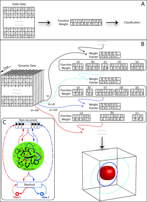
To expand the idea further, let’s revisit some core attributes of cognitive processing. Based on the observations of behavior, higher cognition requires “efficient computation”, “time delay feedback”, the capacity to “retain information” and “contextual” computational properties. Such computational cognitive process surpass the computational capacity of simple RNN-like networks. The essential required properties of a complex cognitive system of such kind are: 1) input should be nonlinearly mapped onto the high-dimensional state, while different inputs map onto different states, 2) slightly different states should map onto identical targets, 3) only recent past should influence the state and network is essentially unaware of remote past, 4) a phase-locked loop should decode information that is already encoded in time and 5) the combination of 1-4, should optimize sensory processing based on the context. The first three attributes of such system have close relevance to constrains and computational properties of higher cortical areas (prefrontal). The same three are also the main features of reservoir computing, namely “separation property”, “approximation property” and “fading memory” (Jaeger,, 2001, 2007; Maass et al.,, 2002, 2003). Interestingly, and RC system can “non-linearly” map a lower dimensional system to a high-dimensional space facilitating classification of the elements of the low-dimensional space. The last two properties match the structure and computational constraints of non-relay thalamic system as a contextual modulator that is phasically changing the input to the RC system. In fact, in an RC model of prefrontal cortex, addition of a phase neuron significantly improved the networks performance in complex cognitive tasks. The phase neuron improves the performance by generating input driven attractor dynamics that best matched the input (Enel et al.,, 2016). This advantageous phase-based bias effect is not limited to the simulation or physiological RC-like neural circuitry. In a recent study, electronic implementation and numerical studies of a limited RC system of a single nonlinear node with delayed feedback has shown efficient information processing (Appeltant et al.,, 2011). Such reservoir’s transient dynamical response follows delay-dynamical systems, and only a limited set of parameters are required to set the rich dynamical properties of delay systems (Ikeda and Matsumoto,, 1987). This system was able to effectively process time-dependent signals.
The phase neuron (Enel et al.,, 2016) and delayed dynamical RC (Appeltant et al.,, 2011) both show properties that resemble the structure-function of MD-like thalamus as discussed here. Specifically, the phasic recruitment of cortical neurons is invoked due to the combination of cortical influence on non-relay thalamic neurons through direct and indirect corticothalamic projections (see reticular nucleus inhibitory influence on thalamic neurons, Fig.LABEL:Fig_xxx). In a given cycle of computation (Fig. 5), cortical feedback leads to the release of GABA (via reticular nucleus) in MD (Kim et al.,, 2011). It has been shown that the increased GABA alters the opening of T-type Ca2+ channels (Crunelli and Leresche,, 1991), which in the case of MD, results in enhanced MD-PFC interaction; yielding mutual drive of the corticothalamic and thalamocortical activity together (Kim et al.,, 2011). Through this calcium-based low-threshold spiking, the gradual synchronization of MD and PFC ensues (Jones,, 2002; Kim et al.,, 2011). As a result, MD units and PFC show strong phase-locked synchrony (Parnaudeau et al.,, 2013). This gradual phase-locking mechanism forms the basis of the temporal dynamics that can nonlinearly (through consecutive cycles of computation) change the cortical activity as a result of novel stimuli or unexpected contextual changes as observed experimentally (Schmitt et al.,, 2017; Bolkan et al.,, 2017). In fact, abnormal activity of T-type Ca2+ channels in MD, leads to hypersynchrony in PFC neurons and frontal lobe-specific seizures(Kim et al.,, 2011). The interactions between non-relay thalamus and cortex, collectively, is neither feedforward, nor locally recurrent, but it has a mixture of non-recurrent phase encoder that keeps copies of the past processing and modulates the sensory input relay and its next step processing (Fig. 5). The distinctive short-term dynamics are well matched with the divergent structure-function relationship of sensory and non-relay MD-like thalamic nuclei. These features further emphasize that the perceptual and cognitive processing can not be solely cortico-centric operations.
Biological constrains and the role of thalamus in computational optimization
Computation and optimization are two sides of the same coin. But how does the brain optimize the computations that would match its required objective, i.e. cognitive processing? There is a current trend of thinking that brain optimizes some arbitrary functions, with the hopes that the future discovery of these unknown functions may guide us to establish a link between brain’s operations and deep learning (Marblestone et al.,, 2016). This line of approach to optimizational (and computational) operations of the brain has few flaws. First, it avoids specifying what function the brain is supposed to optimize (and as a result it remains vague). Second, it refrains from addressing certain limitations that brain has to cope with due to biological constrains. First of these limitations is the importance of using just enough resources to solve the current perceptual problem. Second is the necessity to come up with a solution just in (the needed) time. The importance of “just-enough” and “just-in-time” computation in cortical computation should not be overlooked (Douglas and Martin,, 2007). If the first condition is not met, the organism can not sustain continued activity since the metabolic demand surpasses the dedicated energetic expenditure and the animal can not survive. In fact, the communication in neural networks are highly constrained by number of factors, specifically the energetic demands of the network operations (Laughlin and Sejnowski,, 2003). From estimates of the cost of cortical computation Lennie, (2003), we know that the high cost of spiking forces the brain to rely on sparse communication and using only a small fraction of the available neurons (Shoham et al.,, 2006; Baddeley et al.,, 1997). While, theoretically, cortex can dedicate a large number of neurons (and very high dynamical space) to solve any cognitive task, metabolic demand of such high-energetic neural activity renders such mechanism highly inefficient. As a result, the “law of diminishing returns” dictates that increased energetic cost causing excessive pooling of active neurons to an assembly would be penalized (Niven and Laughlin,, 2008). The penalization for unnecessary high-energetic neural activity, in itself, should be driven by the nature of computation rather than being formulated as a fixed arbitrary threshold imposed by an external observer. On the other hand, a system can resort to low-cost computation at any given time but dedicate long enough time to solve the task on hand. Naturally, such system would not be very relevant to the biological systems since time is of essence. If an animal dedicates a long instance of its computational capacity to solve a problem, the environment has changed before it reaches a solution and the solution becomes obsolete. A deer would never have an advantage for its brain to have fully analyzed the visual scene instead of spotting the approaching wolf and shifting resources to the most-needed task, i.e. escape. As a result, many of the optimization techniques and concepts that may be relevant to artificial neural networks are irrelevant to embodied computational cognition of the brain. The optimization that the brain requires is not aiming for the best possible performance, but rather needs to reach a good mixture of economy and efficiency.
Not surprisingly, these constrains, i.e. efficiency and economy, are cornerstones of homeostasis and are observed across many scales in living systems (Szekely et al.,, 2013). The simple “Integral feedback” acts as the mainstay of control feedback in such homeostatic systems (such as E Coli heat-shock or DNA repair after exposure to gamma radiation) (El-Samad et al.,, 2002, 2005; Krishna et al.,, 2007; Dekel and Alon,, 2005). Change in input leads to change in the output and the proportional change in the controller aiming to reset the output to the desired regime. When the integral feedback is disrupted, the system can no longer reach proper homeostasis and either efficiency or economy (or even both) will be sub-optimal (El-Samad et al.,, 2002, 2005; Szekely et al.,, 2013). Many different etiologies could be behind the integral feedback disruption, but the outcome is loss of robust response in uncertain environments. The presence of feedforward and feedback loops provide the means for robust and fast operation in processing fluctuating incoming inputs. This feedback regulation and operational robustness has an energetic and computational cost for the system. Although for simple systems it is feasible to associate the exact cost of an operation to the overall computational cost of the system, scaling the metabolic cost of feedback regulations to large networks remains a challenge since it will involve multiple feedback loops, nonlinear dynamics and numerous uncertain parameters (Csete and Doyle,, 2002). Specifically in the case of a single neurons, branching architecture, non-uniform ion channel distributions and conduction states of action potentials affect the rate of energy consumption (Ju et al.,, 2016). However, this electrochemical energy of single neuron operation does not linearly scale to the spent energy at networks level (Wang et al.,, 2009; Wang and Wang,, 2014). The total energy function of neural populations will depend not only on the energy function of single neurons and their coupling in a given neural population, but also on the flow of information between different populations (Wang and Wang,, 2014; Wang and Zhu,, 2016). When a large pool of neurons is recruited to form multiple assemblies to perform a certain computation, it is the interactions between the assemblies that will define the collective behavior of the discrete components. Since the coupled processes show additive entropy productions (Demirel,, 2011), the total energetic optimality of the desired function would depend on the feedback loops between the assemblies and how these feedbacks control the intrinsic energy expenditure of a given assembly. These attributes are inline with the general principle of modular composition of biological systems (Hartwell et al.,, 1999; Dehghani,, 2017, 2018). From the dynamical systems’ perspective, to understand the operational principles (here, of large assembly of neurons), we do not need to the strip down the assembly to its individual component level (here, individual neurons) (Dehghani,, 2018). As a result the optimal control at the functional scale of modules where the interaction between the system’s modules take place (Csete and Doyle,, 2002; Dehghani,, 2018).
The constrains that we discussed above, directly translate to the computational operations of thalamocortical system as we discussed. Instead of just trying to deal with one fitness function at a time (where the minima of the landscape would be deemed as “the” optima), the brain has to perform a multi-objective optimization, finding solutions to both metabolic cost (economy) and just-in-time (efficiency) computation. Thus we can infer that a unique solution does not exist for such a problem. Rather, any optimization for computational efficiency will cost us economy and any optimization for economy will cost us efficiency. In such case, a multi-objective optimization pareto frontier is desirable. Pareto frontier of information/cost will be the set of solutions where any other point in the space is objectively worse for both of the objectives (Kung et al.,, 1975; Godfrey et al.,, 2006; Szekely et al.,, 2013). As a result, the optimization mechanism should push the system to this frontier. The iterative dynamical interaction between thalamus and cortex seems to provide an elegant solution for this problem (We discuss this in more details below).
In addition to these theoretical rationals, we also wish to point to some observations that support the emergent optimization in the thalamo-cortical system. For example, metabolic studies have shown that following thalamic injuries, a misbalance in cortical metabolism ensues (Baron et al.,, 1986, 1992; Larson et al.,, 1998; Levasseur et al.,, 1992). Moreover, in healthy humans (and not in mood disorder patients), the metabolic rate of thalamus directly relates to the power of cortical oscillations (Lindgren et al.,, 1999). The misbalance in cortical metabolism have been observed in variety of nuclei damages, but are specially pronounced in mediodorsal, centre median or pulvinar injuries (Baron et al.,, 1986, 1992). In addition, low-frequency and high-frequency stimulation of MD can induce long-term depression/potentiation or in mPFC (medial PFC); however, the exact sign and magnitude of the differential modulation of thalamo-prefrontal functions under low and high input drive depends on the lack or presence of Muscarinic and Nicotinic modulation (Bueno-Junior et al.,, 2012). Just as thalamic injury/modulation can change the cortical activity and metabolism, cortical injuries (due to stroke for example) can cause an attenuation of the excitatory feedback to thalamus and lead to thalamo-cortical dysrhythmia (van Wijngaarden et al.,, 2016). Regardless of where the initial injury has occurred, the disrupted thalamo-cortical interaction is conjoined with a misbalance in metabolism. The resultant out of balance activity leads to cognitive disorders that can happen in form of disrupted information processing due to cortical hypersynchrony as a results of excessive thalamic spiking (Kim et al.,, 2011) or faulty modulation of sensory signals and loss of the normal correlation between glucose metabolism in the thalamus and PFC (Byne et al.,, 2001; Katz et al.,, 1996). The exact celullar/subcellular mechanism that lies beneath the joint fluctuations of firing and metabolic of cortex and thalamus is not very well understood and number of mechanisms may act (not necessarily exclusively). For example, reduced cortical feedback may lead to thalamic hyperpolarization, and the resultant de-inactivation of voltage-gated T-type Ca channels may cause the neurons to switch from tonic spiking to a pathological bursting (van Wijngaarden et al.,, 2016). Or it could be that the thalamic drive of the inhibitory neurons in the cortex not only directly affect the cortical mode of firing (Fan et al.,, 2017) but also change the glycogenolysis in astrocytes through Vasoactive intestinal peptide (VIP) interneurons (Magistretti,, 2006; Magistretti and Allaman,, 2015). Interestingly, and in contrast to the noradrenergic afferent fibers that span horizontally across cortical domains, VIP neurons have a bipolar architecture and therefore their effect is spatially limited (Magistretti and Allaman,, 2015), likely correlated to the size of the functional assemblies that are recruited to perform a computational task. Whichever the exact mechanism at the cellular level is, the collective activity of modulatory thalamus and cortex drives the optimization that inherently can not be controlled by the information available at the scale of single neurons and solely in cortex.
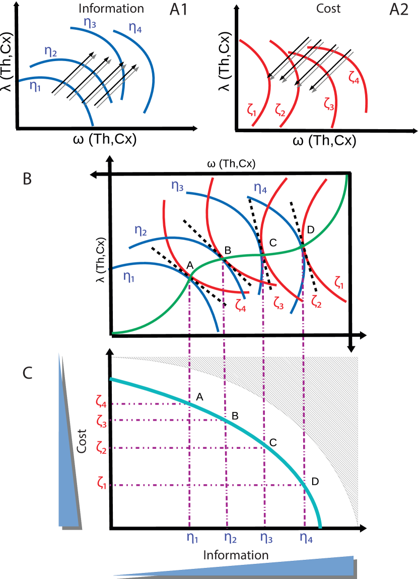
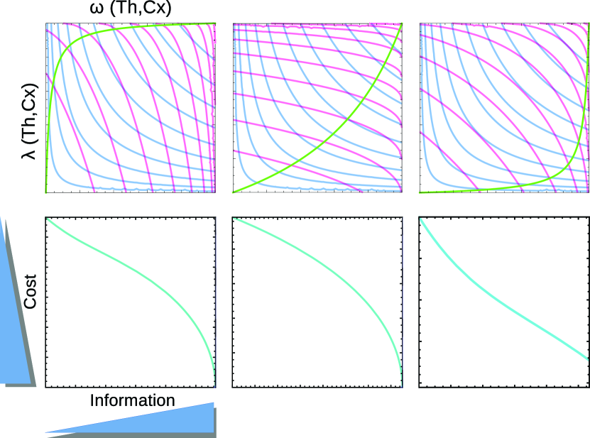
To formalize multiobjective optimization, consider a set of functions, and of (firing rate of thalamic cell) and (firing rate of cortical cells). Uncertainty (or its opposite, information) and computational cost (a mixture of time and metabolic expense) can both be mapped to this functional space of and (Fig. 6A1,2). Let’s define computational cost and information as product and linear sum of cortical and thalamic activity (,; with as coefficients) to reflect the logarithmic nature of information (entropy) and the fact that biological cost is an accelerating function of the cost-inducing variables (Dekel and Alon,, 2005). The hypothetical space of cost/information is depicted in Fig. 6, where top panels show indifference maps of information (A1) and cost (A2). The example simulations and parametric plots of the cost and information functions defined as above are shown in Fig. 7. In each indifference map, along each iso-quant curve, the total functional attribute is the same. For example, anywhere on the curve, the uncertainty (or information) in our computational engine is the same. However, different iso-quant curves represent different levels of the functional attribute. For example, moving outward increases information (reduces uncertainty) as and thus if computational cost was not a constrain, the optimal solution would have existed on or further away (Fig.6 A1). In contrast, moving inward would preserve the cost () if the computational engine did not have the objective of reducing uncertainty (Fig. 6A2). Since information and cost are interdependent and both depend on the interaction between thalamus and cortex, we suggest that information/cost optimization happens through an iterative interaction between thalamus and cortex (note the blackboard analogy and contextual modulation discussed above). Since we defined both information and cost as a set of iso-quant curves in the functional space of and , they can be co-represented in the same space (Fig. 6B). Optimal solutions for information/cost optimizations are simply the solutions to where the tangents of the iso-quant curves are equal (see the tangents [black dashed lines] and points A, B, C and D, in Fig. 6B). These points create a set of optimal solutions for the tradeoff between information and cost (green curve). Mapping of the optimal solutions to the computational efficiency space , gives us the pareto efficient curve (cyan curve, Fig. 6C). Anywhere inside the curve is not pareto efficient (i.e. information gain and computational cost can change in such a way that, collectively, the system can be in a better state (on the pareto curve). Points outside of the pareto efficient curve are not available to the current state of the system due to the coefficients of and . A change in these coefficients can potentially shape a different co-representation of information and cost (see Fig 7, top row for 3 different instances of and based on different values), and thus a different pareto efficient curve (see Fig 7, bottom row). These different possible pareto frontiers can be set based on the prior state of the system and the complexity of the computational problem on hand. For example, the modulatory MD-like thalamic triggering of feedforward inhibition of layer I interneurons and layers II/III pyramidals (Cruikshank et al.,, 2012) may tune the cortical activity to a sustained profile of arousal during wakefulness (Harris and Thiele,, 2011). Or in contrast to relay thalamus where maximum responsiveness to transient signals (such as sensory stimuli onset/offset) is needed (Rose and Metherate,, 2005; Bruno and Sakmann,, 2006), the MD-like modulatory drive may be invoked for tasks where working memory and contextual processing are needed (Parnaudeau et al.,, 2013; Delevich et al.,, 2015). Nonetheless, the computational efficiency of the system can not be infinitely pushed outward because of the system’s intrinsic biophysical constrains (neurons and their wiring). The shaded region in Fig 7, bottom row, shows this non-permissible zone.
In the defined computational efficiency space ,
composed of the two variables information and cost (as the objective
functions, shown in bottom panels of
Fig. 6 and
Fig. 7), solving a computational problem
is represented by a decrease in uncertainty. However, any change in
uncertainty has an associated cost. First derivative of the pareto
frontier shows “marginal rate of substitution” as .
This ratio varies among different points on the pareto efficient curve.
If we take two points on the pareto curve in the computational
efficiency space, such as A and C for example, computational efficiency
of these two points are equal . The change in
efficiency of point A with respect to information and cost, are the
partial derivatives and ,
respectively. As a result,, meaning that there is
constant efficiency along the pareto curve, the tradeoff between
information and cost is not constant. The optimization in this space is
not based on some fixed built-in algorithm or arbitrary thresholds by an
external observer. Rather, information/cost optimization is the result
of back and forth interaction between thalamus and cortex. Based on the
computational perspective that we have portrayed, thalamus seems to be
poised to operate as an optimizer. Thalamus receives a copy of (sensory)
input while relaying it, and receives an efferent copy from the
processor (cortex), while trying to efficiently bind the information
from past and present and sending it back to cortex. The outcome of such
emergent optimization, is a pareto front in the economy-efficiency
landscape
(Fig. 6,7).
If the cortex were to be the sole conductor of cognitive processing, the
dynamics of the relay and cortical processing would meander in the
parameter space and not yielding any optimization that can provide a
feasible solution to economic and just-in-time computation. Such system
is doomed to fail, either due to metabolic costs or due to computational
freeze over time ; thus more or less be a useless cognitive engine. In
contrast, with the help of an optimizer that acts as a contextual
modulator, the acceptable parameters will be confined to a manifold
within the parameter space. Such regime would be a sustainable and
favorable domain for cognitive computing. This property shows another
important facet of a thalamo-cortical computational cognitive system and
the need to move passed the cortico-centric view of cognition.
An important consequence of this formalization is that it provides us testable hypotheses for objectively evaluating information and cost optimization. By careful simultaneous measurements of thalamic and cortical collective activity, during different states and under different neurotransmitter modulatory effects, one should be able to examine the distinctive interaction of cortex and MD-like versus relay thalamic nuclei. Although we wish to emphasize that while information processing is a fundamentally energy-consuming process (Bennett,, 2003; Parrondo et al.,, 2015) and one can drive theoretical estimates of the energetic cost of the activity of a population of neurons, the exact translation of bit to watts in adaptive information processing systems (such as thalamocortical) can only be verified experimentally (Flack,, 2017). Without proper and careful measurements, it is impossible to predict how much more reliable the collective computation could get at the expense of energy (Ay et al.,, 2007). Likewise, the degree to which the energy is traded for accuracy/speed (or their combination) will be a hard challenge for the experimentalists measuring the collective activity (Lan et al.,, 2012).
Concluding remarks: Reframing Thalamic function above and beyond information relay
Lately, new evidence about the possible role of thalamus has started to challenged the cortico-centric view of perception/cognition. Anatomical studies and physiological measurements have begun to unravel the importance of the Cortico-Thalamo-Cortical loops in cognitive processes (Basso et al.,, 2005; Parnaudeau2017). Under this emerging paradigm, thalamus plays two distinctive roles: a) information relay, b) modulation of cortical function (Sherman and Guillery,, 2013), where the neocortex does not work in isolation but is largely dependent on thalamus. In contrast to cortical networks which operate as specialized memory devices via their local recurrent excitatory connections, the thalamus is devoid of local connections, and is instead optimized for capturing state information that is distributed across multiple cortical nodes while animals are engaged in context-dependent task switching (Schmitt et al.,, 2017). This allows the thalamus to explicitly represent task context (corresponding to different combinations of cortical states), and through its unique projection patterns to the cortex, different thalamic inputs modify the effective connections between cortical neurons (Schmitt et al.,, 2017; Bolkan et al.,, 2017).
Here, we started with a brief overview of the architecture of thalamus, the back and forth communication between thalamus and cortex, then we provided the electrophysiological evidence of thalamic modulatory function, and concluded with a computational frame that encapsulates the architectural and functional attributes of the thalamic role in cognition. In such frame, the computational efficiency of the cognitive computing machinery is achieved through iterative interactions between thalamus and cortex embedded in the hierarchical organization (Fig. 4, 5). Under this emergent view, thalamus serves not only as relay, but also as a read/write medium for cortical processing , playing a crucial role in contextual modulation of cognition (Fig. 8). Such multiscale organization of computational processes is a necessary requirement for design of the intelligent systems Dehghani, (2017); Simon, (1969, 1962). Distributed computing in biological systems in most cases operates without central control (Navlakha and Bar-Joseph,, 2014). This is well reflected in the computational perspective that we discussed here. We suggest that through the continuous contextual modulation of cortical activity, thalamus (along with cortex) plays a significant role in emergent optimization of computational efficiency and computational cost. This phenomenon has a deep relation with phase transitions in complex networks. Different states (phases) of the network are associated with the connectivity of the computing elements (see thalamic weight/pointer and cortical function/weight modules in Fig. 5). Interestingly, intrinsic properties of the complex networks do not define the phase transitions in system. Rather, the interplay of the system with its external environment shapes the landscape where phase transitions occur (Seoane and Solé,, 2015). This parallel in well-studied physical systems and neuronal networks of thalamo-cortical system show the importance of the interplay between thalamus and cortex in cognitive computation and optimization. The proposed frame for contextual cognitive computation and the emergent information/cost optimization in thalamo-cortical system can guide us in designing novel AI architecture.
Acknowledgements.
We wish to thank Michael Halassa for helpful discussions.References
- Alcaraz et al., (2018) Alcaraz, F., Fresno, V., Marchand, A. R., Kremer, E. J., Coutureau, E., and Wolff, M. (2018). Thalamocortical and corticothalamic pathways differentially contribute to goal-directed behaviors in the rat. eLife, 7.
- Alelú-Paz and Giménez-Amaya, (2008) Alelú-Paz, R. and Giménez-Amaya, J. M. (2008). The mediodorsal thalamic nucleus and schizophrenia. Journal of psychiatry & neuroscience: JPN, 33(6):489.
- Appeltant et al., (2011) Appeltant, L., Soriano, M. C., Van der Sande, G., Danckaert, J., Massar, S., Dambre, j., Schrauwen, B., Mirasso, C. R., and Fischer, I. (2011). Information processing using a single dynamical node as complex system. Nature Communications, 2:468.
- Ay et al., (2007) Ay, N., Flack, J., and Krakauer, D. C. (2007). Robustness and complexity co-constructed in multimodal signalling networks. Philosophical Transactions of the Royal Society B: Biological Sciences, 362(1479):441–447.
- Baddeley et al., (1997) Baddeley, R., Abbott, L. F., Booth, M. C. A., Sengpiel, F., Freeman, T., Wakeman, E. A., and Rolls, E. T. (1997). Responses of neurons in primary and inferior temporal visual cortices to natural scenes. Proceedings of the Royal Society B: Biological Sciences, 264(1389):1775–1783.
- Baron et al., (1986) Baron, J., D’antona, R., Pantano, P., Serdaru, M., Samson, Y., and Bousser, M. (1986). Effects of thalamic stroke on energy metabolism of the cerebral cortex: a positron tomography study in man. Brain, 109(6):1243–1259.
- Baron et al., (1992) Baron, J., Levasseur, M., Mazoyer, B., Legault-Demare, F., Mauguiere, F., Pappata, S., Jedynak, P., Derome, P., Cambier, J., and Tran-Dinh, S. (1992). Thalamocortical diaschisis: positron emission tomography in humans. Journal of Neurology, Neurosurgery & Psychiatry, 55(10):935–942.
- Basso et al., (2005) Basso, M. A., Uhlrich, D., and Bickford, M. E. (2005). Cortical Function: A View from the Thalamus. Neuron, 45(4):485–488.
- Baxter, (2013) Baxter, M. (2013). Mediodorsal thalamus and cognition in non-human primates. Frontiers in Systems Neuroscience, 7:38.
- Bennett, (2003) Bennett, C. H. (2003). Notes on landauer’s principle, reversible computation, and maxwell’s demon. Studies in History and Philosophy of Science Part B: Studies in History and Philosophy of Modern Physics, 34(3):501 – 510. Quantum Information and Computation.
- Bickford, (2016) Bickford, M. E. (2016). Thalamic Circuit Diversity: Modulation of the Driver/Modulator Framework. Frontiers in Neural Circuits, 9.
- Bolkan et al., (2017) Bolkan, S. S., Stujenske, J. M., Parnaudeau, S., Spellman, T. J., Rauffenbart, C., Abbas, A. I., Harris, A. Z., Gordon, J. A., and Kellendonk, C. (2017). Thalamic projections sustain prefrontal activity during working memory maintenance. Nature Neuroscience, 20(7):987–996.
- Bruno and Sakmann, (2006) Bruno, R. M. and Sakmann, B. (2006). Cortex is driven by weak but synchronously active thalamocortical synapses. Science, 312(5780):1622–1627.
- Bueno-Junior et al., (2012) Bueno-Junior, L. S., Lopes-Aguiar, C., Ruggiero, R. N., Romcy-Pereira, R. N., and Leite, J. P. (2012). Muscarinic and nicotinic modulation of thalamo-prefrontal cortex synaptic pasticity in vivo. PLOS ONE, 7:1–11.
- Byne et al., (2001) Byne, W., Buchsbaum, M. S., Kemether, E., Hazlett, v., Shinwari, A., Mitropoulou, V., and Siever, L. J. (2001). Magnetic resonance imaging of the thalamic mediodorsal nucleus and pulvinar in schizophrenia and schizotypal personality disorder. Archives of General Psychiatry, 58(2):133–140.
- Cardin et al., (2008) Cardin, J. A., Palmer, L. A., and Contreras, D. (2008). Cellular mechanisms underlying stimulus-dependent gain modulation in primary visual cortex neurons in vivo. Neuron, 59:150–160.
- Clasca et al., (2012) Clasca, F., Rubio-Garrido, P., and Jabaudon, D. (2012). Unveiling the diversity of thalamocortical neuron subtypes. Eur J Neurosci, 35:1524–32.
- Collins et al., (2018) Collins, D. P., Anastasiades, P. G., Marlin, J. J., and Carter, A. G. (2018). Reciprocal circuits linking the prefrontal cortex with dorsal and ventral thalamic nuclei. Neuron, 98(2):366–379.e4.
- Corbetta, (1998) Corbetta, M. (1998). Frontoparietal cortical networks for directing attention and the eye to visual locations: identical, independent, or overlapping neural systems? Proceedings of National Academy of Science, 95:831–8.
- Cruikshank et al., (2012) Cruikshank, S. J., Ahmed, O. J., Stevens, T. R., Patrick, S. L., Gonzalez, A. N., Elmaleh, M., and Connors, B. W. (2012). Thalamic control of layer 1 circuits in prefrontal cortex. Journal of Neuroscience, 32(49):17813–17823.
- Cruikshank et al., (2007) Cruikshank, S. J., Lewis, T. J., and Connors, B. W. (2007). Synaptic basis for intense thalamocortical activation of feedforward inhibitory cells in neocortex. Nature Neuroscience, 10.
- Crunelli and Leresche, (1991) Crunelli, V. and Leresche, N. (1991). A role for gabab receptors in excitation and inhibition of thalamocortical cells. Trends in Neurosciences, 14(1):16 – 21.
- Csete and Doyle, (2002) Csete, M. E. and Doyle, J. C. (2002). Reverse engineering of biological complexity. Science, 295(5560):1664–1669.
- Dehghani, (2017) Dehghani, N. (2017). Design of the Artificial: lessons from the biological roots of general intelligence. ArXiv.
- Dehghani, (2018) Dehghani, N. (2018). Theoretical principles of multiscale spatiotemporal control of neuronal networks: A complex systems perspective. Frontiers in Computational Neuroscience, 12:81.
- Dekel and Alon, (2005) Dekel, E. and Alon, U. (2005). Optimality and evolutionary tuning of the expression level of a protein. Nature, 436(7050):588–592.
- Delevich et al., (2015) Delevich, K., Tucciarone, J., Huang, Z. J., and Li, B. (2015). The mediodorsal thalamus drives feedforward inhibition in the anterior cingulate cortex via parvalbumin interneurons. Journal of Neuroscience, 35(14):5743–5753.
- Demirel, (2011) Demirel, Y. (2011). Energy Coupling, pages 419–440. MIT Press.
- Deniau and Chevalier, (1985) Deniau, J. M. and Chevalier, G. (1985). Disinhibition as a basic process in the expression of striatal functions. II. The striato-nigral influence on thalamocortical cells of the ventromedial thalamic nucleus. Brain Research, 334(2):227–233.
- Douglas and Martin, (2007) Douglas, R. J. and Martin, K. A. (2007). Mapping the Matrix: The Ways of Neocortex. Neuron, 56(2):226–238.
- El-Samad et al., (2002) El-Samad, H. J., Goff, J. P., and Khamash, M. H. (2002). Calcium Homeostasis and Parturient Hypocalcemia: An Integral Feedback Perspective. Journal of Theoretical Biology, 214(1):17–29.
- El-Samad et al., (2005) El-Samad, H. J., Kurata, H., Doyle, J. C., Gross, C. A., and Khamash, M. H. (2005). Surviving heat shock: Control strategies for robustness and performance. Proceedings of the National Academy of Sciences, 102(8):2736–2741.
- Enel et al., (2016) Enel, P., Procyk, E., Quilodran, R., and Dominey, P. F. (2016). Reservoir Computing Properties of Neural Dynamics in Prefrontal Cortex. PLOS Computational Biology, 12(6):e1004967.
- Fan et al., (2017) Fan, D., Duan, L., Wang, Q., and Luan, G. (2017). Combined effects of feedforward inhibition and excitation in thalamocortical circuit on the transitions of epileptic seizures. Frontiers in Computational Neuroscience, 11:59.
- Felleman and Essen, (1991) Felleman, D. J. and Essen, D. C. V. (1991). Distributed Hierarchical Processing in the Primate Cerebral Cortex. Cerebral Cortex, 1(1):1–47.
- FitzGibbon et al., (2015) FitzGibbon, T., Eriköz, B., Grünert, U., and Martin, P. R. (2015). Analysis of the lateral geniculate nucleus in dichromatic and trichromatic marmosets. Journal of Comparative Neurology, 523(13):1948–1966.
- Flack, (2017) Flack, J. (2017). Life’s Information Hierarchy, page 283–302. Cambridge University Press.
- Funahashi and Nakamura, (1993) Funahashi, K. and Nakamura, Y. (1993). Approximation of dynamical systems by continuous time recurrent neural networks. Neural Networks, 6(6):801–806.
- Fusi et al., (2016) Fusi, S., Miller, E. K., and Rigotti, M. (2016). Why neurons mix: high dimensionality for higher cognition. Current Opinion in Neurobiology, 37:66–74.
- Gabernet et al., (2005) Gabernet, L., Jadhav, S. P., Feldman, D. E., Carandini, M., and Scanziani, M. (2005). Somatosensory integration controlled by dynamic thalamocortical feed-forward inhibition. Neuron, 48(2):315–327.
- Giguere and Goldman-Rakic, (1988) Giguere, M. and Goldman-Rakic, P. S. (1988). Mediodorsal nucleus: Areal, laminar, and tangential distribution of afferents and efferents in the frontal lobe of rhesus monkeys. Journal of Comparative Neurology, 277(2):195–213.
- Godfrey et al., (2006) Godfrey, P., Shipley, R., and Gryz, J. (2006). Algorithms and analyses for maximal vector computation. The VLDB Journal, 16(1):5–28.
- Goldberg et al., (2013) Goldberg, J. H., Farries, M. A., and Fee, M. S. (2013). Basal ganglia output to the thalamus: still a paradox. Trends in Neurosciences, 36(12):695–705.
- Goldman-Rakic and Porrino, (1985) Goldman-Rakic, P. S. and Porrino, L. J. (1985). The primate mediodorsal (md) nucleus and its projection to the frontal lobe. Journal of Comparative Neurology, 242(4):535–560.
- Grant et al., (2012) Grant, E., Hoerder-Suabedissen, A., and Molnár, Z. (2012). Development of the Corticothalamic Projections. Frontiers in Neuroscience, 6.
- Groh et al., (2014) Groh, A., Bokor, H., Mease, R. A., Plattner, V. M., Hangya, B., Stroh, A., Deschenes, M., and Acsády, L. (2014). Convergence of Cortical and Sensory Driver Inputs on Single Thalamocortical Cells. Cerebral Cortex, 24(12):3167–3179.
- Halassa and Acsády, (2016) Halassa, M. M. and Acsády, L. (2016). Thalamic Inhibition: Diverse Sources Diverse Scales. Trends in Neurosciences, 39(10):680–693.
- Halassa and Kastner, (2017) Halassa, M. M. and Kastner, S. (2017). Thalamic functions in distributed cognitive control. Nature Neuroscience, 20(12):1669–1679.
- Harris and Thiele, (2011) Harris, K. D. and Thiele, A. (2011). Cortical state and attention. Nature Reviews Neuroscience, 12:509 EP –.
- Harth et al., (1987) Harth, E. M., Unnikrishnan, K. P., and Pandya, A. S. (1987). The inversion of sensory processing by feedback pathways: a model of visual cognitive functions. Science, 237(4811):184–187.
- Hartwell et al., (1999) Hartwell, L. H., Hopfield, J. J., Leibler, S., and Murray, A. W. (1999). From molecular to modular cell biology. Nature, 402:C47 EP –.
- Heeger, (2017) Heeger, D. J. (2017). Theory of cortical function. Proceedings of the National Academy of Sciences, 114(8):1773–1782.
- Hubel and Wiesel, (1959) Hubel, D. H. and Wiesel, T. N. (1959). Receptive fields of single neurones in the cat's striate cortex. The Journal of Physiology, 148(3):574–591.
- Hubel and Wiesel, (1962) Hubel, D. H. and Wiesel, T. N. (1962). Receptive fields binocular interaction and functional architecture in the cat's visual cortex. The Journal of Physiology, 160(1):106–154.
- Ikeda and Matsumoto, (1987) Ikeda, K. and Matsumoto, K. (1987). High-dimensional chaotic behavior in systems with time-delayed feedback. Physica D: Nonlinear Phenomena, 29(1-2):223–235.
- Jaeger, (2001) Jaeger, H. (2001). The echo state approach to analysing and training recurrent neural networks. Technical report.
- Jaeger, (2007) Jaeger, H. (2007). Echo state network. Scholarpedia, 2(9):2330.
- Jazayeri and Shadlen, (2015) Jazayeri, M. and Shadlen, M. N. (2015). A Neural Mechanism for Sensing and Reproducing a Time Interval. Current Biology, 25(20):2599–2609.
- Jones, (1981) Jones, E. G. (1981). Functional subdivision and synaptic organization of the mammalian thalamus. Int Rev Physiol, 25:173–245.
- Jones, (1985) Jones, E. G. (1985). Principles of Thalamic Organization. In The Thalamus, pages 85–149. Springer US.
- Jones, (1998) Jones, E. G. (1998). Viewpoint: the core and matrix of thalamic organization. Neuroscience, 85(2):331–345.
- Jones, (2002) Jones, E. G. (2002). Thalamic circuitry and thalamocortical synchrony. Philosophical Transactions of the Royal Society of London B: Biological Sciences, 357(1428):1659–1673.
- Ju et al., (2016) Ju, H., Hines, M. L., and Yu, Y. (2016). Cable energy function of cortical axons. Scientific Reports, 6:29686 EP –.
- Kakei et al., (2001) Kakei, S., Na, J., and Shinoda, Y. (2001). Thalamic terminal morphology and distribution of single corticothalamic axons originating from layers 5 and 6 of the cat motor cortex. The Journal of Comparative Neurology, 437(2):170–185.
- Katz et al., (1996) Katz, M., Buchsbaum, M. S., Siegel Jr, B. V., Wu, J., Haier, R. J., and Bunney Jr, W. E. (1996). Correlational patterns of cerebral glucose metabolism in never-medicated schizophrenics. Neuropsychobiology, 33(1):1–11.
- Khubieh et al., (2016) Khubieh, A., Ratté, S., Lankarany, M., and Prescott, S. A. (2016). Regulation of cortical dynamic range by background synaptic noise and feedforward inhibition. Cerebral Cortex, 26(8):3357–3369.
- Kim et al., (1995) Kim, H. G., Beierlein, M., and Connors, B. W. (1995). Inhibitory control of excitable dendrites in neocortex. Journal of Neurophysiology, 74(4):1810–1814.
- Kim et al., (2011) Kim, J., Woo, J., Park, Y.-G., Chae, S., Jo, S., Choi, J. W., Jun, H. Y., Yeom, Y. I., Park, S. H., Kim, K. H., Shin, H.-S., and Kim, D. (2011). Thalamic t-type ca2+ channels mediate frontal lobe dysfunctions caused by a hypoxia-like damage in the prefrontal cortex. Journal of Neuroscience, 31(11):4063–4073.
- Komura et al., (2013) Komura, Y., Nikkuni, A., Hirashima, N., Uetake, T., and Miyamoto, A. (2013). Responses of pulvinar neurons reflect a subject's confidence in visual categorization. Nature Neuroscience, 16(6):749–755.
- Krettek and Price, (1977) Krettek, J. E. and Price, J. L. (1977). The cortical projections of the mediodorsal nucleus and adjacent thalamic nuclei in the rat. Journal of Comparative Neurology, 171(2):157–191.
- Krishna et al., (2007) Krishna, S., Maslov, S., and Sneppen, K. (2007). UV-Induced Mutagenesis in Escherichia coli SOS Response: A Quantitative Model. PLoS Computational Biology, 3(3):e41.
- Kung et al., (1975) Kung, H.-T., Luccio, F., and Preparata, F. P. (1975). On Finding the Maxima of a Set of Vectors. Journal of the ACM, 22(4):469–476.
- Kuramoto et al., (2017) Kuramoto, E., Pan, S., Furuta, T., Tanaka, Y. R., Iwai, H., Yamanaka, A., Ohno, S., Kaneko, T., Goto, T., and Hioki, H. (2017). Individual mediodorsal thalamic neurons project to multiple areas of the rat prefrontal cortex: A single neuron-tracing study using virus vectors. Journal of Comparative Neurology, 525(1):166–185.
- Kuroda et al., (1998) Kuroda, M., Yokofujita, J., and Murakami, K. (1998). An ultrastructural study of the neural circuit between the prefrontal cortex and the mediodorsal nucleus of the thalamus. Progress in Neurobiology, 54(4):417 – 458.
- Kuroda et al., (2004) Kuroda, M., Yokofujita, J., Oda, S., and Price, J. L. (2004). Synaptic relationships between axon terminals from the mediodorsal thalamic nucleus and -aminobutyric acidergic cortical cells in the prelimbic cortex of the rat. Journal of Comparative Neurology, 477(2):220–234.
- Lan et al., (2012) Lan, G., Sartori, P., Neumann, S., Sourjik, V., and Tu, Y. (2012). The energy–speed–accuracy trade-off in sensory adaptation. Nature Physics, 8:422 EP –.
- Larson et al., (1998) Larson, C. L., Davidson, R. J., Abercrombie, H. C., Ward, R. T., Schaefer, S. M., Jackson, D. C., Holden, J. E., and Perlman, S. B. (1998). Relations between pet-derived measures of thalamic glucose metabolism and eeg alpha power. Psychophysiology, 35(2):162–169.
- Laughlin and Sejnowski, (2003) Laughlin, S. B. and Sejnowski, T. J. (2003). Communication in neuronal networks. Science, 301:1870–4.
- Lennie, (2003) Lennie, P. (2003). The Cost of Cortical Computation. Current Biology, 13(6):493–497.
- Levasseur et al., (1992) Levasseur, M., Baron, J., Sette, G., Legault-Demare, F., Pappata, S., Mauguiere, F., Benoit, N., Dinh, S. T., Degos, J., Laplane, D., et al. (1992). Brain energy metabolism in bilateral paramedian thalamic infarcts: a positron emission tomography study. Brain, 115(3):795–807.
- Lewis et al., (2012) Lewis, D. A., Curley, A. A., Glausier, J. R., and Volk, D. W. (2012). Cortical parvalbumin interneurons and cognitive dysfunction in schizophrenia. Trends in Neurosciences, 35(1):57–67.
- Lin et al., (2017) Lin, H. W., Tegmark, M., and Rolnick, D. (2017). Why Does Deep and Cheap Learning Work So Well? Journal of Statistical Physics, 168(6):1223–1247.
- Lindgren et al., (1999) Lindgren, K. A., Larson, C. L., Schaefer, S. M., Abercrombie, H. C., Ward, R. T., Oakes, T. R., Holden, J. E., Perlman, S. B., Benca, R. M., and Davidson, R. J. (1999). Thalamic metabolic rate predicts eeg alpha power in healthy control subjects but not in depressed patients. Biological Psychiatry, 45(8):943–952.
- Ma and Jazayeri, (2014) Ma, W. J. and Jazayeri, M. (2014). Neural Coding of Uncertainty and Probability. Annual Review of Neuroscience, 37(1):205–220.
- Maass et al., (2003) Maass, W., Natschlaeger, T., and Markram, H. (2003). Computational Models for Generic Cortical Microcircuits. In Computational Neuroscience. Chapman and Hall/CRC.
- Maass et al., (2002) Maass, W., Natschläger, T., and Markram, H. (2002). Real-Time Computing Without Stable States: A New Framework for Neural Computation Based on Perturbations. Neural Computation, 14(11):2531–2560.
- Magistretti, (2006) Magistretti, P. J. (2006). Neuron–glia metabolic coupling and plasticity. Journal of Experimental Biology, 209(12):2304–2311.
- Magistretti and Allaman, (2015) Magistretti, P. J. and Allaman, I. (2015). A cellular perspective on brain energy metabolism and functional imaging. Neuron, 86(4):883 – 901.
- Mante et al., (2013) Mante, V., Sussillo, D., Shenoy, K. V., and Newsome, W. T. (2013). Context-dependent computation by recurrent dynamics in prefrontal cortex. Nature, 503(7474):78–84.
- Marblestone et al., (2016) Marblestone, A. H., Wayne, G., and Kording, K. P. (2016). Toward an Integration of Deep Learning and Neuroscience. Frontiers in Computational Neuroscience, 10.
- Marenco et al., (2012) Marenco, S., Stein, J. L., Savostyanova, A. A., Sambataro, F., Tan, H.-Y., Goldman, A. L., Verchinski, B. A., Barnett, A. S., Dickinson, D., Apud, J. A., Callicott, J. H., Meyer-Lindenberg, A., and Weinberger, D. R. (2012). Investigation of Anatomical Thalamo-Cortical Connectivity and fMRI Activation in Schizophrenia. Neuropsychopharmacology, 37(2):499–507.
- Mathers, (1972) Mathers, L. H. (1972). The synaptic organization of the cortical projection to the pulvinar of the squirrel monkey. The Journal of Comparative Neurology, 146(1):43–59.
- Mesulam, (1990) Mesulam, M. M. (1990). Large-scale neurocognitive networks and distributed processing for attention, language, and memory. Ann Neurol, 28:597–613.
- Mitchell, (2015) Mitchell, A. S. (2015). The mediodorsal thalamus as a higher order thalamic relay nucleus important for learning and decision-making. Neurosci Biobehav Rev, 54:76–88.
- Mitelman et al., (2005) Mitelman, S. A., Byne, W., Kemether, E. M., Hazlett, E. A., and Buchsbaum, M. S. (2005). Metabolic Disconnection Between the Mediodorsal Nucleus of the Thalamus and Cortical Brodmann’s Areas of the Left Hemisphere in Schizophrenia. American Journal of Psychiatry, 162(9):1733–1735.
- Mumford, (1991) Mumford, D. (1991). On the computational architecture of the neocortex. I: The role of the thalamo-cortical loop. Biological Cybernetics, 65(2):135–145.
- Mumford, (1992) Mumford, D. (1992). On the computational architecture of the neocortex. II The role of cortico-cortical loops. Biological Cybernetics, 66(3):241–251.
- Nair et al., (2013) Nair, A., Treiber, J. M., Shukla, D. K., Shih, P., and Müller, R.-A. (2013). Impaired thalamocortical connectivity in autism spectrum disorder: a study of functional and anatomical connectivity. Brain, 136(6):1942–1955.
- Nakajima and Halassa, (2017) Nakajima, M. and Halassa, M. M. (2017). Thalamic control of functional cortical connectivity. Current Opinion in Neurobiology, 44:127–131.
- Navlakha and Bar-Joseph, (2014) Navlakha, S. and Bar-Joseph, Z. (2014). Distributed information processing in biological and computational systems. Communications of the ACM, 58(1):94–102.
- Newell, (1962) Newell, A. (1962). Some problems of basic organization in problem solving programs.
- Nii, (1986) Nii, P. (1986). The Blackboard Model of Problem Solving and the Evolution of Blackboard Architectures. The AI Magazine, 7(2):38–53.
- Niven and Laughlin, (2008) Niven, J. E. and Laughlin, S. B. (2008). Energy limitation as a selective pressure on the evolution of sensory systems. Journal of Experimental Biology, 211(11):1792–1804.
- Ohno et al., (2012) Ohno, S., Kuramoto, E., Furuta, T., Hioki, H., Tanaka, Y., Fujiyama, F., Sonomura, T., Uemura, M., Sugiyama, K., and Kaneko, T. (2012). A morphological analysis of thalamocortical axon fibers of rat posterior thalamic nuclei: a single neuron tracing study with viral vectors. Cereb Cortex, 22:2840–57.
- Parnaudeau et al., (2017) Parnaudeau, S., Bolkan, S. S., and Kellendonk, C. (2017). The Mediodorsal Thalamus: An Essential Partner of the Prefrontal Cortex for Cognition. Biological Psychiatry.
- Parnaudeau et al., (2013) Parnaudeau, S., O’Neill, P.-K., Bolkan, S. S., Ward, R. D., Abbas, A. I., Roth, B. L., Balsam, P. D., Gordon, J. A., and Kellendonk, C. (2013). Inhibition of mediodorsal thalamus disrupts thalamofrontal connectivity and cognition. Neuron, 77(6):1151–1162.
- Parnaudeau et al., (2015) Parnaudeau, S., Taylor, K., Bolkan, S. S., Ward, R. D., Balsam, P. D., and Kellendonk, C. (2015). Mediodorsal Thalamus Hypofunction Impairs Flexible Goal-Directed Behavior. Biological Psychiatry, 77(5):445–453.
- Parrondo et al., (2015) Parrondo, J. M. R., Horowitz, J. M., and Sagawa, T. (2015). Thermodynamics of information. Nature Physics, 11:131 EP –.
- Partlow et al., (1977) Partlow, G. D., Colonnier, M., and Szabo, J. (1977). Thalamic projections of the superior colliculus in the rhesus monkey,Macaca mulatta. A light and electron microscopic study. The Journal of Comparative Neurology, 171(3):285–317.
- Pelzer et al., (2017) Pelzer, P., Horstmann, H., and Kuner, T. (2017). Ultrastructural and functional properties of a giant synapse driving the piriform cortex to mediodorsal thalamus projection. Frontiers in Synaptic Neuroscience, 9:3.
- Pinault, (2004) Pinault, D. (2004). The thalamic reticular nucleus: structure function and concept. Brain Research Reviews, 46(1):1–31.
- Popken et al., (2000) Popken, G. J., Bunney, W. E., Potkin, S. G., and Jones, E. G. (2000). Subnucleus-specific loss of neurons in medial thalamus of schizophrenics. Proceedings of the National Academy of Sciences, 97(16):9276–9280.
- Prescott and De Koninck, (2003) Prescott, S. A. and De Koninck, Y. (2003). Gain control of firing rate by shunting inhibition: Roles of synaptic noise and dendritic saturation. Proceedings of the National Academy of Sciences, 100(4):2076–2081.
- Purushothaman et al., (2012) Purushothaman, G., Marion, R., Li, K., and Casagrande, V. (2012). Gating and control of primary visual cortex by pulvinar. Nat Neurosci, 15:905–12.
- Raczkowski and Fitzpatrick, (1990) Raczkowski, D. and Fitzpatrick, D. (1990). Terminal arbors of individual physiologically identified geniculocortical axons in the tree shrew's striate cortex. The Journal of Comparative Neurology, 302(3):500–514.
- Rigotti et al., (2013) Rigotti, M., Barak, O., Warden, M. R., Wang, X.-J., Daw, N. D., Miller, E. K., and Fusi, S. (2013). The importance of mixed selectivity in complex cognitive tasks. Nature, 497(7451):585–590.
- Rigotti et al., (2010) Rigotti, M., Rubin, D. B. D., Wang, X.-J., and Fusi, S. (2010). Internal representation of task rules by recurrent dynamics: the importance of the diversity of neural responses. Frontiers in Computational Neuroscience, 4.
- Rikhye et al., (2018) Rikhye, R. V., Wimmer, R. D., and Halassa, M. M. (2018). Towards an integrative theory of thalamic function. Nature Neuroscience (In press), xxx(xxx):xxx–xxx.
- Rose and Metherate, (2005) Rose, H. J. and Metherate, R. (2005). Auditory thalamocortical transmission is reliable and temporally precise. Journal of Neurophysiology, 94(3):2019–2030.
- Rotaru et al., (2005) Rotaru, D. C., Barrionuevo, G., and Sesack, S. R. (2005). Mediodorsal thalamic afferents to layer iii of the rat prefrontal cortex: Synaptic relationships to subclasses of interneurons. Journal of Comparative Neurology, 490(3):220–238.
- Rouiller and Welker, (2000) Rouiller, E. M. and Welker, E. (2000). A comparative analysis of the morphology of corticothalamic projections in mammals. Brain Research Bulletin, 53(6):727–741.
- Rovo et al., (2012) Rovo, Z., Ulbert, I., and Acsády, L. (2012). Drivers of the primate thalamus. J Neurosci, 32:17894–908.
- Saalmann, (2014) Saalmann, Y. B. (2014). Intralaminar and medial thalamic influence on cortical synchrony, information transmission and cognition. Frontiers in Systems Neuroscience, 8:83.
- Saalmann and Kastner, (2011) Saalmann, Y. B. and Kastner, S. (2011). Cognitive and perceptual functions of the visual thalamus. Neuron, 71(2):209 – 223.
- Saalmann and Kastner, (2015) Saalmann, Y. B. and Kastner, S. (2015). The cognitive thalamus. Frontiers in Systems Neuroscience, 9:39.
- Schmitt et al., (2017) Schmitt, L. I., Wimmer, R. D., Nakajima, M., Happ, M., Mofakham, S., and Halassa, M. M. (2017). Thalamic amplification of cortical connectivity sustains attentional control. Nature, 545(7653):219–223.
- Schwartz et al., (1991) Schwartz, M. L., Dekker, J. J., and Goldman-Rakic, P. S. (1991). Dual mode of corticothalamic synaptic termination in the mediodorsal nucleus of the rhesus monkey. Journal of Comparative Neurology, 309(3):289–304.
- Scott et al., (2017) Scott, B. B., Constantinople, C., Akrami, A., Hanks, T. D., Brody, C. D., and Tank, D. W. (2017). Fronto-parietal Cortical Circuits Encode Accumulated Evidence with a Diversity of Timescales. Neuron, 95:385–398.e5.
- Selfridge, (1959) Selfridge, O. G. (1959). Pandemonium: a paradigm for learning in . In Blake, D. V. and Uttley, A. M., editors, Proceedings of the Symposium on Mechanisation of Thought Processes, pages 513–526, London. National Physical Laboratory.
- Seoane and Solé, (2015) Seoane, L. F. and Solé, R. (2015). Phase transitions in Pareto optimal complex networks. Physical Review E, 92(3).
- Sherman, (2016) Sherman, S. M. (2016). Thalamus plays a central role in ongoing cortical functioning. Nature Neuroscience, 16(4):533–541.
- Sherman and Guillery, (2002) Sherman, S. M. and Guillery, R. W. (2002). The role of the thalamus in the flow of information to the cortex. Philosophical Transactions of the Royal Society B: Biological Sciences, 357(1428):1695–1708.
- Sherman and Guillery, (2013) Sherman, S. M. and Guillery, R. W. (2013). Functional Connections of Cortical Areas. The MIT Press.
- Shoham et al., (2006) Shoham, S., O’Connor, D. H., and Segev, R. (2006). How silent is the brain: is there a “dark matter” problem in neuroscience? Journal of Comparative Physiology A, 192(8):777–784.
- Simon, (1962) Simon, H. A. (1962). The Architecture of Complexity. Proceedings of the American Philosophical Society, 106(6):467–482.
- Simon, (1969) Simon, H. A. (1969). The Sciences of the Artificial, chapter The Architecture of Complexity. MIT Press.
- Sipper, (1999) Sipper, M. (1999). The emergence of cellular computing. Computer, 32(7):18–26.
- Solé and Macia, (2013) Solé, R. V. and Macia, J. (2013). Expanding the landscape of biological computation with synthetic multicellular consortia. Natural Computing, 12(4):485–497.
- Spacek and Lieberman, (1974) Spacek, J. and Lieberman, A. (1974). Ultrastructure and three-dimensional organization of synaptic glomeruli in rat somatosensory thalamus. Journal of anatomy, 117(Pt 3):487.
- Steriade and Deschenes, (1984) Steriade, M. and Deschenes, M. (1984). The thalamus as a neuronal oscillator. Brain Research Reviews, 8(1):1–63.
- Steriade et al., (1986) Steriade, M., Domich, L., and Oakson, G. (1986). Reticularis thalami neurons revisited: activity changes during shifts in states of vigilance. J Neurosci, 6:68–81.
- Steriade and Llinás, (1988) Steriade, M. and Llinás, R. R. (1988). The functional states of the thalamus and the associated neuronal interplay. Physiological Reviews, 68(3):649–742.
- Steriade and Pare, (2007) Steriade, M. and Pare, D. (2007). Morphology and electroresponsive properties of thalamic neurons. In Gating in Cerebral Networks, pages 1–26. Cambridge University Press.
- Steriade et al., (1984) Steriade, M., Parent, A., and Hada, J. (1984). Thalamic projections of nucleus reticularis thalami of cat: A study using retrograde transport of horseradish peroxidase and fluorescent tracers. The Journal of Comparative Neurology, 229(4):531–547.
- Szekely et al., (2013) Szekely, P., Sheftel, H., Mayo, A., and Alon, U. (2013). Evolutionary Tradeoffs between Economy and Effectiveness in Biological Homeostasis Systems. PLoS Computational Biology, 9(8):e1003163.
- Uhlhaas and Singer, (2010) Uhlhaas, P. J. and Singer, W. (2010). Abnormal neural oscillations and synchrony in schizophrenia. Nature Reviews Neuroscience, 11:100 EP –.
- Urbain and Deschênes, (2007) Urbain, N. and Deschênes, M. (2007). Motor Cortex Gates Vibrissal Responses in a Thalamocortical Projection Pathway. Neuron, 56(4):714–725.
- van Wijngaarden et al., (2016) van Wijngaarden, J. B. G., Zucca, R., Finnigan, S., and Verschure, P. F. M. J. (2016). The impact of cortical lesions on thalamo-cortical network dynamics after acute ischaemic stroke: A combined experimental and theoretical study. PLOS Computational Biology, 12(8):1–16.
- (149) Viaene, A. N., Petrof, I., and Sherman, S. M. (2011a). Properties of the thalamic projection from the posterior medial nucleus to primary and secondary somatosensory cortices in the mouse. Proceedings of the National Academy of Sciences, 108(44):18156–18161.
- (150) Viaene, A. N., Petrof, I., and Sherman, S. M. (2011b). Synaptic properties of thalamic input to the subgranular layers of primary somatosensory and auditory cortices in the mouse. Journal of Neuroscience, 31(36):12738–12747.
- Wang et al., (2018) Wang, J., Narain, D., Hosseini, E. A., and Jazayeri, M. (2018). Flexible timing by temporal scaling of cortical responses. Nature Neuroscience.
- Wang and Wang, (2014) Wang, R. and Wang, Z. (2014). Energy distribution property and energy coding of a structural neural network. Frontiers in Computational Neuroscience, 8:14.
- Wang et al., (2009) Wang, R., Zhang, Z., and Chen, G. (2009). Energy coding and energy functions for local activities of the brain. Neurocomputing, 73(1):139 – 150.
- Wang and Zhu, (2016) Wang, R. and Zhu, Y. (2016). Can the activities of the large scale cortical network be expressed by neural energy? a brief review. Cognitive neurodynamics, 10(1):1–5.
- Woodward et al., (2017) Woodward, N. D., Giraldo-Chica, M., Rogers, B., and Cascio, C. J. (2017). Thalamocortical Dysconnectivity in Autism Spectrum Disorder: An Analysis of the Autism Brain Imaging Data Exchange. Biological Psychiatry: Cognitive Neuroscience and Neuroimaging, 2(1):76–84.
- Yamins and DiCarlo, (2016) Yamins, D. L. K. and DiCarlo, J. J. (2016). Using goal-driven deep learning models to understand sensory cortex. Nature Neuroscience, 19(3):356–365.
- Yamins et al., (2014) Yamins, D. L. K., Hong, H., Cadieu, C. F., Solomon, E. A., Seibert, D., and DiCarlo, J. J. (2014). Performance-optimized hierarchical models predict neural responses in higher visual cortex. Proceedings of the National Academy of Sciences, 111(23):8619–8624.
- Zhou et al., (2016) Zhou, H., Schafer, R. J., and Desimone, R. (2016). Pulvinar-cortex interactions in vision and attention. Neuron, 89(1):209–220.
- Zikopoulos and Barbas, (2007) Zikopoulos, B. and Barbas, H. (2007). Parallel driving and modulatory pathways link the prefrontal cortex and thalamus. PLOS ONE, 2(9):1–19.