In-situ strain-tuning of the metal-insulator-transition of Ca2RuO4 in angle-resolved photoemission experiments
Abstract: We report the evolution of the -space electronic structure of lightly doped bulk Ca2RuO4 with uniaxial strain. Using ultrathin plate-like crystals, we achieve strain levels up to , sufficient to suppress the Mott phase and access the previously unexplored metallic state at low temperature. Angle-resolved photoemission experiments performed while tuning the uniaxial strain reveal that metallicity emerges from a marked redistribution of charge within the Ru shell, accompanied by a sudden collapse of the spectral weight in the lower Hubbard band and the emergence of a well defined Fermi surface which is devoid of pseudogaps. Our results highlight the profound roles of lattice energetics and of the multiorbital nature of Ca2RuO4 in this archetypal Mott transition and open new perspectives for spectroscopic measurements.
Main text:
Mott metal-insulator-transitions are driven by electron-electron interactions but often coincide with structural phase transitions Imada1998 . While the latter were long believed to be a secondary response, as argued originally by N.F. Mott Mott1949 , realistic numerical studies point to a far more important role of structural changes in stabilizing the Mott state of archetypal insulators Pavarini2004 ; Gorelov2010 . This, together with recent theoretical advances, has led to renewed interest in the interplay of lattice energetics and electronic properties near Mott transitions Park2013 ; Leonov2014 ; Han2018 . Hydrostatic and uniaxial pressure is particularly important in the experimental study of Mott transitions and also has a profound effect on other emerging properties of quantum materials Imada1998 ; Mathur1998 ; Schlom2007 ; Drozdov2015 ; Hicks2014 ; Burganov2016 . However, conventional pressure cells are fundamentally incompatible with modern surface sensitive spectroscopies such as angle-resolved photoemission (ARPES). Consequently the evolution of the -space electronic structure in Mott systems as they are tuned across the metal-insulator transition (MIT) has remained largely unkown. In order to overcome this limitation of ARPES, we developed an apparatus which is compatible with modern ARPES facilities and permits in-situ quasi-continuous tuning of uniaxial strain. Here, we use this new capability to investigate the layered perovskite Ca2RuO4, which is of particular scientific interest as a prototypical multiband Mott insulator. Within band theory Ca2RuO4 is a good metal with a nearly uniform distribution of the 4 Ru -electrons over the 3 orbitals. How such a multiband metal with fractional occupation can undergo a Mott transition has been debated intensely, but the lack of data from the metallic state has prevented stringent tests of theoretical models Anisimov2002 ; Liebsch2007 ; Gorelov2010 ; Zhang2017 ; Kubota2005 ; Sutter2017 . More recently the magnetic properties in the insulating state of Ca2RuO4 have attracted much interest Kunkemoller2015 ; Jain2017 ; Zhang2017 following proposals of a state with excitonic magnetism and an exotic doping evolution khaliullin2013 ; Chaloupka2016 as well as the observation of unprecedented diamagnetism in a semimetallic phase induced by dc electric current Sow2017 .
The insulating state of Ca2RuO4 is known to be very sensitive to pressure Nakamura2002 ; Steffens2005 ; Miao2012 ; Dietl2018 , chemical substitution Nakatsuji2000 ; Fukazawa2001 and even electric fields Nakamura2013 ; Sow2017 . To further increase the sensitivity of the insulating ground state of Ca2RuO4 to strain, we have grown a series of La, Nd and Pr doped single crystals. Details of the sample growth and characterization are given in the supplementary information. Despite the slightly different rare earth ionic radii these samples behave qualitatively similarly. We thus chose to concentrate on Ca2-xPrxRuO4 with , 0.03, 0.04, 0.07. Consistent with a previous study on La-doped Ca2RuO4 Fukazawa2001 we find that Pr does not introduce itinerant carriers in the insulating state but suppresses the structural phase transition accompanying the metal insulator transition (MIT) from K for to K at the highest doping level of used in our study (see phase diagram in Fig. 1a). Our single crystal neutron diffraction data show that the structural transition of Ca2-xPrxRuO4 is similar to the one in pure Ca2RuO4. In particular, it is symmetry-preserving for all doping levels and mainly characterized by a flattening of the RuO6 octahedra together with an elongation of the -axis leading to strong orthorhombicity in the insulating phase [see supplementary information]. We will exploit this latter property to tune the MIT by uniaxial strain. Adopting the notation used for pure Ca2RuO4, we call the metallic phase with long -axis and space group L-Pbca and the insulating phase with short axis S-Pbca.
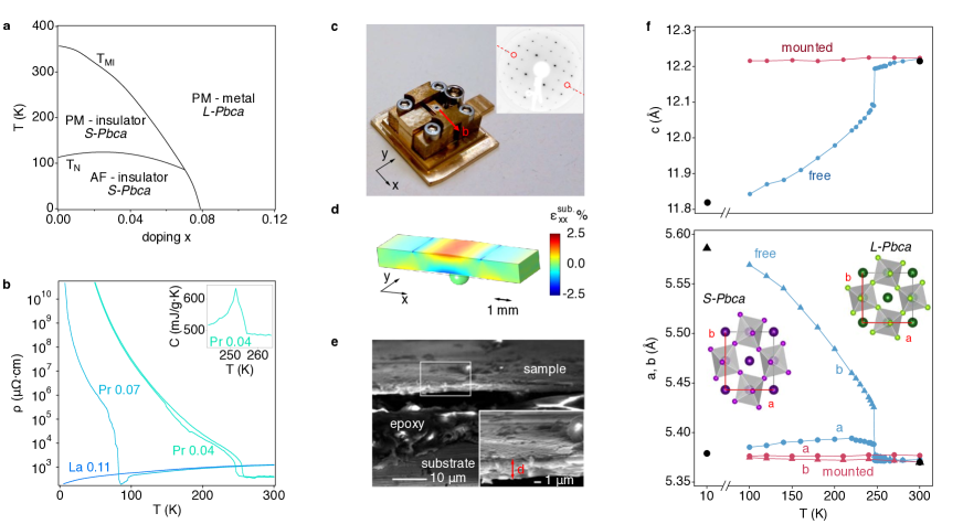
Our in-situ transferable strain apparatus is shown in Fig. 1c. It is actuated mechanically by turning a screw, which causes a lever to press a stainless steel ball from below on a 1 mm thick CuBe substrate. The elastic deformation of the substrate results in tensile strain along the bending direction on the upper surface and a much smaller compressive strain in the orthogonal direction. We calibrate using finite element analysis, as shown in Fig. 1d and supplementary Fig. 2, taking into account the indent in the substrate left by the ball, which we measure at the end of each experiment. For a maximal coupling of in-plane strain to the -axis compression, which putatively drives the MIT Gorelov2010 , we align the crystalline -axis with the bending direction. Since this axis lies in a glide plane of the structure, it can be identified readily in low-energy electron diffraction (LEED) patterns via the extinction of spots at certain energies (inset to Fig. 1c).
Key to our experiment is the exploitation of the initial compressive strain exerted by the large differential thermal contraction as apparatus and sample are cooled to base temperature. Using literature data for the CuBe substrates and our neutron diffraction data for Ca2-xPrxRuO4, we calculate nominal values of for Pr concentrations (0.07) [see supplementary information]. We directly confirm these exceptionally high strain levels for the most challenging case of a sample using X-ray diffraction on cleaved samples mounted on our strain apparatus. From the data shown in Fig. 1f we calculate an initial compressive strain at 100 K, in excellent agreement with the nominal value of at this temperature. These strain levels are achieved by mounting ultrathin plate-like single crystals to minimize strain relaxation. Cross-sectional electron microscopy images of our mounted and cleaved samples indicate typical thicknesses of m for the epoxy layer and m for the single crystals (Fig. 1e). Having confirmed negligible relaxation at the highest strain used in our experiment, we approximate the total strain as , where is compressive and tensile.
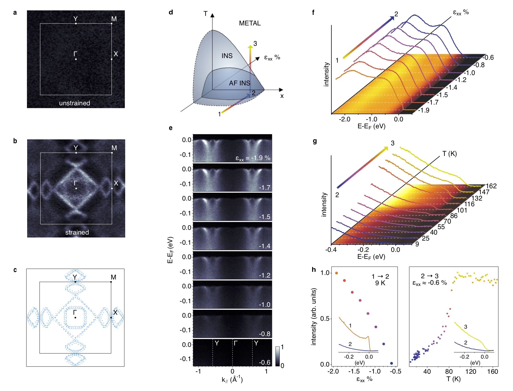
From Fig. 1f it is evident that suitably mounted samples remain in the quasi-tetragonal L-Pbca structure down to base temperature. Bending the substrate in our strain apparatus at low temperature then drives Ca2-xPrxRuO4 towards the orthorhombic S-Pbca ground state. The striking effect of uniaxial strain on the electronic structure of Ca2-xPrxRuO4 is evident from the ARPES Fermi surfaces shown in Fig. 2a,b. For an unstrained sample in the S-Pbca phase we find negligible intensity at the Fermi level and no discernible structure in momentum space consistent with a gapped Mott insulating state. In a fully strained sample with L-Pbca structure, on the other hand, a clear Fermi surface emerges, demonstrating a metallic ground state. Intriguingly, the strain-induced metallic state differs strongly from lightly doped cuprates and iridates Damascelli2003 ; DeLaTorre2015 . In particular, we find no anisotropy in the quasiparticle coherence and no evidence for a pseudogap along the entire Fermi surface within the precision of our experiment of meV. A cut along the Y high-symmetry line (Fig. 2e, first panel) shows well defined, strongly renormalized quasiparticle states at very low energy only indicating a delicate Fermi liquid regime. Beyond a coherence scale of meV, the excitations broaden rapidly and their dispersion increases simultaneously. These high-energy states can be tracked down to eV and thus essentially over the full bare band width (see Fig. 3). Such a coexistence of heavy quasiparticles with ’unrenormalized’ high-energy states was identified as a hallmark of Hund’s metals with profound implications on magnetic susceptibility, thermal and electrical transport Georges2012 .
Subsequently, we use our strain apparatus to tune across the Mott transition. Fig. 2e,f shows the evolution of the near- electronic structure for a sample following the path in the schematic phase diagram of Fig. 2d. Clearly, we achieve a sufficient tuning range to fully recover the characteristic spectrum of insulating Ca2-xPrxRuO4 with an exponential onset of weight. The angle-resolved data show that the Mott transition is approached by a uniform reduction of the spectral weight. Interestingly though, the quasiparticle dispersion is not affected strongly by strain. We can thus exclude that the strain-induced Mott transition is triggered by a divergence of the effective mass predicted in the Brinkmann-Rice model Brinkmann1970 . Raising the temperature (, Fig. 2g), the insulating state undergoes another phase transition close to of the unstrained state and we recover a metallic spectrum with significant weight at . As shown in Fig. 2e,f, the suppression of the spectral weight at during the strain-tuning is gradual. This can either indicate a second order phase transition or a phase coexistence with domains below the lateral dimension of m probed by ARPES. Given the sensitivity of the electronic state of Ca2-xPrxRuO4 to its first order structural phase transition, we consider the latter more likely. Additional evidence for phase coexistence, which was also observed in diffraction experiments on Ca2RuO4 under hydrostatic pressure Steffens2005 , is shown in supplementary information.
The Fermi surface of strained Ca2-xPrxRuO4 is remarkably simple considering the large unit cell containing 4 formula units and 16 electrons in the Ru shell. We find a square hole-like sheet centered at , which encloses a smaller electron-like Fermi surface, and four small lens-shaped sheets at the and points, respectively. The absence of exchange splitting in our experimental data indicates a paramagnetic metallic state, as it is also observed in the L-Pbca phase of undoped Ca2RuO4 and for highly La-doped Ca2RuO4 with L-Pbca structure in the ground state Fukazawa2001 . We thus conclude that the low-temperature metallic state induced by uniaxial strain in our experiments represents the intrinsic metallic phase of Ca2RuO4 but differs from bulk crystals under hydrostatic pressure and from epitaxially strained thin films, where ferromagnetism with an ordered moment of 0.1 0.3 is observed below K Nakamura2002 ; Miao2012 ; Dietl2018 .
Measuring the Fermi surface volume, we find approximate electron-hole compensation, consistent with a localized character of the Pr excess electrons. Doping dependent measurements (not shown), on the other hand indicate a measurable change of the Fermi surface volume with doping , suggesting an at least partial delocalization of the dopants in the metallic L-Pbca phase. Due to the large rotation of the RuO6 octahedra and the sizeable spin-orbit coupling, we cannot uniquely identify the orbital character on the Fermi surface from linear dichroism measurements. However, it appears plausible to interpret the extended straight sections of the experimental Fermi surface as originating predominantly from the quasi-1d , orbitals while the curved sections of the lens-pockets as well as the circular pocket at are likely of dominant character. Based on this interpretation, we estimate the orbital polarization in the metallic state using a simple tight-binding model fitted to the experimental Fermi surface contours. This suggest orbital occupations and , and thus a strongly reduced polarization 0 0.4 with respect to the insulating state characterized in previous work Kubota2005 ; Gorelov2010 ; Sutter2017 ; Han2018 .
Further evidence of a large redistribution of spectral weight at the strain-induced MIT comes from the ARPES data over a larger energy scale. In Fig. 3a,c we compare dispersion plots covering the full width of the shell from two insulating and paramagnetic samples with S-Pbca structure and with a fully strained sample of comparable doping in the L-Pbca structure.
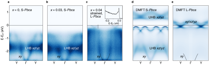
The first important conclusion from this data is that light Pr-doping alone causes minor changes in the electronic structure only. Its main effect is a small shift of the chemical potential. Importantly though, remains in the correlated gap, consistent with the highly insulating nature of crystals in resistivity measurements. We therefore conclude that the additional valence electron of Pr stays fully localized in the insulating S-Pbca structure, possibly due to a Mott transition in the impurity band, as it was proposed for lightly La-doped Sr2IrO4 Battisti2017 . The main spectroscopic features in the insulating phase are a dispersive state with eV bandwidth and an intense non-dispersive peak at -1.7 eV. With reference to our dynamical mean field theory (DMFT) calculations (Fig. 3d) and consistent with Ref. Sutter2017 , we identify these features with the fully occupied orbital and the lower Hubbard band (LHB) of character, respectively. This confirms a basic electronic configuration in the insulating S-Pbca phase with fully occupied orbital and half-filled / bands split into lower and upper Hubbard band, as proposed on the basis of DMFT calculations Liebsch2007 ; Gorelov2010 .
Straining a lightly doped Ca2-xPrxRuO4 sample, we observe a substantial redistribution of spectral weight. Most notably, the intensity in the LHB collapses suddenly across the MIT and coherent quasiparticle states appear at the chemical potential (Fig. 3c). Both of these effects are reproduced by our DMFT calculations shown in Fig. 3e. Interestingly though, the sudden collapse of the LHB is in stark contrast to lightly doped cuprates, where metallicity emerges from a gradual transfer of spectral weight from the LHB to coherent quasiparticle states Shen2004 . We interpret this as a generic manifestation of the additional orbital degree of freedom of multiband Mott insulators. In effective single band systems, such as the cuprates or iridates, the orbital occupancy can only change by the small number of doped carriers, resulting in dominantly half-filled sites retaining a strong memory of the Mott phase. In Ca2-xPrxRuO4, on the other hand, our data show a discontinuous change of the occupancy from 1 to in spite of the only light doping because of substantial interorbital charge transfer across the MIT. The large deviation from half filling causes a sudden collapse of the LHB and renders electronic energies comparable to lattice energies under strain, resulting in a strongly first order nature of the Mott transition.
Our results show that tuning uniaxial strain in ARPES experiments is a promising new method to study phase transitions or, more generally, structure – property relations of quantum materials. Potential applications of our method range from tuning magnetism, to topological phase transitions, two-dimensional van der Waals materials and unconventional superconductors showing large responses to strain, such as Sr2RuO4 Hicks2014 .
Methods:
Single crystals of Ca2-xPrxRuO4 were grown through the floating zone (FZ) technique using a Crystal System Corporation FZ-T-10000-H-VI-VPO-I-HR-PC four mirror optical furnace.
Samples were grown in oxygen pressure, and the initial Ru concentration in the polycrystalline rods was about higher than the nominal value to compensate for evaporation during the growth.
The bulk properties were thoroughly characterized by resistivity, specific heat, magnetization measurements, and single crystal neutron diffraction at the ISIS spallation neutron source Keen2006 .
Doping levels were measured by energy and wavelength dispersive X-ray spectroscopy (EDX/WDX) and were found to be systematically lower by to than in the polycrystalline growth rod.
Angle-resolved photoemission spectroscopy (ARPES) experiments were performed at the I05 Beamline of the Diamond Light Source doi:10.1063/1.4973562 . The presented data were acquired with linearly and circularly polarized light at eV photon energy and an overall resolution of meV / 0.015 Å-1.
Acknowledgements.
We thank E. Giannini, D. McMorrow, D. Pincini, D. Jaccard, C. Renner, M. Spera, V. Pasquier for discussions, J. Teyssier for Raman experiments on Ca2-xPrxRuO4, M. Spera for help with the numerical strain simulations and R. Pellet for machining and aligning the strain apparatus. The experimental work was supported by the Swiss National Science Foundation (200021-153405 and 200020-165791). Theoretical work was supported by the European Research Council grant ERC-319286-QMAC, the NCCR MARVEL of the SNSF and the Simons Foundation (Flatiron Institute). Crystal growth and characterization at UCL was supported by the EPSRC grant EP/N034694/1. We gratefully acknowledge the Science and Technology Facilities Council (STFC) for access to neutron beamtime at ISIS, and also for the support of sample preparation at the UCL crystal growth laboratory. We would like to thank G. Stenning for help on the Smartlab XRD and Quantum Design MPMS instruments in the Materials Characterisation Laboratory at the ISIS Neutron and Muon Source. We acknowledge Diamond Light Source for time on beamline I05 under proposal SI17381.References
- (1) Imada, M., Fujimori, A. & Tokura, Y. Metal-insulator transitions. Rev. Mod. Phys. 70, 1039 (1998). URL http://link.aps.org/abstract/RMP/v70/p1039.
- (2) Mott, N. F. The basis of the electron theory of metals, with special reference to the transition metals. Proceedings of the Physical Society. Section A 62, 416 (1949). URL http://stacks.iop.org/0370-1298/62/i=7/a=303.
- (3) Pavarini, E. et al. Mott transition and suppression of orbital fluctuations in orthorhombic 3d1 perovskites. Phys. Rev. Lett. 92, 176403 (2004). URL http://link.aps.org/doi/10.1103/PhysRevLett.92.176403.
- (4) Gorelov, E. et al. Nature of the Mott transition in Ca2RuO4. Phys. Rev. Lett. 104, 226401 (2010). URL https://link.aps.org/doi/10.1103/PhysRevLett.104.226401.
- (5) Park, H., Millis, A. J. & Marianetti, C. A. Total energy calculations using DFT+ DMFT: Computing the pressure phase diagram of the rare earth nickelates. Phys. Rev. B 89, 245133 (2014). URL https://link.aps.org/doi/10.1103/PhysRevB.89.245133.
- (6) Leonov, I., Anisimov, V. I. & Vollhardt, D. First-principles calculation of atomic forces and structural distortions in strongly correlated materials. Phys. Rev. Lett. 112, 146401 (2014). URL https://link.aps.org/doi/10.1103/PhysRevLett.112.146401.
- (7) Han, Q. & Millis, A. Lattice energetics and correlation-driven metal-insulator transitions: the case of Ca2RuO4. arXiv:1801.06215 1–10 (2018). URL http://arxiv.org/abs/1801.06215.
- (8) Mathur, N. D. et al. Magnetically mediated superconductivity in heavy fermion compounds. Nature 394, 39 (1998). URL http://dx.doi.org/10.1038/27838.
- (9) Schlom, D. G. et al. Strain tuning of ferroelectric thin films. Annual Review of Materials Research 37, 589–626 (2007). URL https://doi.org/10.1146/annurev.matsci.37.061206.113016.
- (10) Drozdov, A. P., Eremets, M. I., Troyan, I. A., Ksenofontov, V. & Shylin, S. I. Conventional superconductivity at 203 kelvin at high pressures in the sulfur hydride system. Nature 525, 73 (2015). URL http://dx.doi.org/10.1038/nature14964.
- (11) Hicks, C. W. et al. Strong increase of Tc of Sr2RuO4 under both tensile and compressive strain. Science 344, 283–285 (2014). URL http://science.sciencemag.org/content/344/6181/283.
- (12) Burganov, B. et al. Strain control of fermiology and many-body interactions in two-dimensional ruthenates. Phys. Rev. Lett. 116, 197003 (2016). URL https://link.aps.org/doi/10.1103/PhysRevLett.116.197003.
- (13) Anisimov, V. I., Nekrasov, I. A., Kondakov, D. E., Rice, T. M. & Sigrist, M. Orbital-selective Mott-insulator transition in Ca2-xSrxRuO4. Eur. Phys. J. B 25, 191–201 (2002). URL https://doi.org/10.1140/epjb/e20020021.
- (14) Liebsch, A. & Ishida, H. Subband Filling and Mott Transition in Ca2-xSrxRuO4. Phys. Rev. Lett. 98, 216403–216404 (2007). URL https://doi.org/10.1103/PhysRevLett.98.216403.
- (15) Zhang, G. & Pavarini, E. Mott transition, spin-orbit effects, and magnetism in . Phys. Rev. B 95, 075145 (2017). URL https://link.aps.org/doi/10.1103/PhysRevB.95.075145.
- (16) Kubota, M. et al. Ferro-type orbital state in the Mott transition system Ca2-xSrxRuO4 studied by the resonant X-ray scattering interference technique. Phys. Rev. Lett. 95, 026401 (2005). URL https://link.aps.org/doi/10.1103/PhysRevLett.95.026401.
- (17) Sutter, D. et al. Hallmarks of Hunds coupling in the Mott insulator Ca2RuO4. Nature Communications 8, 15176 (2017). URL http://dx.doi.org/10.1038/ncomms15176.
- (18) Kunkemöller, S. et al. Highly anisotropic magnon dispersion in : evidence for strong spin orbit coupling. Phys. Rev. Lett. 115, 247201 (2015). URL https://link.aps.org/doi/10.1103/PhysRevLett.115.247201.
- (19) Jain, A. et al. Higgs mode and its decay in a two-dimensional antiferromagnet. Nature Physics 13, 633 EP – (2017). URL http://dx.doi.org/10.1038/nphys4077.
- (20) Khaliullin, G. Excitonic magnetism in Van Vleck-type Mott insulators. Phys. Rev. Lett. 111, 197201 (2013). URL https://link.aps.org/doi/10.1103/PhysRevLett.111.197201.
- (21) Chaloupka, J. & Khaliullin, G. Doping-induced ferromagnetism and possible triplet pairing in mott insulators. Phys. Rev. Lett. 116, 017203 (2016). URL https://link.aps.org/doi/10.1103/PhysRevLett.116.017203.
- (22) Sow, C. et al. Current-induced strong diamagnetism in the Mott insulator Ca2RuO4. Science 358, 1084–1087 (2017). URL http://science.sciencemag.org/content/358/6366/1084.
- (23) Nakamura, F. et al. From Mott insulator to ferromagnetic metal: a pressure study of Ca2RuO4. Phys. Rev. B 65, 220402 (2002). URL https://link.aps.org/doi/10.1103/PhysRevB.65.220402.
- (24) Steffens, P. et al. High-pressure diffraction studies on Ca2RuO4. Phys. Rev. B 72, 094104 (2005). URL https://link.aps.org/doi/10.1103/PhysRevB.72.094104.
- (25) Miao, L. et al. Itinerant ferromagnetism and geometrically suppressed metal-insulator transition in epitaxial thin films of Ca2RuO4. Applied Physics Letters 100, 052401 (2012). URL https://doi.org/10.1063/1.3680250.
- (26) Dietl, C. et al. Tailoring the electronic properties of Ca2RuO4 via epitaxial strain. Applied Physics Letters 112, 031902 (2018). URL https://doi.org/10.1063/1.5007680.
- (27) Nakatsuji, S. & Maeno, Y. Quasi-two-dimensional Mott transition system Ca2-xSrxRuO4. Phys. Rev. Lett. 84, 2666 (2000). URL https://doi.org/10.1103/PhysRevLett.84.2666.
- (28) Fukazawa, H. & Maeno, Y. Filling Control of the Mott Insulator Ca2RuO4. Journal of the Physical Society of Japan 70, 460–467 (2001). URL https://doi.org/10.1143/JPSJ.70.460.
- (29) Nakamura, F. et al. Electric-field-induced metal maintained by current of the Mott insulator Ca2RuO4. Scientific Reports 3, 2536 (2013). URL http://dx.doi.org/10.1038/srep02536.
- (30) Damascelli, A. Angle-resolved photoemission studies of the cuprate superconductors. Review of Modern Physics 75, 473 (2003).
- (31) De La Torre, A. et al. Collapse of the Mott Gap and Emergence of a Nodal Liquid in Lightly Doped Sr2IrO4. Physical Review Letters 115, 2–6 (2015).
- (32) Georges, A., de’ Medici, L. & Mravlje, J. Strong electronic correlations from Hund’s coupling (2012). URL http://arxiv.org/abs/1207.3033{%}0Ahttp://dx.doi.org/10.1146/annurev-conmatphys-020911-125045.
- (33) Brinkman, W. F. & Rice, T. M. Application of gutzwiller’s variational method to the metal-insulator transition. Phys. Rev. B 2, 4302–4304 (1970). URL https://link.aps.org/doi/10.1103/PhysRevB.2.4302.
- (34) Battisti, I. et al. Universality of pseudogap and emergent order in lightly doped mott insulators. Nature Physics 13, 21 (2016). URL http://dx.doi.org/10.1038/nphys3894.
- (35) Shen, K. M. et al. Missing quasiparticles and the chemical potential puzzle in the doping evolution of the cuprate superconductors. Phys. Rev. Lett. 93, 267002 (2004). URL https://doi.org/10.1103/PhysRevLett.93.267002.
- (36) Keen, D. A., Gutmann, M. J. & Wilson, C. C. SXD – the single-crystal diffractometer at the ISIS spallation neutron source. Journal of Applied Crystallography 39, 714–722 (2006). URL https://doi.org/10.1107/S0021889806025921.
- (37) Hoesch, M. et al. A facility for the analysis of the electronic structures of solids and their surfaces by synchrotron radiation photoelectron spectroscopy. Review of Scientific Instruments 88, 013106 (2017). URL https://doi.org/10.1063/1.4973562.