Authors’ Instructions
Context-Aware Learning using Transferable Features for Classification of Breast Cancer Histology Images
Abstract
Convolutional neural networks (CNNs) have been recently used for a variety of histology image analysis. However, availability of a large dataset is a major prerequisite for training a CNN which limits its use by the computational pathology community. In previous studies, CNNs have demonstrated their potential in terms of feature generalizability and transferability accompanied with better performance. Considering these traits of CNN, we propose a simple yet effective method which leverages the strengths of CNN combined with the advantages of including contextual information, particularly designed for a small dataset. Our method consists of two main steps: first it uses the activation features of CNN trained for a patch-based classification and then it trains a separate classifier using features of overlapping patches to perform image-based classification using the contextual information. The proposed framework outperformed the state-of-the-art method for breast cancer classification.
Keywords:
Digital pathology, Convolutional neural network, Context-aware learning, Transferable features, Breast cancer1 Introduction
Breast cancer is the most common type of cancer diagnosed and is the second most common type of cancer with high mortality rate after lung cancer in women [1]. Due to the increased incidence of breast cancer and subjectivity in diagnosis, there is an increasing demand for automated systems. To this end, deep neural networks (DNNs) have been widely used to produce the state-of-the-art results for a variety of histology image analysis tasks such as nuclei detection and classification [2], tissue classification [3, 4] and segmentation [5, 6].
The CAMELYON16 challenge [6] is the best demonstration of using deep learning for automatic tissue analysis, outperforming the pathologists in terms of detection of tumors within the whole slide images (WSIs). The objective of this challenge was to automatically detect the metastasis in haematoxylin and eosin (H&E) stained WSIs of lymph node sections. Cruz-Roa et al. [3] presented a deep learning architecture for automated basal carcinoma detection. This method first learns image representation via autoencoder and then a CNN is applied on this representation to capture both translation invariant features and a compact image representation. Spanhol et al. [7] applied a simple CNN for classifying the BreaKHis database [8] consisting of microscopic images of benign and malignant breast tumor biopsies. Small patches were extracted at different magnification levels to train the network and during inference, final output was produced by combining the predictions of the small patches.
The generalizability property of DNN makes their features transferable to other applications which encouraged the researchers to employ transfer learning for histology images as in [5, 9, 10]. These features have also been used to train separate classifiers for predictions [11, 12, 13, 14], which are particularly useful when there is not enough dataset for training the CNN from scratch. In some recent studies [15, 16], context-aware based learning architecture has been introduced, in which first CNN is trained using high pixel resolution patches to extract features at a cellular level that are then fed to a second CNN, stacked on top of the first for expanding the context from a single patch to a large tissue region. The experimental results of these studies suggest that the contextual information plays a crucial role in identifying abnormalities in heterogeneous tissue structures.
Our contribution in this work is twofold. First, we propose to use CNN features as a generic descriptor for a small dataset, provided as a part of a challenge dataset. We extract transferable features from a number of networks, each trained on a different dataset for the purpose of classification by a separate classifier trained on these features. As our second contribution, we combine these features to learn context of a large patch to improve our classification performance. To this end, we use transferable features for a block of consecutive patches to train a SVM model to classify the H&E stained breast images into normal, benign, carcinoma insitu (CIS) and breast invasive carcinoma (BIC).
2 Dataset and Experimental Setup
We used the dataset provided as a part of the ICIAR 2018 challenge for the classification of breast cancer histology images. This dataset comprises of 400 high resolution images of size 20481536 pixels at 20 magnification, stained with H&E stain. The pixel resolution for these images is 0.42 m. Each image belongs to one of the four classes: normal, benign, insitu carcinoma or invasive carcinoma. The ground truth was provided by the two pathologists. To study the feature transferability of CNN, we experimented with other part of the challenge dataset provided for segmentation task. Ten WSIs with coarse annotations were provided for this task. We extracted patches from these WSIs after manually refining the original annotations.
The challenge dataset for a classification task consists of training images used in [14] along with 151 additional images. To evaluate the effectiveness of our proposed approach, we splitted the challenge dataset for two settings. In the first setting, we use the same images for training and testing which were used in [14] for a fair comparison. We included the additional images in our validation set while training the network. The test dataset contains two set of images, with equal number of images in each class. The testing data is not provided with the challenge data but is made publicly available by the authors in two sets. The first test set contains 20 images while the second set contains 16 images and is referred as ’test extended’ dataset in this paper. In the second setting which is used for submission to the challenge, we combined the whole challenge dataset from task-1 and the test dataset and randomly split them into 75% training and 25% validation set.
Regarding the implementation, we used residual neural network with 50 layers for patch-based classification in Tensorflow. For context-aware image-based classification, support vector machine (SVM) classifier with radial basis function (RBF) was used and implemented in MATLAB. Further details on both these steps are given in the Methods section.
3 Methods
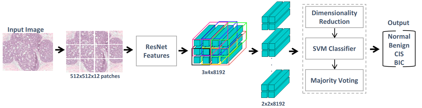
In this paper, we introduce an effective model for the purpose of image-based classification using more context information, particularly for a small dataset. To this end, we design our model in two main steps: patch-based classification and context-aware image-based classification. The overall system architecture is shown in Figure 1.
3.1 Preprocessing
Stain inconsistency of digitized WSIs is a significant issue affecting the performance of machine learning (ML) systems. The dataset provided for this challenge contains images with large stain variation. To this end, we performed stain normalization using the Reinhard method [17], available in our group’s Stain Normalization Toolbox [18]. This method transforms the color distribution of an image to the color distribution of a target image by matching the mean and standard deviation of the source image to that of target image. This transformation is carried out for each channel separately, in the Lab colorspace.
3.2 Patch-based classification
ResNet [19] introduced in 2015 by Microsoft has been shown to outperform several architectures including VGG [20], GoogleNet [21], PReLU-net [22] and BN-inception [23]. This network also outperformed best performing networks with a significant margin for the classification of histopathology colorectal images [24]. The state-of-the-art results of ResNet on different datasets motivated us to use it for our patch-based classification. For our experiments, we used ResNet with 50 layers. For network training, overlapping patches of size 512512 pixels were extracted from the images. The network was trained for 16 epochs with batch size of 12 and the best trained network was selected for further processing. The training was done using stochastic gradient descent with momentum set to 0.95. The learning rate was initially set to 0.001 and was decremented after each update. Due to the very small dataset and also to make our network robust to feature transformation, we performed data augmentation involving random rotation (90 to 360 degrees with step of 90 degrees) and flipping during the training stage.
3.3 Context-aware image-wise classification
The above patch-based classification network learns a limited contextual representation for each class by using small patches of size 512512 pixels. To train a classifier with larger context, we divided each image into twelve non-overlapping patches and for each patch, we then extracted 8192 dimensional feature vector from the last layer of our patch-based network. We then trained an SVM classifier with the flattened features of 22 overlapping blocks of patches which is equivalent to training the classifier with the features of patch 10241024 pixels in size. Principal component analysis (PCA) was performed to reduce the dimensionality. The image level label was then decided by majority voting on the labels of overlapping blocks. This approach has been shown to improve the results in previous studies by training a CNN using the features of bigger blocks of patches. Due to the limited availability of dataset for training, we chose to train an SVM classifier rather than a CNN. To increase the number of samples for SVM training, we augmented the training samples using rotation and flipping.
4 Experimental Results
For the evaluation of our proposed method, we experimented with different configurations to show the significance of contextual information, effect of feature transferability using networks trained on different datasets and also to compare our method with the results of [14].
Firstly, we experimented with contextual information captured from the varying size of block of patches. We trained SVM with the context of 11 block (512512 pixels), 22 block (10241024 pixels) and 33 block (15361536 pixels) of patches. As shown in Figure 3 (a), we obtained better accuracy with 22 block as compared to the 11 block but we observed a decline in accuracy for 33 block of context. It is due to the fact that the increase in context using the large block of patches reduces the amount of data for training SVM. Otherwise, incorporating a larger context could have improved our results if we had enough dataset. For our further experiments, we used 22 block of context for classification.
To study the feature transferability, we used activation features from three networks, each trained on a different dataset: training images used in [14], challenge dataset (WSIs) for segmentation task and the Camelyon dataset [6]. To enrich the SVM training, we added half of the additional challenge dataset to our training set. Our results demonstrate that the features learned by the first network tend to provide the most appropriate representation for training the SVM. The network trained on the WSIs has been shown to be the second best feature descriptor as the dataset used to train this network is similar to the evaluation data. While our third network, since it is trained on a completely different dataset did not perform well as compared to the other networks.
For comparative analysis of our proposed method, we compared our results with the results of previously published work of the organizers of BACH challenge [14]. For a fair comparison, we used the same dataset as in [14] for training and testing. Along with 4-class classification, we also present comparison using 2-class classification, for which we grouped normal and benign images in one class while CIS and BIC were grouped in the other class. The 2-class and 4-class comparative results are shown in Figure 3 (c) and (d) respectively. Our method achieved higher accuracy compared to [14] which demonstrates the capability of the contextual information for discriminating different classes.

5 Conclusion
In this paper, we proposed a context-aware network for automated classification of breast cancer histology images. The proposed method leverages the power of CNNs to encode the representation of a patch into high dimensional space and uses traditional machine method (SVM) to aggregate the contextual information from the high dimensional features while having a limited dataset. Our proposed approach outperformed the existing methods proposed for the same task. The proposed method is not limited to breast cancer classification task. It could be applied to other problems where both high resolution and contextual information are required to make an optimal prediction.
References
- [1] R. L. Siegel, K. D. Miller, and A. Jemal, “Cancer statistics, 2016,” CA: a cancer journal for clinicians, vol. 66, no. 1, pp. 7–30, 2016.
- [2] K. Sirinukunwattana, S. E. A. Raza, Y.-W. Tsang, D. R. Snead, I. A. Cree, and N. M. Rajpoot, “Locality sensitive deep learning for detection and classification of nuclei in routine colon cancer histology images,” IEEE transactions on medical imaging, vol. 35, no. 5, pp. 1196–1206, 2016.
- [3] A. Cruz-Roa, A. Basavanhally, F. González, H. Gilmore, M. Feldman, S. Ganesan, N. Shih, J. Tomaszewski, and A. Madabhushi, “Automatic detection of invasive ductal carcinoma in whole slide images with convolutional neural networks,” in SPIE medical imaging, vol. 9041, pp. 904103–904103, International Society for Optics and Photonics, 2014.
- [4] D. Wang, A. Khosla, R. Gargeya, H. Irshad, and A. H. Beck, “Deep learning for identifying metastatic breast cancer,” arXiv preprint arXiv:1606.05718, 2016.
- [5] H. Chen, X. Qi, L. Yu, and P.-A. Heng, “Dcan: Deep contour-aware networks for accurate gland segmentation,” in Proceedings of the IEEE conference on Computer Vision and Pattern Recognition, pp. 2487–2496, 2016.
- [6] B. E. Bejnordi, M. Veta, P. J. van Diest, B. van Ginneken, N. Karssemeijer, G. Litjens, J. A. van der Laak, M. Hermsen, Q. F. Manson, M. Balkenhol, et al., “Diagnostic assessment of deep learning algorithms for detection of lymph node metastases in women with breast cancer,” Jama, vol. 318, no. 22, pp. 2199–2210, 2017.
- [7] F. A. Spanhol, L. S. Oliveira, C. Petitjean, and L. Heutte, “Breast cancer histopathological image classification using convolutional neural networks,” in Neural Networks (IJCNN), 2016 International Joint Conference on, pp. 2560–2567, IEEE, 2016.
- [8] F. A. Spanhol, L. S. Oliveira, C. Petitjean, and L. Heutte, “A dataset for breast cancer histopathological image classification,” IEEE Transactions on Biomedical Engineering, vol. 63, no. 7, pp. 1455–1462, 2016.
- [9] N. Bayramoglu and J. Heikkilä, “Transfer learning for cell nuclei classification in histopathology images,” in Computer Vision–ECCV 2016 Workshops, pp. 532–539, Springer, 2016.
- [10] Z. Han, B. Wei, Y. Zheng, Y. Yin, K. Li, and S. Li, “Breast cancer multi-classification from histopathological images with structured deep learning model,” Scientific reports, vol. 7, no. 1, p. 4172, 2017.
- [11] Y. Xu, Z. Jia, L.-B. Wang, Y. Ai, F. Zhang, M. Lai, I. Eric, and C. Chang, “Large scale tissue histopathology image classification, segmentation, and visualization via deep convolutional activation features,” BMC bioinformatics, vol. 18, no. 1, p. 281, 2017.
- [12] M. Valkonen, K. Kartasalo, K. Liimatainen, M. Nykter, L. Latonen, and P. Ruusuvuori, “Dual structured convolutional neural network with feature augmentation for quantitative characterization of tissue histology,” in Proceedings of the IEEE Conference on Computer Vision and Pattern Recognition, pp. 27–35, 2017.
- [13] Y. Xu, Z. Jia, Y. Ai, F. Zhang, M. Lai, I. Eric, and C. Chang, “Deep convolutional activation features for large scale brain tumor histopathology image classification and segmentation,” in Acoustics, Speech and Signal Processing (ICASSP), 2015 IEEE International Conference on, pp. 947–951, IEEE, 2015.
- [14] T. Araújo, G. Aresta, E. Castro, J. Rouco, P. Aguiar, C. Eloy, A. Polónia, and A. Campilho, “Classification of breast cancer histology images using convolutional neural networks,” PloS one, vol. 12, no. 6, p. e0177544, 2017.
- [15] A. Agarwalla, M. Shaban, and N. M. Rajpoot, “Representation-aggregation networks for segmentation of multi-gigapixel histology images,” arXiv preprint arXiv:1707.08814, 2017.
- [16] B. E. Bejnordi, G. Zuidhof, M. Balkenhol, M. Hermsen, P. Bult, B. van Ginneken, N. Karssemeijer, G. Litjens, and J. van der Laak, “Context-aware stacked convolutional neural networks for classification of breast carcinomas in whole-slide histopathology images,” arXiv preprint arXiv:1705.03678, 2017.
- [17] E. Reinhard, M. Adhikhmin, B. Gooch, and P. Shirley, “Color transfer between images,” IEEE Computer graphics and applications, vol. 21, no. 5, pp. 34–41, 2001.
- [18] A. M. Khan, N. Rajpoot, D. Treanor, and D. Magee, “A nonlinear mapping approach to stain normalization in digital histopathology images using image-specific color deconvolution,” IEEE Transactions on Biomedical Engineering, vol. 61, no. 6, pp. 1729–1738, 2014.
- [19] K. He, X. Zhang, S. Ren, and J. Sun, “Deep residual learning for image recognition,” in Proceedings of the IEEE conference on computer vision and pattern recognition, pp. 770–778, 2016.
- [20] K. Simonyan and A. Zisserman, “Very deep convolutional networks for large-scale image recognition,” arXiv preprint arXiv:1409.1556, 2014.
- [21] C. Szegedy, W. Liu, Y. Jia, P. Sermanet, S. Reed, D. Anguelov, D. Erhan, V. Vanhoucke, and A. Rabinovich, “Going deeper with convolutions,” in Proceedings of the IEEE conference on computer vision and pattern recognition, pp. 1–9, 2015.
- [22] K. He, X. Zhang, S. Ren, and J. Sun, “Delving deep into rectifiers: Surpassing human-level performance on imagenet classification,” in Proceedings of the IEEE international conference on computer vision, pp. 1026–1034, 2015.
- [23] S. Ioffe and C. Szegedy, “Batch normalization: Accelerating deep network training by reducing internal covariate shift,” in International Conference on Machine Learning, pp. 448–456, 2015.
- [24] B. Korbar, A. M. Olofson, A. P. Miraflor, K. M. Nicka, M. A. Suriawinata, L. Torresani, A. A. Suriawinata, and S. Hassanpour, “Deep-learning for classification of colorectal polyps on whole-slide images,” arXiv preprint arXiv:1703.01550, 2017.