Protein motion in the nucleus: from anomalous diffusion to weak interactions
Abstract
Understanding how transcription factors (TFs) regulate mammalian gene expression in space and time is a central topic in biology. To activate a gene, a TF has first to diffuse in the available space of the nucleus until it reaches a target DNA sequence or protein (target site). This eventually results in the recruitment of the whole transcriptional machinery.
All these processes take place in the mammalian nucleoplasm, a highly organized and dynamic environment, in which some complexes transiently assemble and break apart, whereas others appear more stable. This diversity of dynamic behaviors arises from the number of biomolecules that make up the nucleoplasm and their pairwise interactions. Indeed, interactions energies that span several orders of magnitude, from covalent bounds to transient and dynamic interactions can shape nuclear landscapes. Thus, the nuclear environment determines how frequently and how fast a TF contacts its target site, and indirectly gene expression. How exactly transient interactions are involved in the regulation of TF diffusion is unclear, but are reflected by live cell imaging techniques such as fluorescence correlation spectroscopy, fluorescence recovery after photobleaching or single-particle tracking. Overall, the macroscopic result of these microscopic interactions is almost always anomalous diffusion, a phenomenon widely studied and modeled.
Here, we review the connections between the anomalous diffusion of a TF and the microscopic organization of the nucleus, including recently described topologically associated domains and dynamic phase-separated compartments. We propose that anomalous diffusion found in single particle tracking (SPT) data result from weak and transient interactions with dynamic nuclear substructures, and that SPT data analysis would benefit form a better description of such structures.
1 Introduction
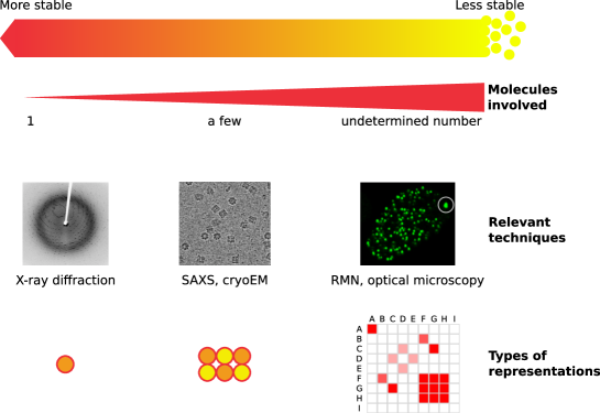
Mammalian gene expression and its regulation take place in the nucleus, a highly complex and sub-compartmented organelle. Interactions strengths between nuclear constituents span several orders of magnitude, from covalent bounds to ”strong” non-covalent interactions. These interactions lead to the formation of macromolecular structures, either stable (Figure 1-left; for instance double-stranded DNA or biochemically purifiable macromolecular complexes such as the ones involved in gene expression) or transient but specific, leading to preferential associations of classes of proteins (Figure 1-right).
The regulation of transcription is of utmost interest as it is central not only to developmental biology, but also to cancer biology, drug screening, etc. To express a mRNA, a macromolecular complexes constituted of several subunits and dozens of proteins, the preinitiation complex, has first to assemble at the promoter of a gene in a time and space-specific manner [1]. This complex is able to robustly integrate transient signals such as the ones mediated by proteins binding to cis-regulatory sequences such as enhancers [2].
More mechanistically, the assembly of such a complex can be characterized by a set of chemical reactions describing the progressive recruitment of factors and subunits. A kinetic rate can be associated to each of these reactions. Furthermore, traditional biochemistry and in vitro experiments have been the methods of choice to investigate such complex processes. Most biochemical techniques involve purification steps allowing to reveal strong, non covalent interactions such as the ones occurring in a stably-assembled complex [3, 4] (Figure 1-left). Then, further quantification of stoichiometry and affinity constants became possible, progressively building a network of interacting proteins, usually represented as a graph with nodes linked with arrows.
Within this framework, the understanding of gene expression regulation reduces to elucidating how external factors (including TFs) affect the kinetic constants . Although it can be assumed that kinetic rates are characterized only by the nature and concentration of enzyme, substrate and cofactors, it was shown in 1906 by Marian Smoluchowki [5] that the kinetic rate of a well-mixed reaction can be decomposed as . Thus, the kinetic rate is a function of both the cross-section of interaction (reflecting the chemical properties of the partners and usually studied by biochemical approaches) and the diffusion constant of the species.
Since is determined by the local environment, this finding is striking in the context of gene expression regulation: now the kinetics of one reaction depend on the whole nuclear structure. More specifically, any factor that affects diffusion in any specific or non-specific way will ultimately influence reaction rates. Indeed, interactions resulting in facilitated diffusion on a substructure (such as a TF on DNA, [6, 7, 8]) or segregation inside a membrane-less compartment in a phase-separated manner [9] can all be seen under the unifying framework of diffusion on a surface of reduced dimensionality. Diffusion on surfaces of reduced dimensionality yields kinetics that are qualitatively different than in free, 3D diffusion and leads to potentially dramatically increased reaction rates.
Anomalous diffusion, a phenomenon occurring when a molecule explores a volume lower than predicted by diffusion, affects all proteins inside a cell. Numerous physical models can describe anomalous diffusion [10], and several have been applied to the motion of nuclear proteins. However, many of them only provide a phenomenological description of diffusion, rather than mechanistic insights, and radically distinct models can often fit the available data equally well.
In light of these considerations, it is worthwhile to examine the recently published discoveries describing either stable subnuclear compartments or their more transient, weak-interaction induced counterparts to highlight their influence on the diffusion of factors through dimensionality reduction. This includes TADs, LADs, nucleoli, ncRNAs, transcription factories, phase-separated domains, etc. They constitute substructures with a high valency amenable to weak interactions that can qualitatively influence diffusion and target search.
Here, we first review anomalous diffusion models applied to a protein motion and link them with a potential physical generative model. Then, we emphasize recent advances in the characterization of regions of reduced dimensionality in mammalian nuclei, both aspecific through volume exclusion and specific through transient, weak-but-specific interactions. Finally, we propose that these weak interactions shape TF dynamics, and that single particle tracking (SPT) analysis would greatly benefit from a better understanding of the pairwise interaction map between nuclear proteins.
2 Most anomalous diffusion models reflect underlying networks of weak interactions
The technique of choice to investigate protein motion in the nucleus of live cells is light microscopy of fluorescently tagged proteins. Different imaging and modeling modalities have been applied, including fluorescence recovery after photobleaching (FRAP), fluorescence correlation microscopy (FCS) or single-particle tracking (SPT).
In solution, the diffusion coefficient of a protein is inversely proportional to the hydrodynamic radius of the protein () and the viscosity of the medium () through the Stokes-Einstein relationship where reflects thermal agitation, with the Boltzmann constant and the absolute temperature. This description, however, is too simplistic in the complex cellular environment. Indeed, with the exception of inert tracers of small molecular weight [11, 12], it is well acknowledged that (a) macromolecules in a cell diffuse much slower than in a medium of comparable viscosity, (b) that complexes of high molecular weight can diffuse faster than small proteins, and (c) most molecules exhibit anomalous diffusion.
Thus, the diffusion of TFs cannot be described by simple friction/viscosity relationships, and their behavior, perhaps unsurprisingly, has to be seen from the angle of transient interactions with a dense matrix of interactants. In the context of this review, we define transient (or ”weak”) interactions as interactions that are usually too short-lived to be captured by traditional biochemistry techniques, that typically involve one or several wash step, during which proteins interacting specifically but transiently get diluted and washed out.
Furthermore, diffusion of many factors is highly anomalous (more specifically, subdiffusive; Figure 2), meaning that the space explored over time by one factor is lower than expected by free diffusion (reviewed in [13, 10]). Anomalous diffusion is usually characterized by a sublinear growth of the mean squared displacement (MSD) as a function of time (Figure 2). Nonetheless, some anomalous diffusion processes can have a MSD identical to the MSD of free diffusion, and other characterizations are needed (Figure 2b). Phenomenological models have been fitted to it with success, and include continuous time random walks (CTRW; Figure 2d) [14, 15], fractional Brownian motion (Figure 2e) [16, 17, 18, 19], diffusion in fractal media (Figure 2f) [20, 21, 22]. Although useful as phenomenological descriptions, these models are often agnostic regarding the underlying reality of the process. In any case, the explanation of diffusion has to rely on physics and chemistry of the nucleus.

From a physical perspective, proteins can adsorb and diffuse on nuclear substructures. When this happens, the exploration properties of the protein are universally given by two parameters: first, the dimension of the random walk , and second, the dimension of the space available to diffuse . can be integer ( for instance for sliding on DNA without jumps), or non-integer, a feature that characterizes self-similar structures, that is, fractals (Figure 2f). For the sake of this review, we will denote structures of as structures of reduced dimensionality. Depending on and , the motion of the protein then falls into two universal categories, termed compact and non-compact [23, 24, 25]. In a compact exploration (), exploration is local and distance-dependent and a given site is explored repeatedly over time, in a highly recurrent manner. Conversely, in a non-compact exploration (), the exploration is global, and every site on the structure has a constant probability to be explored (distance independence); the exploration is non-recurrent (transient). For instance, a particle freely diffusing has a of 2. When diffusion takes place in a 3D space () the particle tends not to never revisit sites, adopting a non-compact exploration (indeed, ). Conversely, a particle in free, Brownian diffusion () constrained to diffuse in 1D (; hypothetically along a DNA fiber) will repeatedly sample the same sites (compact exploration, ). Consequently, target search times are decreased and reaction rates are increased in the compact case.
Structures of reduced dimensionality, including fractals, emerge naturally from various processes, including diffusion-limited aggregation and hierarchical assembly of macromolecular scaffolds, such as the multi-scale organization of chromatin. The goal of the next sections is to highlight a few structures of reduced dimensionality in the nucleus and how they influence kinetics of TFs.
3 Steric hindrance in the nucleus
Far from constituting a homogeneous medium, the nucleus is a highly organized and subcompartmentalized organelle. The main organizing structure, chromatin, constitutes approximately 10-30% of the nuclear volume [26, 27] and likely accounts for a significant part of the diffusion slowdown [28]. Since every molecule has to slalom around a dense and heterogeneous chromatin environment, diffusion is impaired. Note that, however, similar diffusion coefficients are usually observed in the cytoplasm and the nucleoplasm [29], suggesting that protein crowding can also account for diffusion slowdown ([30, 31] and [21] for a discussion).
Over the past years, organizing principles of chromatin have emerged: at large scale, the genome is segregated in chromosome territories and regions of heterochromatin/euchromatin, lamina-associated domains at the periphery and nucleoli lying more at the center. At higher magnification, chromatin is organized in areas of preferential interactions such as A/B compartments and topologically associated domains (TADs) that reflect the functional organization of chromatin (Figure 3a) [32].
Overall, although highly heterogeneous, chromatin in the mammalian nucleus is well described by a self-similar, fractal structure that occupies a non-zero volume. This was initially postulated [33], and later evidenced by spectroscopic [34, 35], genomic [36, 37] and imaging techniques [21, 38, 18, 39, 40].
As a consequence, factors diffusing in the available volume are constrained by this structure [41], possibly experiencing diffusion in a medium of reduced dimensionality, as evidenced by numerous reports [21, 42]. In a model where only volume exclusion happens, proteins of the same size and shape should have the same diffusion coefficient. Thus, the embedding structure of the nucleus only sets a lower bound on the level of anomalous diffusion that can be observed.
Several lines of argument, however, point to the fact that steric-hindrance-induced anomalous diffusion is mild. Indeed, FRAP experiments performed with protein or non-protein tracers of increasing molecular weights suggest that low molecular weight tracers diffuse almost freely in the nucleus, allowing to infer a viscosity close to the one of water [11]. At higher molecular weights, anomalous diffusion becomes more and more prominent [29, 12], eventually leading to particles being trapped in the chromatin mesh. This effect is consistent with the relatively limited volume occupied by chromatin [26, 27]. Second, FRAP and FCS measurements have shown that the degree of anomalous diffusion for higher molecular weight tracers is moderate [21].
In conclusion, although volume exclusion by chromatin and other nuclear constituents is real, it affects all proteins of the same size in a similar manner. In contrast, a protein weakly interacting with such a structure (for instance, TFs sliding/hopping on DNA [43, 6, 44, 45, 46])) will immediately show a much higher level of anomalous diffusion. Furthermore, even without considering a fractal structure, simple dimensionality reduction to 1D or 2D can yield non-traditional kinetics (fractal kinetics [47, 48]). For instance, fractal kinetics in 2D could occur by weak interaction with the nuclear lamina, Figure 2b. All in all, weak and transient interactions shape the nuclear landscape and can give rise to emergent structures and properties, as exemplified in the next section.
4 Weak interactions in the nucleus
Unlike inert tracers whose diffusion is only determined by volume exclusion, proteins have both a relevant shape and electrostatic interaction pattern that determine their interaction landscape and thus their diffusive properties. These non-covalent interactions are obviously crucial to form biochemically stable complexes such as the transcription preinitiation complex or the spliceosome (Figure 1-left), but also to form dynamic emergent structures of reduced dimensionality upon which TFs can transiently adsorb and diffuse. Under this model, proteins do not form stable complexes anymore, but rather have a high number of weakly-interacting partners. The traditional representation of protein-protein interaction networks as graphs and arrows is not relevant, and can be replaced by representations such as pairwise interaction matrices (Figure 1-right) [49]. Indeed, simulation studies have shown that molecules can naturally undergo soft matter processes yielding structures of reduced dimensionality under very minimal hypotheses [50, 51]. Furthermore, the list of proteins exhibiting phase separation in vitro or in vivo is quickly growing, supporting the vision that the emergence of structures of reduced dimensionality is closer to a general organizing principle than an anecdotal biophysical phenomenon. Such processes include aggregation, complex coacervation, demixing and phase transition, some of them linked with transcriptional regulation [52, 53, 54, 55].
First, structures of reduced dimensionality, such as aggregates, phase separated domains or subnuclear compartments require at least one multivalent partner, that can nucleate the aggregation. As such, many abundant constituents of the mammalian nucleus have been shown to nucleate a structure of reduced dimensionality, in a manner very much akin to heterogeneous catalysis in chemistry [56]. These constituents include low complexity protein domains, that constitute the majority of the mammalian proteome [57, 58], especially TFs [59], repeated DNA [60] or RNA sequences [61, 62, 63] or small amphiphilic molecules [64, 65].
Second, the partners have to exhibit compatible interactions: it is chemically unlikely that both highly charged and hydrophobic proteins will coexist in the same structure without the help of additional compounds acting as counterions [66], setting the basis of a “grammar of interactions” [67], that is being progressively deciphered [68, 69, 70, 54, 65, 71].
Third, structures of reduced dimensionality emerging from weak interactions exhibit the following properties: (1) they usually exist as an extremely dynamic equilibrium rather than a stable structure [9, 72, 63], and can thus be at the same time prevalent in the nucleus and hard to purify by traditional biochemistry that preferentially capture stable interactions. (2) Moreover, they emerge from a dynamic mesh of pairwise chemical interactions. They can show a high level of specificity, and several structures of reduced dimensionality can coexist in the same nucleus without intermixing [55, 73, 66, 53, 60]. Furthermore, the number and spatial relationships of such structures is only limited by the combinatorics of chemical interactions. (3) Finally, these structures can be regulated by the well-studied post-translational machinery of eukaryotic cells. For instance, phosphorylation of one of the proteins involved in such structure can trigger the timely disassembly of the whole structure and free all the factors interacting with it [74, 75, 53]. All those factors will then exhibit a dramatically different dynamics and target search properties, potentially switching from a compact exploration mode to a non-compact one. As such, a specific (and potentially functional) group of factors can be regulated at once by modulation of the post-translational modifications of one “architectural” protein [62, 60].
The characterization of structures of reduced dimensionality emerging from weak interactions is still in its infancy, but appears more and more strongly as a clear organizing principle of mammalian nuclei. These structures create the matrix upon which fast-diffusing factors can specifically and transiently bind, diffuse and unbind, thus dynamically shaping the “diffusion landscape” of the whole transcriptional machinery.
Even though live imaging approaches specifically characterize the behavior of one single factor, they are blind to all these substructures. Indeed, SPT reflects the dynamics of proteins transiently interacting with those structures of reduced dimensionality and one TF potentially visits several of them in the span of a few tens of milliseconds. Such complex behavior therefore appears macroscopically as various kinds of anomalous diffusion.
5 Perspectives: seeing beyond the dots
In a complex mammalian nucleus, the diffusion of a TF is ruled by transient interactions with underlying structures of reduced dimensionality, such as detailed in the two previous sections. From a more general perspective, the question arises of how gene expression regulation processes relate to the multiplicity of structures of reduced dimensionality?
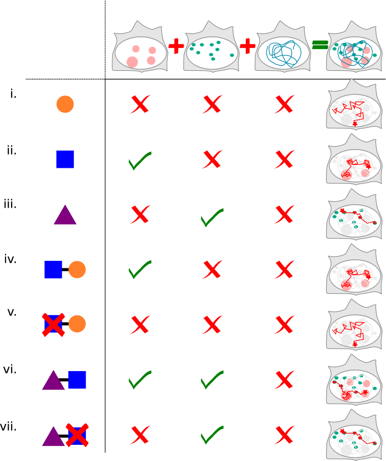
Proteins often harbor several domains, holding the potential to interact alternatively and repeatedly with multiple classes of structures of reduced dimensionality. Thus, depending on its interaction domains, a TF will “see” a different landscape and will interact with some structures whereas other factors will either be excluded or cross them without any additional interactions than limited steric hindrance (Figure 3). In this respect, the nucleus can be described as a “multiverse”, in which some factors coexist in the same physical space but exhibit radically distinct dynamics and interactions (Figure 3).
Furthermore, structures of reduced dimensionality have been proven to be functionally relevant. For example, the dynamic and regulated switching of a TF between structures of reduced dimensionality determines its function. It has been shown that TF exhibit radically different dynamics before/after a post-translational modification [76], or an artificial deletion of a domain (Figure 3), [8, 77, 78, 79, 80, 81]. In that case, the observed diffusion will be arising from the remaining interactions from the other interaction domains, or ultimately from simple volume exclusion [82].
Although theoretical and experimental support for the importance of weak interactions as an architectural principle of the nucleus and gene expression regulation is being actively investigated, several questions remain unaddressed:
First, how many distinct types of structures of reduced dimensionality exist? Since the numbers of types of low-complexity domains is likely to be limited, one can expect that a limited number of such structures actually coexist at a given time in a nucleus [70]. This implies that the SPT dynamics of TFs will fall in a limited number of categories, which in turn is determined by their combinatorial interactions with one or several of these structures. To take into consideration such processes paves the way the way for a higher-order understanding of gene expression regulation and key transitions occurring for instance during mitosis or development.
Second, can we determine the pairwise interaction matrix between low-complexity protein domains, which would allow to derive predictive dynamics of a given TF modification? Ideally, such matrix will encompass all known low-complexity domains, but also abundant multivalent RNAs and DNA sequences, and each element of this matrix will reflect the affinity between two domains under physiological conditions (Figure 1).
Third, how much detail is required to describe these structures of reduced dimensionality? Is the pairwise interaction between protein domains a good approximation of the properties of the nucleus? Conversely, one can imagine substructures of reduced dimensionality arising from interactions more complex than simple pairwise-interactions. Indeed, it is widely known that cooperativity plays a role in the assembly of many more or less stable macromolecular structures [83], including some phase-separated domains [66].
Fourth, how do key biological transitions such as differentiation intertwine with these structures of reduced dimensionality? In a similar way as pluripotency or cell-type specific TF networks have been identified, can pluripotency or cell-type specific structures of reduced dimensionality be evidenced, integrating the expression levels of TFs and providing a framework to better understand such key processes?
To answer those questions, our understanding of nuclear processes need to be drastically expanded. Hitherto, a dynamic picture of spatially segregated factors, together with their interaction matrix, is currently missing. Promising tools to access those parameters include quantitative FRET [84], in cell NMR [85, 86, 87], low-photons SPT [88], tracking FCS [89], spatially resolved FCS [90] and computational methods [71, 91].
6 Conclusion
Although the so far identified key players in gene expression regulation are biochemically stable complexes that can be purified using traditional methods, increasing evidence suggest that higher-order, weaker-interaction structures, acting as structures of reduced dimensionality, play a central role in transcriptional regulation. They do so by providing a remarkably versatile way of specifically and timely regulating TF target search dynamics and thus gene expression. All in all, the functional properties of the nucleus emerge more and more as a continuum of weak, seemingly random interactions, rather than from an unstructured assembly of structured macromolecular complexes. In this context, the saying from Heraclitus makes probably more sense than ever: ”The fairest order in the world is alike a heap of random sweepings”.
7 Acknowledgements
We apologize to our colleagues whose work could not be cited due to limited space. We thank Elena Rensen and Anders S. Hansen for critical feedback on the manuscript. Work in the Darzacq lab is supported by National Institutes of Health (UO1-EB021236) and California Institute for Regenerative Medicine (LA1-08013).We are very thankful to the Sci-Hub database, without which the writing of this review would have been impossible.
References
- Sarah et al. [2015] Sainsbury Sarah, Bernecky Carrie, and Cramer Patrick. Structural basis of transcription initiation by RNA polymerase II. Nature Reviews Molecular Cell Biology, 16(3):129–143, 2015. doi: 10.1038/nrm3952. URL http://www.nature.com/doifinder/10.1038/nrm3952.
- François [2016] Spitz François. Gene regulation at a distance: From remote enhancers to 3d regulatory ensembles. Seminars in Cell & Developmental Biology, 57:57–67, 2016. doi: 10.1016/j.semcdb.2016.06.017. URL http://linkinghub.elsevier.com/retrieve/pii/S1084952116301781.
- Robert K. et al. [2016] Louder Robert K., He Yuan, López-Blanco José Ramón, Fang Jie, Chacón Pablo, and Nogales Eva. Structure of promoter-bound TFIID and model of human pre-initiation complex assembly. Nature, 531(7596):604–609, 2016. doi: 10.1038/nature17394. URL http://www.nature.com/nature/journal/v531/n7596/full/nature17394.html.
- Kenji et al. [2015] Murakami Kenji, Tsai Kuang-Lei, Kalisman Nir, Bushnell David A., Asturias Francisco J., and Kornberg Roger D. Structure of an RNA polymerase II preinitiation complex. Proceedings of the National Academy of Sciences, 112(44):13543–13548, 2015. doi: 10.1073/pnas.1518255112. URL http://www.pnas.org/lookup/doi/10.1073/pnas.1518255112.
- M. [1906] von Smoluchowski M. Zur kinetischen theorie der brownschen molekularbewegung und der suspensionen. Annalen der Physik, 326(14):756–780, 1906. doi: 10.1002/andp.19063261405. URL http://doi.wiley.com/10.1002/andp.19063261405.
- Petter et al. [2012] Hammar Petter, Leroy Prune, Mahmutovic Anel, Marklund E. G., Berg Otto G, and Elf Johan. The lac repressor displays facilitated diffusion in living cells. Science, 336(6088):1595–1598, 2012.
- Davide et al. [2015] Normanno Davide, Boudarène Lydia, Dugast-Darzacq Claire, Chen Jiji, Richter Christian, Proux Florence, Bénichou Olivier, Voituriez Raphaël, Darzacq Xavier, and Dahan Maxime. Probing the target search of DNA-binding proteins in mammalian cells using TetR as model searcher. Nature Communications, 6:7357, 2015. doi: 10.1038/ncomms8357. URL http://www.nature.com/doifinder/10.1038/ncomms8357.
- Anders S. et al. [2017] Hansen Anders S., Pustova Iryna, Cattoglio Claudia, Tjian Robert, and Darzacq Xavier. CTCF and cohesin regulate chromatin loop stability with distinct dynamics. Elife, 6, 2017.
- Amy R. et al. [2017] Strom Amy R., Emelyanov Alexander V., Mir Mustafa, Fyodorov Dmitry V., Darzacq Xavier, and Karpen Gary H. Phase separation drives heterochromatin domain formation. Nature, 547(7662):241–245, 2017. doi: 10.1038/nature22989. URL http://www.nature.com/doifinder/10.1038/nature22989.
- Ralf et al. [2014] Metzler Ralf, Jeon Jae-Hyung, Cherstvy Andrey G., and Barkai Eli. Anomalous diffusion models and their properties: non-stationarity, non-ergodicity, and ageing at the centenary of single particle tracking. Phys. Chem. Chem. Phys., 16(44):24128–24164, 2014. doi: 10.1039/C4CP03465A. URL http://xlink.rsc.org/?DOI=C4CP03465A.
- Olivier et al. [1997] Seksek Olivier, Biwersi Joachim, and Verkman A. S. Translational diffusion of macromolecule-sized solutes in cytoplasm and nucleus. The Journal of cell biology, 138(1):131–142, 1997. URL http://jcb.rupress.org/content/138/1/131.abstract.
- Matthias et al. [2004] Weiss Matthias, Elsner Markus, Kartberg Fredrik, and Nilsson Tommy. Anomalous subdiffusion is a measure for cytoplasmic crowding in living cells. Biophysical Journal, 87(5):3518–3524, 2004. doi: 10.1529/biophysj.104.044263. URL http://linkinghub.elsevier.com/retrieve/pii/S0006349504738163.
- Felix and Thomas [2013] Höfling Felix and Franosch Thomas. Anomalous transport in the crowded world of biological cells. Reports on Progress in Physics, 76(4):046602, 2013. doi: 10.1088/0034-4885/76/4/046602. URL http://stacks.iop.org/0034-4885/76/i=4/a=046602?key=crossref.d72b9b76244e9222284e073065248855.
- Haim et al. [1989] Weissman Haim, Weiss George H., and Havlin Shlomo. Transport properties of the continuous-time random walk with a long-tailed waiting-time density. Journal of Statistical Physics, 57(1):301–317, 1989.
- Michael J. [2007] Saxton Michael J. A biological interpretation of transient anomalous subdiffusion. i. qualitative model. Biophysical Journal, 92(4):1178–1191, 2007. doi: 10.1529/biophysj.106.092619. URL http://linkinghub.elsevier.com/retrieve/pii/S0006349507709293.
- Vincent et al. [2010] Tejedor Vincent, Bénichou Olivier, Voituriez Raphael, Jungmann Ralf, Simmel Friedrich, Selhuber-Unkel Christine, Oddershede Lene B., and Metzler Ralf. Quantitative analysis of single particle trajectories: Mean maximal excursion method. Biophysical Journal, 98(7):1364–1372, 2010. doi: 10.1016/j.bpj.2009.12.4282. URL http://linkinghub.elsevier.com/retrieve/pii/S0006349509060974.
- Gernot and Matthias [2008] Guigas Gernot and Weiss Matthias. Sampling the cell with anomalous diffusion—the discovery of slowness. Biophysical Journal, 94(1):90–94, 2008. doi: 10.1529/biophysj.107.117044. URL http://linkinghub.elsevier.com/retrieve/pii/S0006349508707677.
- Soya et al. [2016] Shinkai Soya, Nozaki Tadasu, Maeshima Kazuhiro, and Togashi Yuichi. Dynamic nucleosome movement provides structural information of topological chromatin domains in living human cells. PLOS Computational Biology, 12(10):e1005136, 2016. doi: 10.1371/journal.pcbi.1005136. URL http://dx.plos.org/10.1371/journal.pcbi.1005136.
- Rajarshi P et al. [2017] Ghosh Rajarshi P, Franklin Matthew J, Draper Will E, Shi Quanming, and Liphardt Jan T. A fluorogenic array tag for temporally unlimited single molecule tracking. bioRxiv, 2017. doi: 10.1101/159004. URL http://biorxiv.org/content/early/2017/07/03/159004.abstract.
- Daniel and Shlomo [2000] Ben-Avraham Daniel and Havlin Shlomo. Diffusion and reactions in fractals and disordered systems. Cambridge University Press, 2000. ISBN 978-0-521-62278-3.
- Aurélien et al. [2009] Bancaud Aurélien, Huet Sébastien, Daigle Nathalie, Mozziconacci Julien, Beaudouin Joël, and Ellenberg Jan. Molecular crowding affects diffusion and binding of nuclear proteins in heterochromatin and reveals the fractal organization of chromatin. The EMBO Journal, 28(24):3785–3798, 2009. doi: 10.1038/emboj.2009.340. URL http://emboj.embopress.org/cgi/doi/10.1038/emboj.2009.340.
- Ignacio et al. [2014] Izeddin Ignacio, Récamier Vincent, Bosanac Lana, Cissé Ibrahim I, Boudarene Lydia, Dugast-Darzacq Claire, Proux Florence, Bénichou Olivier, Voituriez Raphaël, Bensaude Olivier, Dahan Maxime, and Darzacq Xavier. Single-molecule tracking in live cells reveals distinct target-search strategies of transcription factors in the nucleus. eLife, 3, 2014. doi: 10.7554/eLife.02230. URL http://www.ncbi.nlm.nih.gov/pmc/articles/PMC4095940/.
- P. G. [1982] de Gennes P. G. Kinetics of diffusion-controlled processes in dense polymer systems. I. Nonentangled regimes. The Journal of Chemical Physics, 76(6):3316–3321, 1982. doi: 10.1063/1.443328. URL http://aip.scitation.org/doi/10.1063/1.443328.
- S. et al. [2007] Condamin S., Bénichou O., Tejedor V., Voituriez R., and Klafter J. First-passage times in complex scale-invariant media. Nature, 450(7166):77–80, 2007. doi: 10.1038/nature06201. URL http://www.nature.com/nature/journal/v450/n7166/full/nature06201.html.
- O. et al. [2010] Bénichou O., Chevalier C., Klafter J., Meyer B., and Voituriez R. Geometry-controlled kinetics. Nature Chemistry, 2(6):472–477, 2010. doi: 10.1038/nchem.622. URL http://www.nature.com/doifinder/10.1038/nchem.622.
- Ron and Rob [2016] Milo Ron and Phillips Rob. Cell biology by the numbers. Garland Science, Taylor & Francis Group, 2016. ISBN 978-0-8153-4537-4.
- Horng D. et al. [2017] Ou Horng D., Phan Sébastien, Deerinck Thomas J., Thor Andrea, Ellisman Mark H., and O’Shea Clodagh C. ChromEMT: Visualizing 3d chromatin structure and compaction in interphase and mitotic cells. Science, 357(6349):eaag0025, 2017. doi: 10.1126/science.aag0025. URL http://www.sciencemag.org/lookup/doi/10.1126/science.aag0025.
- Hiroaki et al. [2014] Matsuda Hiroaki, Putzel Gregory Garbès, Backman Vadim, and Szleifer Igal. Macromolecular crowding as a regulator of gene transcription. Biophysical Journal, 106(8):1801–1810, 2014. doi: 10.1016/j.bpj.2014.02.019. URL http://linkinghub.elsevier.com/retrieve/pii/S0006349514002252.
- Gernot et al. [2007] Guigas Gernot, Kalla Claudia, and Weiss Matthias. The degree of macromolecular crowding in the cytoplasm and nucleoplasm of mammalian cells is conserved. FEBS Letters, 581(26):5094–5098, 2007. doi: 10.1016/j.febslet.2007.09.054. URL http://doi.wiley.com/10.1016/j.febslet.2007.09.054.
- Tadashi and Jeffrey [2010] Ando Tadashi and Skolnick Jeffrey. Crowding and hydrodynamic interactions likely dominate in vivo macromolecular motion. Proceedings of the National Academy of Sciences, 107(43):18457–18462, 2010. doi: 10.1073/pnas.1011354107. URL http://www.pnas.org/content/107/43/18457.
- Sean R. and Adrian H. [2010] McGuffee Sean R. and Elcock Adrian H. Diffusion, crowding & protein stability in a dynamic molecular model of the bacterial cytoplasm. PLoS Computational Biology, 6(3):e1000694, 2010. doi: 10.1371/journal.pcbi.1000694. URL http://dx.plos.org/10.1371/journal.pcbi.1000694.
- R. et al. [2016] Pueschel R., Coraggio F., and Meister P. From single genes to entire genomes: the search for a function of nuclear organization. Development, 143(6):910–923, 2016. doi: 10.1242/dev.129007. URL http://dev.biologists.org/cgi/doi/10.1242/dev.129007.
- A. et al. [1993] Grosberg A., Rabin Y., Havlin S., and Neer A. Crumpled globule model of the three-dimensional structure of DNA. EPL (Europhysics Letters), 23(5):373, 1993.
- D.V. et al. [2005] Lebedev D.V., Filatov M.V., Kuklin A.I., Islamov A.Kh., Kentzinger E., Pantina R., Toperverg B.P., and Isaev-Ivanov V.V. Fractal nature of chromatin organization in interphase chicken erythrocyte nuclei: DNA structure exhibits biphasic fractal properties. FEBS Letters, 579(6):1465–1468, 2005. doi: 10.1016/j.febslet.2005.01.052. URL http://doi.wiley.com/10.1016/j.febslet.2005.01.052.
- D. V. et al. [2008] Lebedev D. V., Filatov M. V., Kuklin A. I., Islamov A. Kh., Stellbrink J., Pantina R. A., Denisov Yu. Yu., Toperverg B. P., and Isaev-Ivanov V. V. Structural hierarchy of chromatin in chicken erythrocyte nuclei based on small-angle neutron scattering: Fractal nature of the large-scale chromatin organization. Crystallography Reports, 53(1):110–115, 2008. doi: 10.1134/S1063774508010136. URL http://link.springer.com/10.1134/S1063774508010136.
- E. et al. [2009] Lieberman-Aiden E., van Berkum N. L., Williams L., Imakaev M., Ragoczy T., Telling A., Amit I., Lajoie B. R., Sabo P. J., Dorschner M. O., Sandstrom R., Bernstein B., Bender M. A., Groudine M., Gnirke A., Stamatoyannopoulos J., Mirny L. A., Lander E. S., and Dekker J. Comprehensive mapping of long-range interactions reveals folding principles of the human genome. Science, 326(5950):289–293, 2009. doi: 10.1126/science.1181369. URL http://www.sciencemag.org/cgi/doi/10.1126/science.1181369.
- A. Yu. [2012] Grosberg A. Yu. How two meters of DNA fit into a cell nucleus: Polymer models with topological constraints and experimental data. Polymer Science Series C, 54(1):1–10, 2012. doi: 10.1134/S1811238212070028. URL http://link.springer.com/10.1134/S1811238212070028.
- Vincent et al. [2014] Récamier Vincent, Izeddin Ignacio, Bosanac Lana, Dahan Maxime, Proux Florence, and Darzacq Xavier. Single cell correlation fractal dimension of chromatin. Nucleus, 5(1):75–84, 2014. doi: 10.4161/nucl.28227. URL https://doi.org/10.4161/nucl.28227.
- Soya et al. [2017] Shinkai Soya, Nozaki Tadasu, Maeshima Kazuhiro, and Togashi Yuichi. Bridging the dynamics and organization of chromatin domains by mathematical modeling. Nucleus, pages 1–7, 2017. doi: 10.1080/19491034.2017.1313937. URL https://www.tandfonline.com/doi/full/10.1080/19491034.2017.1313937.
- S. et al. [2016] Wang S., Su J.-H., Beliveau B. J., Bintu B., Moffitt J. R., Wu C.-t., and Zhuang X. Spatial organization of chromatin domains and compartments in single chromosomes. Science, 2016. doi: 10.1126/science.aaf8084. URL http://www.sciencemag.org/cgi/doi/10.1126/science.aaf8084.
- Mark and Sanford M. [2000] Goulian Mark and Simon Sanford M. Tracking single proteins within cells. Biophysical journal, 79(4):2188–2198, 2000.
- Hansen et al. [2018] Anders S. Hansen, Maxime Woringer, Jonathan B. Grimm, Luke D. Lavis, Robert Tjian, and Xavier Darzacq. Robust model-based analysis of single-particle tracking experiments with spot-on. eLife, 7:e33125, 2018. doi: 10.7554/eLife.33125. URL https://elifesciences.org/articles/33125.
- Zeba and Leonid A. [2008] Wunderlich Zeba and Mirny Leonid A. Spatial effects on the speed and reliability of protein–DNA search. Nucleic Acids Research, 36(11):3570–3578, 2008. doi: 10.1093/nar/gkn173. URL https://academic.oup.com/nar/article-lookup/doi/10.1093/nar/gkn173.
- M. et al. [2004] Coppey M., Bénichou O., Voituriez R., and Moreau M. Kinetics of target site localization of a protein on DNA: a stochastic approach., kinetics of target site localization of a protein on DNA: A stochastic approach. Biophysical journal, Biophysical Journal, 87, 87(3):1640,1640–1649, 2004. doi: 10.1529/biophysj.104.045773,10.1529/biophysj.104.045773.
- Junji et al. [2006] Iwahara Junji, Zweckstetter Markus, and Clore G. Marius. NMR structural and kinetic characterization of a homeodomain diffusing and hopping on nonspecific DNA. Proceedings of the National Academy of Sciences, 103(41):15062–15067, 2006. doi: 10.1073/pnas.0605868103. URL http://www.pnas.org/content/103/41/15062.
- Michaeleen and G. Marius [2008] Doucleff Michaeleen and Clore G. Marius. Global jumping and domain-specific intersegment transfer between DNA cognate sites of the multidomain transcription factor oct-1. Proceedings of the National Academy of Sciences, 105(37):13871–13876, 2008.
- Raoul [1986] Kopelman Raoul. Rate processes on fractals: theory, simulations, and experiments. Journal of Statistical Physics, 42(1):185–200, 1986.
- Hugues [2002] Berry Hugues. Monte carlo simulations of enzyme reactions in two dimensions: Fractal kinetics and spatial segregation. Biophysical Journal, 83(4):1891–1901, 2002. doi: 10.1016/S0006-3495(02)73953-2. URL http://linkinghub.elsevier.com/retrieve/pii/S0006349502739532.
- Louis-Philippe et al. [2018] Bergeron-Sandoval Louis-Philippe, Safaee Nozhat, and Michnick Stephen W. Mechanisms and consequences of macromolecular phase separation. Cell, 165(5):1067–1079, 2018. doi: 10.1016/j.cell.2016.05.026. URL http://www.sciencedirect.com/science/article/pii/S0092867416305748.
- Dino and Yitzhak [2016] Osmanović Dino and Rabin Yitzhak. Effect of non-specific interactions on formation and stability of specific complexes. The Journal of Chemical Physics, 144(20):205104, 2016. doi: 10.1063/1.4952981. URL http://scitation.aip.org/content/aip/journal/jcp/144/20/10.1063/1.4952981.
- Lenin S. and Yitzhak [2016] Shagolsem Lenin S. and Rabin Yitzhak. Particle dynamics in fluids with random interactions. The Journal of Chemical Physics, 144(19):194504, 2016. doi: 10.1063/1.4949546. URL http://scitation.aip.org/content/aip/journal/jcp/144/19/10.1063/1.4949546.
- Albert et al. [2017] Tsai Albert, Muthusamy Anand K., Alves Mariana RP, Lavis Luke D., Singer Robert H., Stern David L., and Crocker Justin. Nuclear microenvironments modulate transcription from low-affinity enhancers. eLife, 6:e28975, 2017. doi: 10.7554/eLife.28975. URL https://elifesciences.org/articles/28975.
- Ilmin et al. [2013] Kwon Ilmin, Kato Masato, Xiang Siheng, Wu Leeju, Theodoropoulos Pano, Mirzaei Hamid, Han Tina, Xie Shanhai, Corden Jeffry L., and McKnight Steven L. Phosphorylation-regulated binding of RNA polymerase II to fibrous polymers of low-complexity domains. Cell, 155(5):1049–1060, 2013. doi: 10.1016/j.cell.2013.10.033. URL http://linkinghub.elsevier.com/retrieve/pii/S0092867413013512.
- Kathryn P. et al. [2017] Sherry Kathryn P., Das Rahul K., Pappu Rohit V., and Barrick Doug. Control of transcriptional activity by design of charge patterning in the intrinsically disordered RAM region of the notch receptor. Proceedings of the National Academy of Sciences, 114(44):E9243–E9252, 2017. doi: 10.1073/pnas.1706083114. URL http://www.pnas.org/content/114/44/E9243.
- Shasha et al. [2017] Chong Shasha, Dugast-Darzacq Claire, Liu Zhe, Dong Peng, Dailey Gina, Banala Sambashiva, Lavis Luke, Darzacq Xavier, and Tjian Robert. Dynamic and selective low-complexity domain interactions revealed by live-cell single-molecule imaging. bioRxiv, page 208710, 2017. doi: 10.1101/208710. URL https://www.biorxiv.org/content/early/2017/10/25/208710.
- Maxime et al. [2014] Woringer Maxime, Darzacq Xavier, and Izeddin Ignacio. Geometry of the nucleus: a perspective on gene expression regulation. Current Opinion in Chemical Biology, 20:112–119, 2014. doi: 10.1016/j.cbpa.2014.05.009. URL http://linkinghub.elsevier.com/retrieve/pii/S1367593114000647.
- Sarah L [2017] Shammas Sarah L. Mechanistic roles of protein disorder within transcription. Current Opinion in Structural Biology, 42:155–161, 2017. doi: 10.1016/j.sbi.2017.02.003. URL http://linkinghub.elsevier.com/retrieve/pii/S0959440X1730026X.
- Masato et al. [2012] Kato Masato, Han Tina W., Xie Shanhai, Shi Kevin, Du Xinlin, Wu Leeju C., Mirzaei Hamid, Goldsmith Elizabeth J., Longgood Jamie, Pei Jimin, Grishin Nick V., Frantz Douglas E., Schneider Jay W., Chen She, Li Lin, Sawaya Michael R., Eisenberg David, Tycko Robert, and McKnight Steven L. Cell-free formation of RNA granules: Low complexity sequence domains form dynamic fibers within hydrogels. Cell, 149(4):753–767, 2012. doi: 10.1016/j.cell.2012.04.017. URL http://www.cell.com/cell/abstract/S0092-8674(12)00514-4.
- Jiangang et al. [2006] Liu Jiangang, Perumal Narayanan B., Oldfield Christopher J., Su Eric W., Uversky Vladimir N., and Dunker A. Keith. Intrinsic disorder in transcription factors. Biochemistry, 45(22):6873–6888, 2006. doi: 10.1021/bi0602718. URL http://dx.doi.org/10.1021/bi0602718.
- Timothy J. et al. [2015] Nott Timothy J., Petsalaki Evangelia, Farber Patrick, Jervis Dylan, Fussner Eden, Plochowietz Anne, Craggs Timothy D., Bazett-Jones David P., Pawson Tony, Forman-Kay Julie D., and Baldwin Andrew J. Phase transition of a disordered nuage protein generates environmentally responsive membraneless organelles. Molecular Cell, 57(5):936–947, 2015. doi: 10.1016/j.molcel.2015.01.013. URL http://www.cell.com/molecular-cell/abstract/S1097-2765(15)00014-3.
- Ankur and Ronald D. [2017] Jain Ankur and Vale Ronald D. RNA phase transitions in repeat expansion disorders. Nature, 546(7657):243–247, 2017. doi: 10.1038/nature22386. URL http://www.nature.com/doifinder/10.1038/nature22386.
- Pilong et al. [2012] Li Pilong, Banjade Sudeep, Cheng Hui-Chun, Kim Soyeon, Chen Baoyu, Guo Liang, Llaguno Marc, Hollingsworth Javoris V., King David S., Banani Salman F., Russo Paul S., Jiang Qiu-Xing, Nixon B. Tracy, and Rosen Michael K. Phase transitions in the assembly of multivalent signalling proteins. Nature, 483(7389):336–340, 2012. doi: 10.1038/nature10879. URL http://www.nature.com/doifinder/10.1038/nature10879.
- Amandine et al. [2015] Molliex Amandine, Temirov Jamshid, Lee Jihun, Coughlin Maura, Kanagaraj Anderson P., Kim Hong Joo, Mittag Tanja, and Taylor J. Paul. Phase separation by low complexity domains promotes stress granule assembly and drives pathological fibrillization. Cell, 163(1):123–133, 2015. doi: 10.1016/j.cell.2015.09.015. URL http://www.cell.com/cell/abstract/S0092-8674(15)01176-9.
- Avinash et al. [2017] Patel Avinash, Malinovska Liliana, Saha Shambaditya, Wang Jie, Alberti Simon, Krishnan Yamuna, and Hyman Anthony A. ATP as a biological hydrotrope. Science, 356(6339):753–756, 2017. URL http://science.sciencemag.org/content/356/6339/753.abstract.
- Avinash et al. [2015] Patel Avinash, Lee Hyun O., Jawerth Louise, Maharana Shovamayee, Jahnel Marcus, Hein Marco Y., Stoynov Stoyno, Mahamid Julia, Saha Shambaditya, Franzmann Titus M., Pozniakovski Andrej, Poser Ina, Maghelli Nicola, Royer Loic A., Weigert Martin, Myers Eugene W., Grill Stephan, Drechsel David, Hyman Anthony A., and Alberti Simon. A liquid-to-solid phase transition of the ALS protein FUS accelerated by disease mutation. Cell, 162(5):1066–1077, 2015. doi: 10.1016/j.cell.2015.07.047. URL http://linkinghub.elsevier.com/retrieve/pii/S0092867415009630.
- Chi W. et al. [2016] Pak Chi W., Kosno Martyna, Holehouse Alex S., Padrick Shae B., Mittal Anuradha, Ali Rustam, Yunus Ali A., Liu David R., Pappu Rohit V., and Rosen Michael K. Sequence determinants of intracellular phase separation by complex coacervation of a disordered protein. Molecular Cell, 63(1):72–85, 2016. doi: 10.1016/j.molcel.2016.05.042. URL http://www.cell.com/molecular-cell/abstract/S1097-2765(16)30228-3.
- Mario [2006] Gimona Mario. Protein linguistics — a grammar for modular protein assembly? Nature Reviews Molecular Cell Biology, 7(1):68–73, 2006. doi: 10.1038/nrm1785. URL http://www.nature.com/doifinder/10.1038/nrm1785.
- Jacob P. et al. [2017] Brady Jacob P., Farber Patrick J., Sekhar Ashok, Lin Yi-Hsuan, Huang Rui, Bah Alaji, Nott Timothy J., Chan Hue Sun, Baldwin Andrew J., Forman-Kay Julie D., and Kay Lewis E. Structural and hydrodynamic properties of an intrinsically disordered region of a germ cell-specific protein on phase separation. Proceedings of the National Academy of Sciences, 114(39):E8194–E8203, 2017. doi: 10.1073/pnas.1706197114. URL http://www.pnas.org/lookup/doi/10.1073/pnas.1706197114.
- Sean E. et al. [2017] Reichheld Sean E., Muiznieks Lisa D., Keeley Fred W., and Sharpe Simon. Direct observation of structure and dynamics during phase separation of an elastomeric protein. Proceedings of the National Academy of Sciences, 114(22):E4408–E4415, 2017. doi: 10.1073/pnas.1701877114. URL http://www.pnas.org/lookup/doi/10.1073/pnas.1701877114.
- Rahul K et al. [2015] Das Rahul K, Ruff Kiersten M, and Pappu Rohit V. Relating sequence encoded information to form and function of intrinsically disordered proteins. Current Opinion in Structural Biology, 32:102–112, 2015. doi: 10.1016/j.sbi.2015.03.008. URL http://linkinghub.elsevier.com/retrieve/pii/S0959440X15000354.
- Felipe García and Ashutosh [2015] Quiroz Felipe García and Chilkoti Ashutosh. Sequence heuristics to encode phase behaviour in intrinsically disordered protein polymers. Nature Materials, 14(11):1164–1171, 2015. doi: 10.1038/nmat4418. URL http://www.nature.com/doifinder/10.1038/nmat4418.
- Ming-Tzo et al. [2017] Wei Ming-Tzo, Elbaum-Garfinkle Shana, Holehouse Alex S., Chen Carlos Chih-Hsiung, Feric Marina, Arnold Craig B., Priestley Rodney D., Pappu Rohit V., and Brangwynne Clifford P. Phase behaviour of disordered proteins underlying low density and high permeability of liquid organelles. Nature Chemistry, 9(11):1118–1125, 2017. doi: 10.1038/nchem.2803. URL https://www.nature.com/nchem/journal/v9/n11/full/nchem.2803.html.
- Yaron et al. [2005] Shav-Tal Yaron, Blechman Janna, Darzacq Xavier, Montagna Cristina, Dye Billy T., Patton James G., Singer Robert H., and Zipori Dov. Dynamic sorting of nuclear components into distinct nucleolar caps during transcriptional inhibition. Molecular Biology of the Cell, 16(5):2395–2413, 2005. doi: 10.1091/mbc.E04-11-0992. URL http://www.molbiolcell.org/content/16/5/2395.
- Won-Ki et al. [2016] Cho Won-Ki, Jayanth Namrata, Mullen Susan, Tan Tzer Han, Jung Yoon J., and Cissé Ibrahim I. Super-resolution imaging of fluorescently labeled, endogenous RNA polymerase II in living cells with CRISPR/cas9-mediated gene editing. Scientific Reports, 6(1), 2016. doi: 10.1038/srep35949. URL http://www.nature.com/articles/srep35949.
- I. I. et al. [2013] Cisse I. I., Izeddin I., Causse S. Z., Boudarene L., Senecal A., Muresan L., Dugast-Darzacq C., Hajj B., Dahan M., and Darzacq X. Real-time dynamics of RNA polymerase II clustering in live human cells. Science, 341(6146):664–667, 2013. doi: 10.1126/science.1239053. URL http://www.sciencemag.org/cgi/doi/10.1126/science.1239053.
- Loffreda et al. [2017] Alessia Loffreda, Emanuela Jacchetti, Sofia Antunes, Paolo Rainone, Tiziana Daniele, Tatsuya Morisaki, Marco E. Bianchi, Carlo Tacchetti, and Davide Mazza. Live-cell p53 single-molecule binding is modulated by c-terminal acetylation and correlates with transcriptional activity. Nature Communications, 8(1):313, 2017. doi: 10.1038/s41467-017-00398-7. URL https://www.nature.com/articles/s41467-017-00398-7.
- J. et al. [2007] Elf J., Li G.-W., and Xie X. S. Probing transcription factor dynamics at the single-molecule level in a living cell. Science, 316(5828):1191–1194, 2007. doi: 10.1126/science.1141967. URL http://www.sciencemag.org/cgi/doi/10.1126/science.1141967.
- D. et al. [2012] Mazza D., Abernathy A., Golob N., Morisaki T., and McNally J. G. A benchmark for chromatin binding measurements in live cells. Nucleic Acids Research, 40(15):e119–e119, 2012. doi: 10.1093/nar/gks701. URL https://academic.oup.com/nar/article-lookup/doi/10.1093/nar/gks701.
- T. et al. [2009] Sekiya T., Muthurajan U. M., Luger K., Tulin A. V., and Zaret K. S. Nucleosome-binding affinity as a primary determinant of the nuclear mobility of the pioneer transcription factor FoxA. Genes & Development, 23(7):804–809, 2009. doi: 10.1101/gad.1775509. URL http://genesdev.cshlp.org/cgi/doi/10.1101/gad.1775509.
- Karen et al. [2017] Clauß Karen, Popp Achim P., Schulze Lena, Hettich Johannes, Reisser Matthias, Escoter Torres Laura, Uhlenhaut N. Henriette, and Gebhardt J. Christof M. DNA residence time is a regulatory factor of transcription repression. Nucleic Acids Research, 2017. doi: 10.1093/nar/gkx728. URL https://academic.oup.com/nar/article/doi/10.1093/nar/gkx728/4085842.
- Chao Yu et al. [2016] Zhen Chao Yu, Tatavosian Roubina, Huynh Thao Ngoc, Duc Huy Nguyen, Das Raibatak, Kokotovic Marko, Grimm Jonathan B., Lavis Luke D., Lee Jun, Mejia Frances J., Li Yang, Yao Tingting, and Ren Xiaojun. Live-cell single-molecule tracking reveals co-recognition of h3k27me3 and DNA targets polycomb cbx7-PRC1 to chromatin. eLife, 5:e17667, 2016. doi: 10.7554/eLife.17667. URL https://elifesciences.org/articles/17667.
- S. A. et al. [2011] Isaacson S. A., McQueen D. M., and Peskin Charles S. The influence of volume exclusion by chromatin on the time required to find specific DNA binding sites by diffusion. Proceedings of the National Academy of Sciences, 108(9):3815–3820, 2011. doi: 10.1073/pnas.1018821108. URL http://www.pnas.org/content/108/9/3815.
- Constantinos et al. [2017] Chronis Constantinos, Fiziev Petko, Papp Bernadett, Butz Stefan, Bonora Giancarlo, Sabri Shan, Ernst Jason, and Plath Kathrin. Cooperative binding of transcription factors orchestrates reprogramming. Cell, 168(3):442–459.e20, 2017. doi: 10.1016/j.cell.2016.12.016. URL http://linkinghub.elsevier.com/retrieve/pii/S0092867416317305.
- Shahar et al. [2017] Sukenik Shahar, Ren Pin, and Gruebele Martin. Weak protein-protein interactions in live cells are quantified by cell-volume modulation. Proceedings of the National Academy of Sciences, page 201700818, 2017. doi: 10.1073/pnas.1700818114. URL http://www.pnas.org/lookup/doi/10.1073/pnas.1700818114.
- Andres Y. et al. [2011] Maldonado Andres Y., Burz David S., and Shekhtman Alexander. In-cell NMR spectroscopy. Progress in nuclear magnetic resonance spectroscopy, 59(3):197–212, 2011. doi: 10.1016/j.pnmrs.2010.11.002. URL https://www.ncbi.nlm.nih.gov/pmc/articles/PMC3175053/.
- Darón I. and Philipp [2014] Freedberg Darón I. and Selenko Philipp. Live cell NMR. Annual Review of Biophysics, 43(1):171–192, 2014. doi: 10.1146/annurev-biophys-051013-023136. URL http://www.annualreviews.org/doi/10.1146/annurev-biophys-051013-023136.
- Francois-Xavier et al. [2016] Theillet Francois-Xavier, Binolfi Andres, Bekei Beata, Martorana Andrea, Rose Honor May, Stuiver Marchel, Verzini Silvia, Lorenz Dorothea, van Rossum Marleen, Goldfarb Daniella, and Selenko Philipp. Structural disorder of monomeric -synuclein persists in mammalian cells. Nature, 530(7588):45–50, 2016. doi: 10.1038/nature16531. URL http://www.nature.com/doifinder/10.1038/nature16531.
- Francisco et al. [2017] Balzarotti Francisco, Eilers Yvan, Gwosch Klaus C., Gynnå Arvid H., Westphal Volker, Stefani Fernando D., Elf Johan, and Hell Stefan W. Nanometer resolution imaging and tracking of fluorescent molecules with minimal photon fluxes. Science, 355(6325):606–612, 2017. doi: 10.1126/science.aak9913. URL http://www.sciencemag.org/lookup/doi/10.1126/science.aak9913.
- Charles et al. [2017] Limouse Charles, Bell Jason C., Fuller Colin J., Straight Aaron F., and Mabuchi Hideo. Intramolecular dynamics of single molecules in free diffusion. bioRxiv, page 120311, 2017. URL http://biorxiv.org/content/early/2017/03/24/120311.abstract.
- Anand P. et al. [2017] Singh Anand P., Galland Rémi, Finch-Edmondson Megan L., Grenci Gianluca, Sibarita Jean-Baptiste, Studer Vincent, Viasnoff Virgile, and Saunders Timothy E. 3d protein dynamics in the cell nucleus. Biophysical Journal, 112(1):133–142, 2017. doi: 10.1016/j.bpj.2016.11.3196. URL http://linkinghub.elsevier.com/retrieve/pii/S0006349516342655.
- Tyler S. et al. [2016] Harmon Tyler S., Crabtree Michael D., Shammas Sarah L., Posey Ammon E., Clarke Jane, and Pappu Rohit V. GADIS: Algorithm for designing sequences to achieve target secondary structure profiles of intrinsically disordered proteins. Protein Engineering Design and Selection, 29(9):339–346, 2016. doi: 10.1093/protein/gzw034. URL https://academic.oup.com/peds/article-lookup/doi/10.1093/protein/gzw034.