Recent studies on single-shot diagnostics for plasma accelerators at SPARC_LAB
Abstract
Plasma wakefield acceleration is the most promising acceleration technique for compact and cheap accelerators, thanks to the high accelerating gradients achievable. Nevertheless, this approach still suffers of shot-to-shot instabilities, mostly related to experimental parameters fluctuations. Therefore, the use of single shot diagnostics is needed to properly understand the acceleration mechanism. In this work, we present two diagnostics to probe electron beams from laser-plasma interactions, one relying on Electro Optical Sampling (EOS) for laser-solid matter interactions, the other one based on Optical Transition Radiation (OTR) for single shot measurements of the transverse emittance of plasma accelerated electron beams, both developed at the SPARC_LAB Test Facility.
keywords:
High power laser , plasma acceleration , high brightness electron beam , electron diagnostics1 Introduction
The possibility to reach power densities larger than at femtosecond level, thanks to the important achievements in laser technology, has opened the way for future compact accelerators [1, 2]. Even though compact accelerating systems have been already demonstrated [3, 4, 5, 6, 7, 8, 9] and new x-ray radiation sources has been developed [10, 11, 12], the produced charged particle beams are still affected by shot-by-shot instabilities. Moreover, the physical mechanism for ion acceleration, exploiting the interaction of high power lasers with solid matter, is not clear yet. Therefore, single-shot diagnostics play an important role in order to achieve a better control in laser-plasma experiments.
In this work, we present two diagnostics tools, developed at SPARC_LAB Test Facility [13], suitable to investigate the properties of electrons produced by high intensity laser - matter interaction. One relying on Electro Optical Sampling (EOS) has been employed to probe the fast electron temporal profile, produced during the interaction between the high intensity laser FLAME and solid state matter. On the other hand, we have studied the possibility to exploit Optical Transition Radiation (OTR) for a single-shot transverse emittance measurement of plasma accelerated electron beams. In particular, it is possible to evaluate the rms emittance correlation term by means of a microlens array [14, 15, 16].
2 Longitudinal profile measurement of fast electron electric field
Thin foils irradiated by high-intensity short-pulse lasers produce a large amount of particles, e.g. ions and protons with energies in multi-MeV range [8, 9, 17]. A key role in the acceleration process is played by the most energetic electrons, amoung those directly accelerated by the laser and passing through the target, who escape from its rear surface. Indeed, these particles, called fast electrons, leave an electrostatic potential onto the target, due to the unbalanced positive charge left on it [18]. In turn, such potential generates an electric field that ionizes and accelerates surface ions in a process called Target Normal Sheath Acceleration (TNSA) [2].
In this work, we report about our diagnostics for temporal measurement of the electric field carried by fast electrons, exploiting a diagnostics based on Electro-Optical Sampling (EOS) [19, 20, 21]. The experiment has been performed with the FLAME laser [22] at the SPARC_LAB test-facility [13]. This system can deliver ultrashort pulses down to fs (FWHM), with energy up to J on target at nm central wavelength and at Hz repetition rate. Thanks to a off-axis parabolic mirror with focal length m, it is possible to reach peak intensities of about . Moreover, an ancillary laser line is directly split from the main laser and it is employed as test beam in pump-probe experiments. The synchronization between the two lasers is achieved by performing an optical autocorrelation: this represents our reference time.
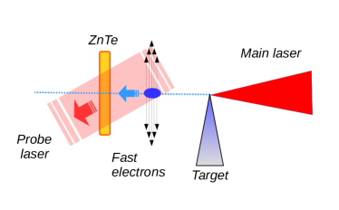
Figure 1 shows the experimental set-up, based on EOS spatial decoding technique [23], with the probe laser entering into the crystal at an incidence angle . In this way, the temporal charge profile of the emitted electrons is spatially imprinted along the transverse profile of the probe laser [24]. Being mm (FWHM) its transverse spot size, the effective time window is ps, where is the speed of light in vacuum, with a resolution of about fs.
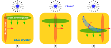
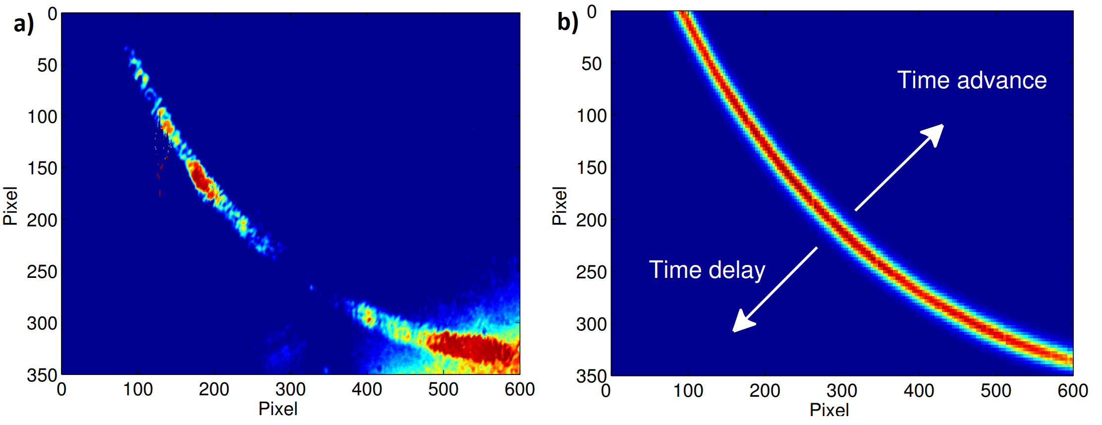
Figure 3 shows some typical results, compared with simulations. Due to our set-up geometry, with the electrons moving normally to the ZnTe crystal and the probe laser propagating laterally from right to left, as in Fig. 2, the expected signal is curved. This has been confirmed by simulations and experiment (Fig. 3).
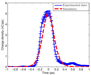
By measuring the relative delay with respect to the reference time, this diagnostics can also estimate the energy of the emitted electrons [24], providing measurements resolved in time. Moreover, since the electric field, inducing the electro-optic effect, is proportional to the bunch charge, this can be estimated from the signal intensity [20], properly calibrated with respect to the probe laser energy. Once the electron mean energy and charge are determined, the width of the measured signal is proportional to the bunch duration. Figure 4 shows a typical longitudinal profile measurement. Here, it is reported the average of different line-outs performed along the time direction, parallel to the temporal window diagonal. In detail, in this shot, an electron bunch with nC charge, MeV energy and about fs duration has been detected.
3 Diagnostics for single-shot emittance measurements
High gradient plasma structures provide accelerators at mm-scale. Nevertheless, the accelerated electron beams are still characterized by a large energy spread, around , and shot-to-shot instabilities. These issues prevent the use of conventional, multi-shot diagnostics for the emittance measurements, such as the quadrupole scan technique [25, 26].
Our idea relies on Optical Transition Radiation (OTR), emitted when a charged particle crosses the boundary between two media with different refractive indices [27]. Indeed, its angular distribution in the far field is sensitive to the beam divergence. In particular, starting from the angular distribution for a single particle [27] and considering the convolution with a Gaussian distribution in angle, we obtain a higher central minimum, directly correlated with the rms beam divergence [28]. Despite previous work aiming to exploit OTR to measure electron beam emittance [29], our method relies on the analysis of the OTR angular distribution in the focal plane of a microlens array to measure also the rms emittance correlation term. Indeed, this optical element, focusing different portions of the radiation in different points, directly correlates electron transverse momentum with the spatial position. The principle is similar to the pepper-pot emittance measurement [30], but, at the same time, our technique is more flexible. Indeed, it does not need a thick mask to be put on the electron beam cutting its phase space and, moreover, there is not a dependence on the screen position, that in our case is fixed in the microlens focal plane.
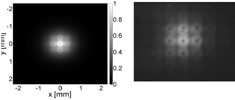
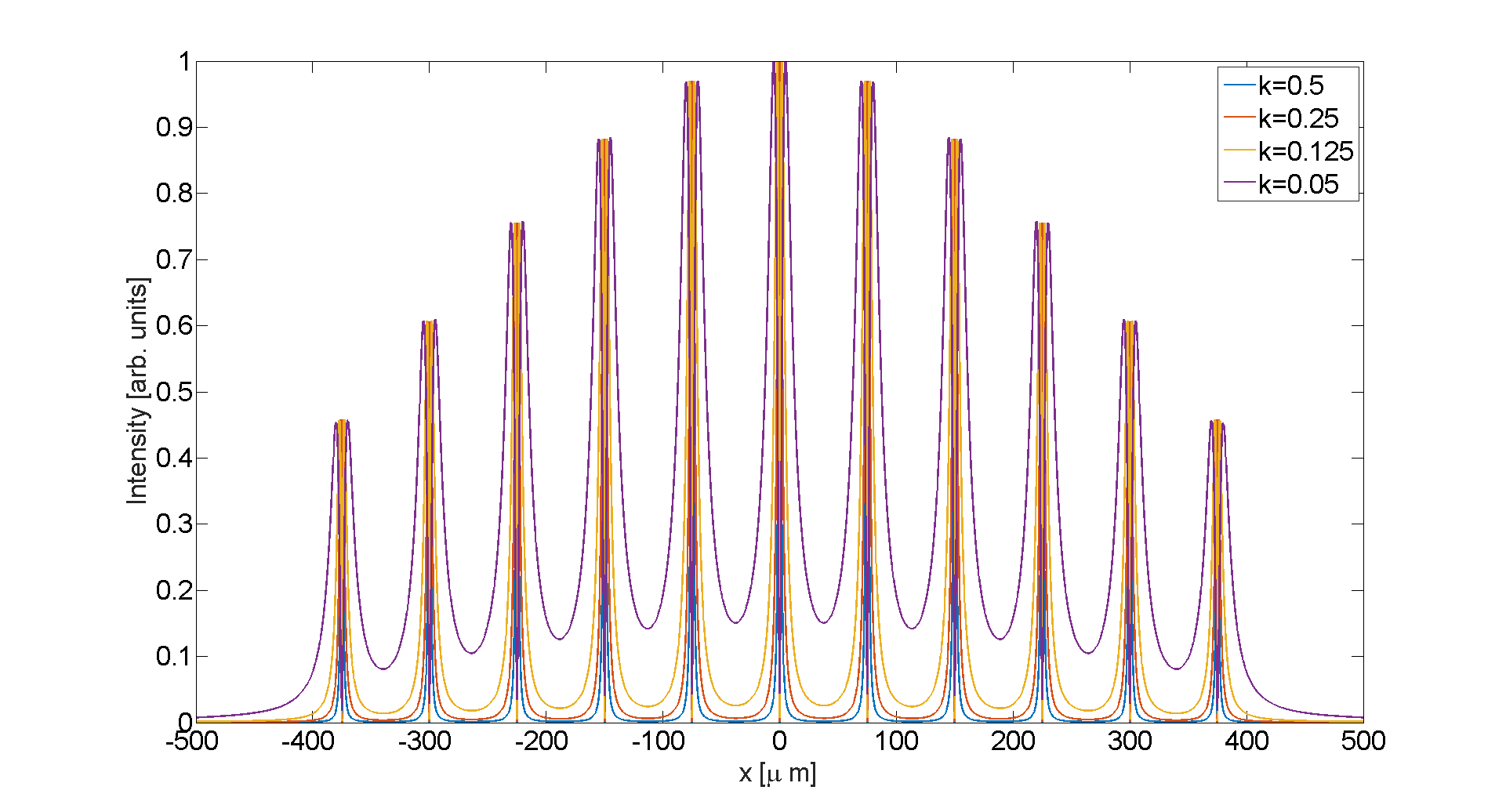
Several simulations have been performed to prove the feasiblity of this technique, firstly to understand the behaviour of the OTR field with a microlens array. For this purpose, a custom code in Zemax has been created to simulate this radiation as produced from a whole electron bunch [31]. Simulation results are shown in Fig. 5. The angular distributions coming from each microlens are clearly distinguishable. Moreover, a design analysis of the microlens array to be used has been performed. In particular, effects due to the diffraction with microlens aperture and to the overlap between adiacent angular distribution have been considered. Concerning the first one, microlenses with a diameter are needed to avoid diffraction, while the overlap between their different focal planes is minimized when , i.e. when the lobes of two different angular distributions are far enough (see Fig. 6). The microlens array commercially available at that time satisfied the first requirement, but not the second one. Indeed, the focal length was too long to avoid the overlap between different angular distributions.
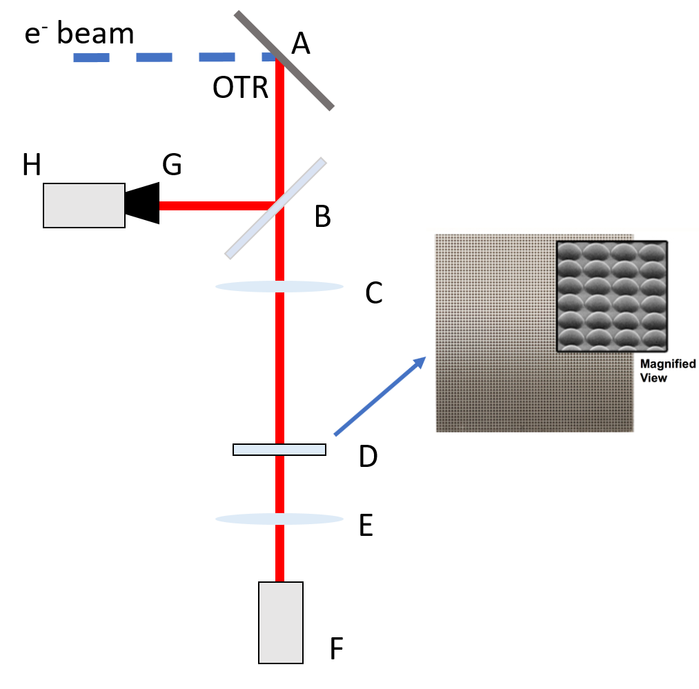
A preliminary experiment has been performed with SPARCLAB photoinjector [13], using bunch charge at . Figure 7 shows the experimental set-up layout: the OTR coming from an aluminium-coated silicon screen (A) is divided by means of a beam splitter (B); an optical line hosts a high quantum efficiency CCD camera (G) to image the source transverse profile, while a mm achromatic doublet (C) in the second arm transfers the image of source on a microlens array (D); a second mm achromatic doublet (E) is used to image the microlens focal plane onto an intensified CCD camera (F). Although we could not measure the electron bunch emittance, since its energy has limited our angular resolution [16], these preliminary results (Fig. 5) have shown that it is possible to produce the OTR angular distribution from different portions of the beam, as expected from our simulations.
4 Conclusions
We presented two single-shot diagnostics for electron beams generated from ultra intense laser-plasma interactions developed at SPARC_LAB Test Facility [13]. One, relying on Electro Optical Sampling (EOS), has allowed to measure the longitudinal profile of electric field carried by fast electrons generated during the interaction of a high intensity laser with solid matter. The characterization of these electrons can help to better understand the process of ion acceleration occurring in this kind of interaction. As reported in Fig. 3, the signals have been well reproduced and electron properties, in terms of charge, current and mean energy, have been retrieved. Besides the first measurement of longitudinal profile of the fast electrons, shown in Fig. 4, this diagnostics has been exploited to study the influence of geometrical shape of the target. In particular, we have shown that faster electrons and with more charge, resulting in a stronger electric field, are emitted from structured target [32]. Therefore, it is possible to boost the ion acceleration by using structured targets exploiting the consequently electric field enhancement.
On the other hand, the properties of Optical Transition Radiation (OTR) have been studied to design a single-shot transverse emittance measurement. In particular, our idea relies on using a microlens array, analyzing the OTR angular distribution coming from each microlens, placed in a specified position. In this way, by measuring also the transverse spot size (see Fig. 7), an emittance measurement is achievable in one shot. This kind of diagnostics represents a very useful tool to fully characterize an electron beam from LWFA despite its typical shot-to-shot instabilities and relative large energy spread. Even though some preliminary tests, obtained with the SPARC_LAB photo-injector, did not allow to measure the emittance because of poor angular resolution (due to a low electron energy), they have shown the possibility to measure the OTR angular distribution produced by different portions of the electron beam, as foreseen by our simulations. In the next future, a measurement on a plasma-accelerated electron beam at FLAME Facility is still under design, with both self-injection and external injection [33, 34] scheme, as well as a new experimental campaing at the SPARC_LAB photoinjector, working at the nominal energy, will be performed.
References
- [1] T. Tajima, J. Dawson, Laser electron accelerator, Physical Review Letters 43 (4) (1979) 267.
- [2] S. Wilks, A. Langdon, T. Cowan, M. Roth, M. Singh, S. Hatchett, M. Key, D. Pennington, A. MacKinnon, R. Snavely, Energetic proton generation in ultra-intense laser–solid interactions, Physics of plasmas 8 (2) (2001) 542–549.
- [3] C. Geddes, C. Toth, J. Van Tilborg, E. Esarey, C. Schroeder, D. Bruhwiler, C. Nieter, J. Cary, W. Leemans, High-quality electron beams from a laser wakefield accelerator using plasma-channel guiding, Nature 431 (7008) (2004) 538–541.
- [4] S. P. Mangles, C. Murphy, Z. Najmudin, A. G. R. Thomas, et al., Monoenergetic beams of relativistic electrons from intense laser-plasma interactions, Nature 431 (7008) (2004) 535.
- [5] W. Leemans, B. Nagler, A. Gonsalves, C. Tóth, K. Nakamura, C. Geddes, E. Esarey, C. Schroeder, S. Hooker, Gev electron beams from a centimetre-scale accelerator, Nature physics 2 (10) (2006) 696–699.
- [6] A. Maksimchuk, S. Gu, K. Flippo, D. Umstadter, V. Y. Bychenkov, Forward ion acceleration in thin films driven by a high-intensity laser, Physical Review Letters 84 (18) (2000) 4108.
- [7] A. Macchi, M. Borghesi, M. Passoni, Ion acceleration by superintense laser-plasma interaction, Reviews of Modern Physics 85 (2) (2013) 751.
- [8] E. Clark, K. Krushelnick, M. Zepf, F. Beg, M. Tatarakis, A. Machacek, M. Santala, I. Watts, P. Norreys, A. Dangor, Energetic heavy-ion and proton generation from ultraintense laser-plasma interactions with solids, Physical Review Letters 85 (8) (2000) 1654. doi:10.1103/physrevlett.85.1654.
- [9] R. Snavely, M. Key, S. Hatchett, T. Cowan, M. Roth, T. Phillips, M. Stoyer, E. Henry, T. Sangster, M. Singh, Intense high-energy proton beams from petawatt-laser irradiation of solids, Physical Review Letters 85 (14) (2000) 2945.
- [10] A. Curcio, M. Anania, F. Bisesto, E. Chiadroni, A. Cianchi, M. Ferrario, F. Filippi, D. Giulietti, A. Marocchino, F. Mira, et al., First measurements of betatron radiation at flame laser facility, Nuclear Instruments and Methods in Physics Research Section B: Beam Interactions with Materials and Atoms.
- [11] A. Rousse, K. T. Phuoc, R. Shah, A. Pukhov, E. Lefebvre, V. Malka, S. Kiselev, F. Burgy, J.-P. Rousseau, D. Umstadter, et al., Production of a kev x-ray beam from synchrotron radiation in relativistic laser-plasma interaction, Physical review letters 93 (13) (2004) 135005.
- [12] M. Fuchs, Laser-driven soft-x-ray undulator source, Ph.D. thesis, lmu (2010).
- [13] M. Ferrario, D. Alesini, M. Anania, A. Bacci, M. Bellaveglia, O. Bogdanov, R. Boni, M. Castellano, E. Chiadroni, A. Cianchi, et al., Sparc_lab present and future, Nuclear Instruments and Methods in Physics Research Section B: Beam Interactions with Materials and Atoms 309 (2013) 183–188.
- [14] F. Bisesto, M. Anania, E. Chiadroni, A. Cianchi, G. Costa, A. Curcio, M. Ferrario, M. Galletti, R. Pompili, E. Schleifer, et al., Innovative single-shot diagnostics for electrons accelerated through laser-plasma interaction at flame, in: SPIE Optics+ Optoelectronics, International Society for Optics and Photonics, 2017, pp. 102400K–102400K.
- [15] F. Bisesto, M. P. Anania, E. Chiadroni, A. Cianchi, A. Curcio, M. Ferrario, R. Pompili, A. Zigler, Innovative single-shot diagnostics for electrons from laser wakefield acceleration at flame, in: 8th Int. Particle Accelerator Conf.(IPAC’17), Copenhagen, Denmark, 14â 19 May, 2017, JACOW, Geneva, Switzerland, 2017, pp. 1727–1730.
- [16] A. Cianchi, M. Anania, M. Bellaveglia, F. Bisesto, M. Castellano, E. Chiadroni, D. Di Giovenale, G. Di Pirro, M. Ferrario, G. Gatti, et al., Transverse emittance diagnostics for high brightness electron beams, Nuclear Instruments and Methods in Physics Research Section A: Accelerators, Spectrometers, Detectors and Associated Equipment 865 (2017) 63–66.
- [17] A. Mackinnon, Y. Sentoku, P. Patel, D. Price, S. Hatchett, M. Key, C. Andersen, R. Snavely, R. Freeman, Enhancement of proton acceleration by hot-electron recirculation in thin foils irradiated by ultraintense laser pulses, Physical Review Letters 88 (21) (2002) 215006.
- [18] A. Poyé, S. Hulin, M. Bailly-Grandvaux, J.-L. Dubois, J. Ribolzi, D. Raffestin, M. Bardon, F. Lubrano-Lavaderci, E. D’Humières, J. J. Santos, Physics of giant electromagnetic pulse generation in short-pulse laser experiments, Physical Review E 91 (4) (2015) 043106. doi:10.1103/physreve.91.043106.
- [19] I. Wilke, A. M. MacLeod, W. Gillespie, G. Berden, G. Knippels, A. Van Der Meer, Single-shot electron-beam bunch length measurements, Physical Review Letters 88 (12) (2002) 124801. doi:10.1103/physrevlett.88.124801.
- [20] R. Pompili, M. P. Anania, M. Bellaveglia, F. Bisesto, E. Chiadroni, A. Cianchi, A. Curcio, D. Di Giovenale, G. Di Pirro, M. Ferrario, et al., Electro-optical methods for multipurpose diagnostics, in: 5th Int. Beam Instrumentation Conf.(IBIC’16), Barcelona, Spain, Sept. 13-18, 2016, JACOW, Geneva, Switzerland, 2017, pp. 291–294.
- [21] B. Steffen, V. Arsov, G. Berden, W. Gillespie, S. Jamison, A. M. MacLeod, A. Van Der Meer, P. Phillips, H. Schlarb, B. Schmidt, Electro-optic time profile monitors for femtosecond electron bunches at the soft x-ray free-electron laser flash, Physical Review Special Topics-Accelerators and Beams 12 (3) (2009) 032802. doi:10.1103/physrevstab.12.032802.
- [22] F. B. et al., The flame laser at sparclab, these proceedings.
- [23] A. L. Cavalieri, D. Fritz, S. Lee, P. Bucksbaum, D. Reis, J. Rudati, D. Mills, P. Fuoss, G. Stephenson, C. Kao, Clocking femtosecond x rays, Physical Review Letters 94 (11) (2005) 114801. doi:10.1103/physrevlett.94.114801.
- [24] R. Pompili, M. Anania, F. Bisesto, M. Botton, M. Castellano, E. Chiadroni, A. Cianchi, A. Curcio, M. Ferrario, M. Galletti, et al., Sub-picosecond snapshots of fast electrons from high intensity laser-matter interactions, Optics Express 24 (26) (2016) 29512–29520.
- [25] F. Löhl, S. Schreiber, M. Castellano, G. Di Pirro, L. Catani, A. Cianchi, K. Honkavaara, Measurements of the transverse emittance at the flash injector at desy, Physical Review Special Topics-Accelerators and Beams 9 (9) (2006) 092802.
- [26] A. Mostacci, M. Bellaveglia, E. Chiadroni, A. Cianchi, M. Ferrario, D. Filippetto, G. Gatti, C. Ronsivalle, Chromatic effects in quadrupole scan emittance measurements, Physical Review Special Topics-Accelerators and Beams 15 (8) (2012) 082802.
- [27] M. L. Ter-Mikaelian, High energy electromagnetic processes in condensed media.
- [28] F. Bisesto, M. P. Anania, M. Botton, E. Chiadroni, A. Cianchi, A. Curcio, M. Ferrario, M. Galletti, R. Pompili, E. Schleifer, et al., Novel single-shot diagnostics for electrons from laser-plasma interaction at sparc_lab, Quantum Beam Science 1 (3) (2017) 13.
- [29] R. Feldman, A. Lumpkin, D. Rule, R. Fiorito, Developments in on-line, electron-beam emittance measurements using optical-transition radiation techniques, Nuclear Instruments and Methods in Physics Research Section A: Accelerators, Spectrometers, Detectors and Associated Equipment 296 (1) (1990) 193–198.
- [30] M. Zhang, Emittance formula for slits and pepper-pot measurement, Tech. rep., Fermi National Accelerator Lab., Batavia, IL (United States) (1996).
- [31] E. C. F. Bisesto, A. Castellano, A. Cianchi, Zemax simulations describing collective effects in transition and diffraction radiation, Opt. Exp. (to be published).
- [32] R. Pompili, M. Anania, F. Bisesto, M. Botton, M. Castellano, E. Chiadroni, A. Cianchi, A. Curcio, M. Ferrario, M. Galletti, et al., Femtosecond dynamics of energetic electrons in high intensity laser-matter interactions, Scientific Reports 6.
- [33] F. Bisesto, M. Anania, A. Bacci, M. Bellaveglia, E. Chiadroni, A. Cianchi, A. Curcio, D. Di Giovenale, G. Di Pirro, M. Ferrario, et al., Laser–capillary interaction for the exin project, Nuclear Instruments and Methods in Physics Research Section A: Accelerators, Spectrometers, Detectors and Associated Equipment 829 (2016) 309–313.
- [34] A. Rossi, M. Anania, A. Bacci, M. Belleveglia, F. Bisesto, E. Chiadroni, A. Cianchi, A. Curcio, A. Gallo, D. Di Giovenale, et al., Stability study for matching in laser driven plasma acceleration, Nuclear Instruments and Methods in Physics Research Section A: Accelerators, Spectrometers, Detectors and Associated Equipment 829 (2016) 67–72.