Growth and characterization of a Li2Mg2(MoO4)3 scintillating bolometer
Abstract
Lithium magnesium molybdate (Li2Mg2(MoO4)3) crystals were grown by the low-thermal-gradient Czochralski method. Luminescence properties of the material (emission spectra, thermally stimulated luminescence, dependence of intensity on temperature, phosphorescence) have been studied under X-Ray excitation in the temperature interval from 8 K to 400 K, while at the same being operated as a scintillating bolometer at 20 mK for the first time. We demonstrated that Li2Mg2(MoO crystals are a potentially promising detector material to search for neutrinoless double beta decay of 100Mo.
keywords:
Double beta decay , 100Mo , Cryogenic scintillating bolometer , Li2Mg2(MoO4)3 crystal , Crystal growth , Luminescence1 Introduction
Neutrinoless double beta () decay is of essential interest to particle physics since this process violates lepton number and requires the neutrino to be a massive Majorana particle [1, 2, 3, 4]. Generally speaking, decay can be mediated by many other beyond the Standard Model effects [5, 6].
Significant efforts have been made to observe the decay, however; the decay have yet to be observed (see the reviews [7, 8, 9, 10, 11] and recent results [12, 13, 14, 15, 16]). The experiments have set limits on the half-life of this process in the range of yr for various nuclei, allowing us to restrict the effective Majorana neutrino mass at the level of eV. The broad range of the neutrino mass limits results from the uncertainty of the nuclear matrix elements calculations [17].
The experimental sensitivity should be further improved to explore the inverted hierarchy of the neutrino mass ( eV) corresponding to yr even for the most promising nuclei candidates [4, 17]. Experiments able to detect so rare decays should operate hundreds of kg of an isotope of interest, and have high detection efficiency and energy resolution, and a very low (ideally zero) background. Low temperature scintillating bolometers have a great potential to realize large-scale high-sensitivity decay experiments with several nuclei. The isotope 100Mo is one of the most promising candidates thanks to the high endpoint energy of decay keV [18], a high isotopic abundance [19] (and possibility of enrichment in large amount by gas-centrifugation), and a high decay probability [20, 21, 22, 23].
There are several molybdate crystals have already been tested as scintillating bolometers: ZnMoO4 [24, 25, 26], Li2MoO4 [26, 27], Li2Zn2(MoO4)3 [28], CaMoO4 [29] with their main properties reported in Table 1.
| Properties | Li2MoO4 [27] | Li2Mg2(MoO4)3 | Li2Zn2(MoO4)3 [28] | CaMoO4 [30] | ZnMoO4 [31] |
| Density (g/cm3) | 3.89 [32] | 4.38 | 4.3 | ||
| 3.798 [33] | |||||
| 3.82 (present work) | |||||
| Melting point (∘C) | 701 | 1033 [32] | 890 | 1003 | |
| Structural type | Phenacite | Pnma [33, 34, 35] | Orthorhombic | Scheelite | Triclinic, |
| Hardness (Mohs) | 3.5 | ||||
| (nm) | 590 | 585 (present work) | 610 | 520 | 625 |
| Refractive index | 1.44 | 2.0 | 1.98 | ||
| Hygroscopicity | Weak | No | No | No | No |
| (atoms/cm3) |
In addition to the technical requirements described above, a crystal scintillator for a large-scale high-sensitivity decay experiment should meet some practical requirements: low material cost, the capability for mass production, possibility of enriched molybdenum recycling to minimize loss of the enriched isotope, and to produce more scintillation elements. In a case of large scale cryogenic experiments (e.g., in the CUPID project [36, 37]), one should place a maximum number of the nuclei of interest in a restricted cryostat volume. From this point of view, the Li2Mg2(MoO4)3 crystal looks to be one of the most appropriate materials for cryogenic experiments with 100Mo. Absence of hygroscopicity (in contrast to weak hygroscopicity of Li2MoO4) is also an advantage of the material.
In the present work, a Li2Mg2(MoO4)3 crystal has been investigated as possible low temperature scintillator for double beta decay experiments with 100Mo. Crystal growth of the compound is reported in Sec. 2, the luminescent properties of the material are summarized in Sec. 3, and the first test of a Li2Mg2(MoO4)3 crystal sample as scintillating bolometer is described in Sec. 4.
2 Growth of Li2Mg2(MoO4)3 crystals
The main difficulty in the production of Li2Mg2(MoO4)3 crystals is the incongruent melting of the compound [32, 33]. This property makes it difficult to grow large volume crystals by the ordinary Czochralski technique from a stoichiometric compound. In the present study, Li2Mg2(MoO4)3 crystals were grown by using the low-thermal-gradient Czochralski method [38, 39]. The Li2Mg2(MoO4)3 compound for the crystal growth has been synthesized by mixing Li2CO3, MgO and MoO3 powders in the ratio Li2Mg2(MoO : Li2MoO. In the first stage of the crystal R&D, a molybdenum oxide of 99.9% purity grade has been used, replaced by a 99.999% high purity MoO3 in later development. The powders mixture was placed in a platinum crucible mm covered by a tight platinum lid with a thin pipe, heated to a temperature degrees higher than the melting temperature. The melt homogeneity was achieved mixing the melt by a platinum roller unit over hours. The HX620 crystal growing set-up utilized a three-zone resistance furnace with weight monitoring. Several crystal boules have been grown from a crystal seed of [010] orientation with a pulling rate mm per day [40]. Two Li2Mg2(MoO4)3 crystal boules produced from 99.9% and 99.999% purity MoO3 are shown in Fig. 1.


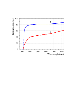
The higher optical quality of the Li2Mg2(MoO4)3 crystals produced from higher purity MoO3 was confirmed by transmittance measurements performed with a Shimadzu UV-3101PC spectrometer. The results are presented in Fig. 2. The main cause of the crystals coloration is the contamination of the raw materials (particularly of the molybdenum oxide) by transition metals. The most abundant impurities of the 99.9% purity molybdenum oxide are K on the level of 0.01 wt%, and Si, Fe, Ti, Ni, Ca, Cr ( 0.001 wt%). Presence of Fe, Ti, Ni, Cr can lead to coloration of the crystal produced from the molybdenum oxide of the 99.9% purity grade. The effect of crystals coloration by transition metals is well known, and was also observed in molybdate crystals (see e.g. investigation of the raw-materials purity level on the optical quality of zinc molybdate crystal scintillators in [41]).
Li2Mg2(MoO4)3 crystals are isotypic with lithium zinc
molybdate (Li2Zn2
(MoO4)3) [35].
Therefore, one can assume that Li+ and Mg2+ ions are
distributed over the , and sites (see Fig. 4 in
[35]). Taking into account that the
composition of Li2Mg2(MoO4)3 crystals may depend on
the synthesis temperature (as observed for
Li2Zn2(MoO4)3 [33]), one can expect
that the Li2Mg2(MoO4)3 crystal stoichiometry varies
due to the substitution/subtraction process 2Li Mgvacancy. Therefore, the real crystal
composition is more accurately described by
Li2-2xMg2+x(MoO, where .
However; the use of Li2Mg2(MoO4)3 compound in double
beta decay or dark matter experiments would require a large number
of identical crystals, with well-known compositions. It should be
stressed that the composition of the produced
Li2Mg2(MoO4)3 crystals is most strongly defined by the
composition of the powder used for the crystal growth. And
furthermore, it has been demonstrated in potassium gadolinium
tungstate (KGd(WO4)2, a similar compound from the point of
view of crystal growth), the low-thermal-gradient Czochralski
method allowed to produce large volume (up to 200 cm3),
optically uniform KGd(WO4)2 crystals for laser applications
[42]. R&D is already underway to produce high
quality Li2Mg2(MoO4)3 crystal scintillators.
3 Luminescence of Li2Mg2(MoO4)3 crystal under X-Ray irradiation
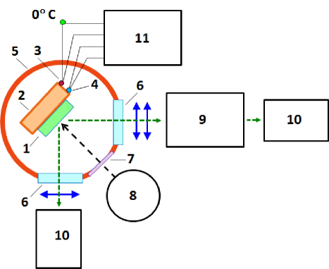
The luminescence of a Li2Mg2(MoO4)3 crystal sample mm3 was investigated under X-Ray Irradiation (X-Ray Luminescence, XRL) in a wide temperature range from 8 K to 400 K. The sample was mounted on a copper holder inside a vacuum cryostat (a simplified scheme of the set-up is presented in Fig. 3). The temperature of the copper holder was controlled by a semiconductor silicon sensor WAD305 (in the temperature interval 8 K – 85 K), and with a chromel-copel thermocouple (in the temperature interval 85 K – 450 K). We showed in separate measurements, that the temperature difference between a crystal sample and the support does not exceed 0.2 K (0.4 K upon linear heating of the sample). The crystal sample was irradiated through a beryllium window in the cryostat by X-Rays from an X-Ray tube BHV-7 with a rhenium anode operated at 20 kV, 25 mA with a flux of 0.635 mW/cm2. The luminescence was measured in two spectral channels: in a wide wavelength interval (integral mode) and at a selected wavelength 548 nm (spectral mode). In the integral mode, the luminescence light was focused with the help of a quartz lens on the photocathode of a photomultiplier tube FEU-106 with extended sensitivity in the wavelength region of nm. A high-transmission monochromator MDR-2 (with a 600 mm-1 diffraction grating) was used in the spectral mode. The emission spectra were then corrected for the spectral sensitivity of the registration system. The temperature dependencies of the XRL intensity was obtained by cooling the sample to avoid effect of Thermally Stimulated Luminescence (TSL). Phosphorescence measurements were carried out after irradiation at temperatures of 8 K and 85 K over minutes (the exposure dose was J/cm2). The TSL of the sample was measured after the phosphorescence measurements while heating of the samples with a rate K/s.
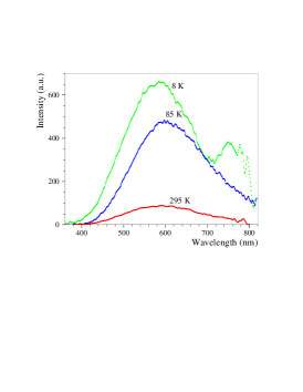
The emission spectra of Li2Mg2(MoO4)3 crystal measured under X-Ray irradiation at 8 K, 85 K and 295 K are shown in Fig. 4. The main broad emission band with a maximum at nm is observed at room and liquid nitrogen temperatures, while the position of the maximum is slightly shifted to a shorter wavelength nm at 8 K. An additional infrared band appeared at nm with cooling of the sample to the liquid helium temperature. The measured emission spectra are slightly different from those observed under ultraviolet excitation with the maximum at 520 nm [43]. It should be stressed that the luminescence emission wavelength range of Li2Mg2(MoO4)3 crystal is suitable for application of the material as low temperature scintillating bolometer as discussed in Sec. 4. Indeed, the wide spectral sensitivity of germanium photodetectors ( nm [44]) covers the emission spectral range of the Li2Mg2(MoO4)3 crystal.
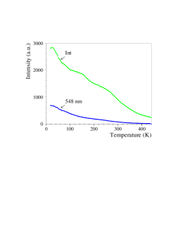
The dependencies of the Li2Mg2(MoO4)3 luminescence intensity on temperature measured in the integral and spectral modes are presented in Fig. 5. The luminescence intensity increases by more than three times by cooling the sample. Similar properties were observed in Li2MoO4 [27] and Li2Zn2(MoO4)3 [28]. Other molybdate crystals (as, e.g., CaMoO4 [45], ZnMoO4 [31], SrMoO4 [46]) are also characterized by an increasing of luminescence and scintillation efficiency as they are cooled down to near liquid helium temperatures.
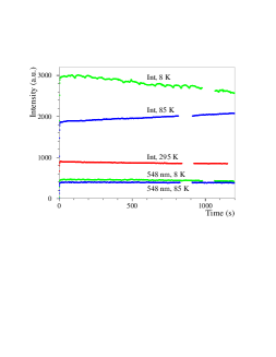
The luminescence intensity during X-Ray excitation in Li2Mg2(MoO4)3 crystal is almost constant at room temperature (see Fig. 6). A rather weak increase of the XRL intensity at 77 K by % with a decrease at 8 K by % after irradiation over 20 minutes were observed. A systematic error of the measurements does not exceeds . It should be noted that after heating the sample up to 450 K and then cooling again (without irradiation during the cooling) to low temperatures (8 K or 85 K), the dose dependencies of the XRL intensity remain the same. Taking into account the constant XRL intensity at 295 K, one could assume that the change in the XRL intensity at low temperatures is due to the processes of charge carriers accumulation in deep traps and recharging of recombination centers [47]. This hypothesis is also confirmed by the presence of phosphorescence and TSL described below. In general, the observed independence of the Li2Mg2(MoO4)3 luminescence on the dose (that is proportional to the duration of X-Ray irradiation) indicates a high radiation resistance of the material.
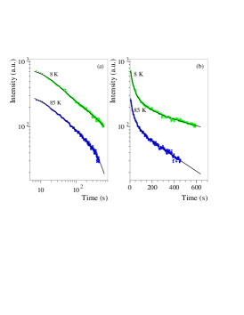
The Li2Mg2(MoO4)3 crystal showed a substantial phosphorescence, as one can see in Fig. 7, where the phosphorescence decay curves recorded at 8 K and 85 K are shown. As it was demonstrated for ZnMoO4 crystals, long term phosphorescence can be described by three exponential functions better than by the Becquerel hyperbolic formula [47]. The fits of the Li2Mg2(MoO4)3 phosphorescence decay curves are presented in Fig. 7. E.g., for the phosphorescence at 85 K the decay times are s, s and s, while at 8 K the decay times are s, s and s. The represents the effects of free charge recombination coupled with charge carriers delocalized from shallow traps, and stem from delocalization from phosphorescent traps and deep traps, respectively. The phosphorescence shows the recombination character of luminescence in Li2Mg2(MoO4)3. Similar phosphorescence properties were observed for ZnMoO4 [47] and Li2MoO4 [27] crystals. The long term phosphorescence indicates that the material tends to have some afterglow, a well known effect in scintillators. However; the intensity of the signal due to afterglow after s of energy release in the Li2Mg2(MoO4)3 crystal (the time to analyze scintillation pulse profiles in low temperature scintillating bolometers is typically shorter) decreases by two orders of amplitude, approaching the photodetectors noise. Moreover; further improvement of the crystals quality should reduce the phosphorescence and possible afterglow effects.
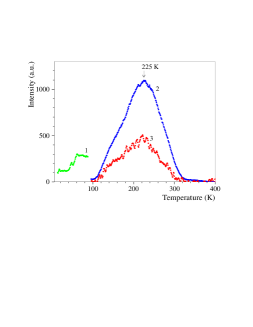
The TSL of Li2Mg2(MoO4)3 crystal measured after X-Ray irradiation at 8 K and 85 K are presented in Fig. 8. It should be noted that use of two excitation temperatures allows more accurate investigations of TSL, since accumulation of charge carriers on the traps depends on the temperature of the excitation. The most intensive TSL peak is observed at K (similar to Li2Zn2(MoO4)3 [28]). At room temperature, the traps responsible for the TSL become shallow resulting in a stronger radiation resistance of the material (see Fig. 6).
Both the intensive phosphorescence and the TSL prove the presence of traps in the Li2Mg2(MoO4)3 crystal sample due to defects, which indicates that there is still room to improve the material.
4 Performance tests at milli-Kelvin temperature
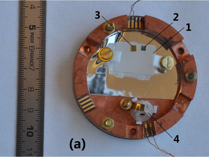
A Li2Mg2(MoO4)3 crystal sample (10.24 g, cm3) was equipped with a Neutron Transmutation Doped (NTD) Ge thermistor and a silicon heater (for stabilization), which was than mounted in a copper frame (see Fig. 9 (a)) to run as bolometer.
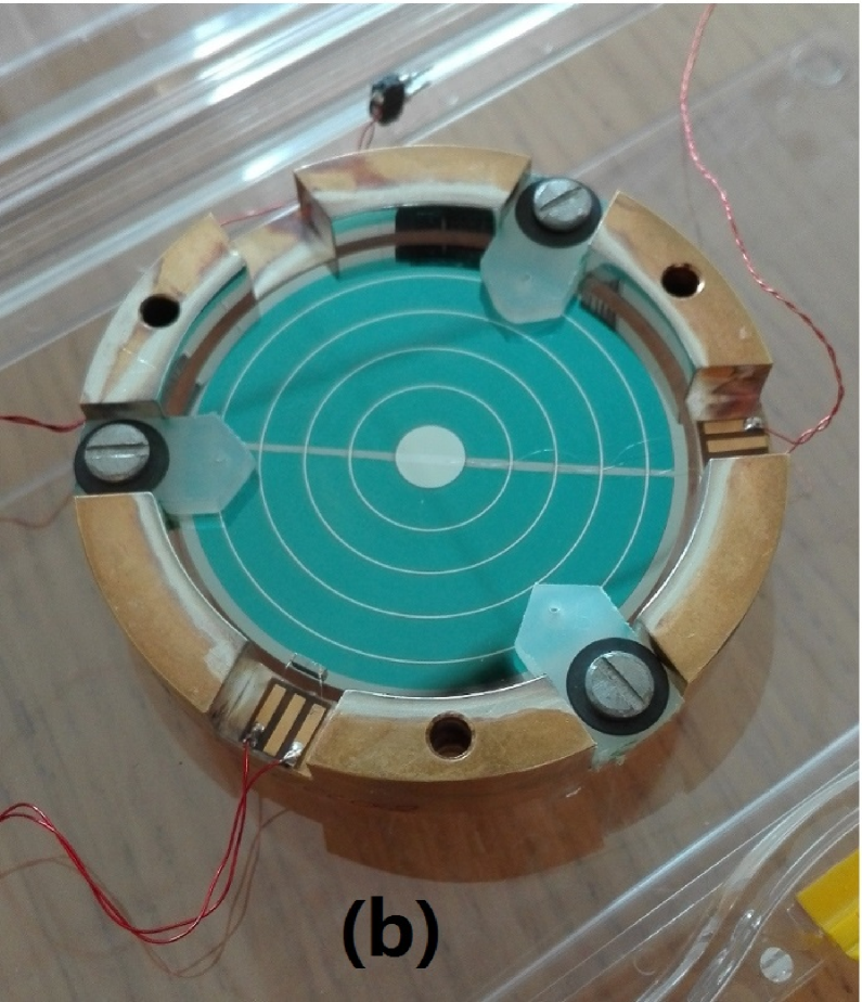
Scintillation signals from the Li2Mg2(MoO4)3 crystal were recorded with the help of a photodetector operated at 60 V in Neganov-Luke mode [48, 49]. The photodetector (see Fig. 9 (b)) consists of a germanium disk 44 mm in diameter with a set of concentric annular Al electrodes with a pitch 3.7 mm and coated with a SiO anti-reflective film 70 nm thick (cyan color area on the picture). 60 V voltage was applied between the electrodes to amplify the scintillation signals. Heat signals after absorption of scintillation photons are read-out by an NTD germanium thermistor (visible in the lower part of the picture, attached at the germanium disk near the edge).
The detector module was run in a pulse-tube dilution refrigerator [50]. The set-up is located aboveground at the CSNSM (Orsay, France). A weak 210Po alpha source was installed in the detector module to irradiate the crystal. The cryostat is surrounded by a massive low-activity lead shield with 10 cm maximum thickness to reduce environmental gamma background and the soft component of cosmic rays. The data were acquired in stream mode with a 5 kHz sampling rate.
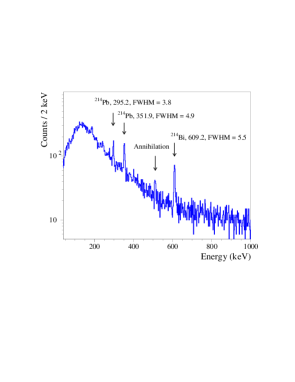
A background energy spectrum was gathered over 88 h with the Li2Mg2(MoO4)3 scintillating bolometer at 20 mK as shown in in Fig. 10. Several gamma peaks (gamma quanta of 214Pb and 214Bi, daughters of environmental 226Ra) are visible in the spectrum which allowed us to calibrate the detector and estimate its energy resolution (full width at half maximum, FWHM) as shown in the Fig. 10. For instance, the energy resolution of the detector was measured as FWHM keV at 609 keV (gamma quanta of 214Bi), while the baseline energy resolution of the detector module was 3.8 keV. The spectrometric characteristics of a Li2Mg2(MoO4)3-based detector could be further improved operating the device in lower-level background conditions and at lower temperature.
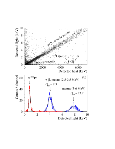
The particle discrimination capability of the detector (an important characteristic for application in rare-event searches) is demonstrated by Fig. 11 with data accumulated over 88 h with the Li2Mg2(MoO scintillating bolometer. The , , and cosmic muon events are clearly separated from the -triton and events111The events between the bands can be explained by pulses pile-ups due to comparatively slow response of bolometric detectors (the time window of Li2Mg2(MoO4)3 heat pulses is s). The pile-up events nature was checked by their pulse profile analysis..
The particle discrimination capability (denoted here as ) to discriminate particles and quanta ( particles, muons) can be determined by the following formula:
| (1) |
where () are average values (standard deviations) of the scintillation signals distributions for particles and quanta (or other particles such as betas and muons). We obtained for 5.3 MeV particles of 210Po and (, muons) events in the energy intervals MeV ( MeV). The distributions are shown in Fig. 11 (b). However, the discrimination power should be estimated in the same energy interval for particles and quanta ( particles) in the energy region of the peak of 100Mo. Unfortunately, the statistic of events with energy MeV in our data is rather poor. We are going to utilize an source emitting energy-degraded particles to investigate the discrimination power of Li2Mg2(MoO4)3-based scintillating bolometers in our further studies of the detector material. Nevertheless, the achieved discrimination power is high enough to discriminate and events in a double beta experiment with 100Mo.
Moreover; some differences were observed in the heat-pulse shapes of (, cosmic muons) and events. For this analysis, we applied a parameter (pulse-shape parameter) that is a ratio between the fitted amplitude (a value of signal maximum obtained by the pulse fit) to the filtered amplitude (a signal maximum after applying the optimum filter [51, 52]). A background scatter plot of the pulse-shape parameter versus the heat signal amplitudes accumulated with the Li2Mg2(MoO4)3 scintillating bolometer over 88 h is presented in Fig. 12 (a). The distributions of the pulse-shape parameter for , , muon events with energy MeV, and events of 210Po are shown in Fig. 12 (b). The discrimination power for the distributions calculated by equation (1) is .

The scintillation light yield of the Li2Mg2(MoO4)3 crystal scintillator (the detected light energy per particle energy measured by the deposited heat) is estimated to be 1.3 keV/MeV. The estimation was done thanks to the calibration of the photodetector by using 5.9 keV X-rays from a weak 55Fe source. The value is comparable to the light yield observed with ZnMoO4 ( keV/MeV) and slightly exceeds the light yield of Li2MoO4 crystal scintillators ( keV/MeV) [26].
The quenching factor for particles in the Li2Mg2(MoO4)3 scintillator was estimated as the ratio between the detected scintillation signals amplitudes for particles of 210Po (in the energy interval around the peak keV) and the scintillation signals amplitudes for cosmic muons ( quanta, particles) in the same energy interval as 0.105(2).
An energy spectrum of the -triton and events accumulated by the Li2Mg2(MoO4)3 scintillating bolometer was built by using the difference in scintillation yield presented in Fig. 11. The obtained energy spectrum is shown in Fig. 13. There are two clearly visible groups of events in the data: -triton peak with energy 4784 keV caused by thermal neutrons capture on 6Li, and two peaks of 210Po. One peak is from the 210Po alpha source that irradiated the crystal (the peak is labelled in Fig. 13 as “210Po ext”), while the second weak peak is due to the bulk contamination of the crystal by 210Po (labelled on the Figure as “210Po int”). The energy resolution is FWHM keV for the -triton peak, and FWHM keV for the internal 210Po peak.
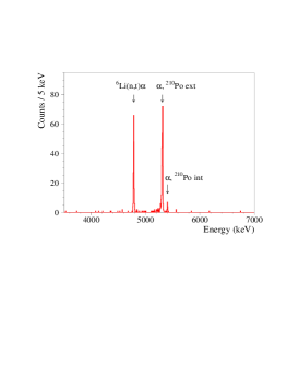
Analysis of the alpha spectrum allowed us to evaluate radioactive contamination of the Li2Mg2(MoO4)3 crystal. Activity of the bulk 210Po can be estimated as mBq/kg, which is rather low taking into account that no particular efforts were made to obtain radiopure material. It should be stressed that we cannot conclude whether the 210Po is result of the crystal contamination by 210Pb or it is 210Po itself. If it is polonium contamination then the activity should decrease substantially within a few years, due to the comparatively short half-life of 210Po: d. In contrast, the activity of 210Po could even increase (see, e.g., Fig. 7 in [53]) in the case of led (210Pb) contamination (the half-life of 210Pb is yr, the crystal was produced approximately 9 months before the test). Due to the absence of other peaks we were only able to set limits () on the active alpha members of the 235U, 238U and 232Th chains with the equation (2):
| (2) |
where is the detection efficiency (assumed to be 100% due to a very short path length of alpha particles in the Li2Mg2(MoO4)3 crystal), is the particle emission probability, is the mass of the crystal sample, is the measuring time, and is the number of events that can be excluded at a given confidence level (C.L., all the limits here are given with 90% C.L.). Values of were estimated by using the Feldman-Cousins procedure for no effect observed on an estimated background [54]. The measured numbers of events were calculated in the energy intervals keV around the of the expected peaks (the interval is wider than 3 sigma for all expected peaks, therefore, the detection efficiency remains almost 100%), while the background counting rate was estimated in the energy intervals MeV and MeV, where no particles of U/Th are expected. For instance, in the energy interval keV (where a peak of 232Th with energy 4081.6 keV is expected) 1 event was detected, while the estimated background is 1.32 counts. Therefore, according to [54] we should exclude counts, that lead to the limit on 232Th activity in the crystal mBq/kg. Other limits were obtained in a similar way. A summary of the Li2Mg2(MoO4)3 crystal radioactive contamination is presented in Table 2.
| Chain | Sub-chain | Activity (mBq/kg) |
|---|---|---|
| 232Th | 232Th | |
| 228Th | ||
| 238U | 238U | |
| 234U | ||
| 226Ra | ||
| 210Po | ||
| 235U | 235U | |
| 223Ra |
5 Conclusions
Large volume optically-clear quality Li2Mg2(MoO4)3 crystals were grown with the help of the low-thermal-gradient Czochralski method. The luminescence of a Li2Mg2(MoO4)3 crystal sample was studied under X-Ray excitation in the K temperature range. Luminescence with a maximum at nm is observed at 8 K. The luminescence intensity increases by more than a factor 3 when cooling the crystal sample from room temperature to 8 K. Intense phosphorescence and thermally stimulated luminescence indicate presence of traps due to defects in the crystal sample, which points to that fact that the material quality can be further improved.
Low temperature measurements (at 20 mK) of a 10 g sample of Li2Mg2(MoO4)3 scintillating bolometer were carried out over 88 h, demonstrating energy resolution as good as 5.5 keV (FWHM, at 609 keV), despite the comparatively high baseline noise (3.8 keV, whereas it is typically on the level of keV). The scintillator light yield is estimated as keV/MeV. The Li2Mg2(MoO4)3 scintillating bolometer showed an excellent particle discrimination capability with a discrimination power (see equation 1) for MeV (, muons) events and 5.3 MeV particles of 210Po. We have observed also a weak difference in the heat signal shapes of (, cosmic muons) and particles. Despite no special efforts to obtain radiopure material, the radioactive contamination of the crystal was measured to be quite low. The activity of 210Po is mBq/kg, while only limits on the mBq/kg level were estimated for other active members of the 235U, 238U and 232Th families. These bolometric measurements have demonstrated that Li2Mg2(MoO is a potentially promising detector material for double beta decay experiments with molybdenum as well as other rare-event searches.
In particular, we would like to stress that Li2Mg2(MoO scintillator contains a variety of elements with different atomic masses , 16, 24, 96 (Li, O, Mg and Mo, respectively) which can be exploited in dark matter low temperature scintillating bolometers (see CRESST [55]) to probe new areas of parameter space, particularly of spin-dependent dark matter.
6 Acknowledgements
These studies were supported in part by the project “Investigation of neutrino and weak interaction in double beta decay of 100Mo” in the framework of the Programme “Dnipro” based on Ukraine-France Agreement on Cultural, Scientific and Technological Cooperation, and by the IDEATE International Associated Laboratory (LIA). F.A. Danevich gratefully acknowledges the support from the “Jean d’Alembert” Grants program (Project CYGNUS) of the University of Paris-Saclay. A.S. Zolotarova is supported by the “IDI 2015” project funded by the IDEX Paris- Saclay, ANR-11-IDEX-0003-02. The authors are grateful to Alexander Leder from the Massachusetts Institute of Technology for a careful reading of the manuscript and helpful corrections.
References
- [1] J. Barea, J. Kotila, F. Iachello, Limits on Neutrino Masses from Neutrinoless Double- Decay, Phys. Rev. Lett. 109 (2012) 042501.
- [2] W. Rodejohann, Neutrino-less double beta decay and particle physics, J. Phys. G 39 (2012) 124008.
- [3] S. Dell’Oro, S. Marcocci, M. Viel, F. Vissani, Neutrinoless Double Beta Decay: 2015 Review, AHEP 2016 (2016) 2162659.
- [4] J.D. Vergados, H. Ejiri, F. Šimkovic, Neutrinoless double beta decay and neutrino mass, Int. J. Mod. Phys. E 25 (2016) 1630007.
- [5] F.F. Deppisch, M. Hirsch, H. Ps, Neutrinoless double-beta decay and physics beyond the standard model, J. Phys. G 39 (2012) 124007.
- [6] S.M. Bilenky, C. Giunti, Neutrinoless double-beta decay: A probe of physics beyond the Standard Model, Int. J. Mod. Phys. A 30 (2015) 1530001.
- [7] S.R. Elliott, Recent progress in double beta decay, Mod. Phys. Lett. A 27 (2012) 123009.
- [8] A. Giuliani, A. Poves, Neutrinoless Double-Beta Decay, AHEP 2012 (2012) 857016.
- [9] O. Cremonesi, M. Pavan, Challenges in Double Beta Decay, AHEP 2014 (2014) 951432.
- [10] J.J. Gmez-Cadenas, J. Martn-Albo, Phenomenology of Neutrinoless Double Beta Decay, Proc. of Sci. (GSSI14) 004 (2015) 1.
- [11] X. Sarazin, Review of Double Beta Experiments, J. Phys.: Conf. Ser. 593 (2015) 012006.
- [12] J.B. Albert et al. (The EXO-200 Collaboration), Search for Majorana neutrinos with the first two years of EXO-200 data, Nature 510 (2014) 229.
- [13] R. Arnold et al., Results of the search for neutrinoless double- decay in 100Mo with the NEMO-3 experiment, Phys. Rev. D 92 (2015) 072011.
- [14] K. Alfonso et al. (CUORE Collaboration), Search for Neutrinoless Double-Beta Decay of 130Te with CUORE-0, Phys. Rev. Lett. 115 (2015) 102502.
- [15] M. Agostini et al., Background-free search for neutrinoless double- decay of 76Ge with GERDA, Nature 544 (2017) 47.
- [16] A. Gando et al. (KamLAND-Zen Collaboration), Search for Majorana Neutrinos Near the Inverted Mass Hierarchy Region with KamLAND-Zen, Phys. Rev. Lett. 117 (2016) 082503.
- [17] J. Engel, J. Menéndez, Status and future of nuclear matrix elements for neutrinoless double-beta decay: a review, Rep. Prog. Phys. 80 (2017) 046301.
- [18] M. Wang et al., The AME2016 atomic mass evaluation, Chin. Phys. C 41 (2017) 030003.
- [19] J. Meija et al., Isotopic compositions of the elements 2013 (IUPAC Technical Report), Pure Appl. Chem. 88 (2016) 293.
- [20] T.R. Rodryguez, G. Martynez-Pinedo, Energy Density Functional Study of Nuclear Matrix Elements for Neutrinoless Decay, Phys. Rev. Lett. 105 (2010) 252503.
- [21] F. Šimkovic, V. Rodin, A. Faessler, P. Vogel, and nuclear matrix elements, quasiparticle random-phase approximation, and isospin symmetry restoration, Phys. Rev. C 87 (2013) 045501.
- [22] J. Hyvarinen, J. Suhonen, Nuclear matrix elements for decays with light or heavy Majorana-neutrino exchange, Phys. Rev. C 91 (2015) 024613.
- [23] J. Barea, J. Kotila, F. Iachello, and nuclear matrix elements in the interacting boson model with isospin restoration, Phys. Rev. C 91 (2015) 034304.
- [24] J.W. Beeman et al., A next-generation neutrinoless double beta decay experiment based on ZnMoO4 scintillating bolometers, Phys. Lett. B 710 (2012) 318.
- [25] J.W. Beeman et al., ZnMoO4: A promising bolometer for neutrinoless double beta decay searches, Astropart. Phys. 35 (2012) 813.
- [26] E. Armengaud et al., Development of 100Mo-containing scintillating bolometers for a high-sensitivity neutrinoless double-beta decay search, Eur. Phys. J. C 77 (2017) 785.
- [27] T.B. Bekker et al., Aboveground test of an advanced Li2MoO4 scintillating bolometer to search for neutrinoless double beta decay of 100Mo, Astropart. Phys. 72 (2016) 38.
- [28] N.V. Bashmakova et al., Li2Zn2(MoO as a potential detector for 100Mo search, Functional Materials 16 (2009) 266.
- [29] G.B. Kim et al., A CaMoO4 Crystal Low Temperature Detector for the AMoRE Neutrinoless Double Beta Decay Search, AHEP 2015 (2015) 817530.
- [30] A.N. Annenkov et al., Development of CaMoO4 crystal scintillators for a double beta decay experiment with 100Mo, Nucl. Instrum. Meth. A 584 (2008) 334.
- [31] D.M. Chernyak et al., Optical, luminescence and thermal properties of radiopure ZnMoO4 crystals used in scintillating bolometers for double beta decay search, Nucl. Instrum. Meth. A 729 (2013) 856.
- [32] V.G. Penkova, P.V. Klevtsov, Synthesis of crystals of double lithium molybdates with Mg, Ni, Co, Fe and Zn divalent metals, J. Inorg. Chem. 22 (1977) 1713 (in Russian).
- [33] L. Sebastian et al., Synthesis, structure and lithium-ion conductivity of Li2-2xMg2+x(MoO and Li3M(MoO (M Cr, Fe), J. Mater. Chem. 13 (2003) 1797.
- [34] R.F. Klevtsova, S.A. Magarill, Crystal Structure of lithium iron molybdates Li3Fe∙∙∙(MoO and Li3Fe2∙∙(MoO, Crystallography Rep. 15 (1970) 710 (in Russian).
- [35] S.F. Solodovnikov et al., Revised phase diagram of Li2MoO4–ZnMoO4 system, crystal structure and crystal growth of lithium zinc molybdate, J. Solid State Chem. 182 (2009) 1935.
- [36] G. Wang et al., CUPID: CUORE (Cryogenic Underground Observatory for Rare Events) Upgrade with Particle IDentification, arXiv:1504.03599v1 [physics.ins-det].
- [37] G. Wang et al., R&D towards CUPID (CUORE Upgrade with Particle IDentification), arXiv:1504.03612v1 [physics.ins-det].
- [38] A.A. Pavlyuk et al., Low Thermal Gradient technique and method for large oxide crystals growth from melt and flux, in the proceedings of the APSAM-92 (Asia Pacific Society for Advanced Materials), Shanghai, China, 26-29 April 1992, p. 164.
- [39] V.A. Trifonov et al., Growth and spectroscopic characteristics of Li2Mg2(MoO4)3 and Li2Mg2(MoO4)3:Co2+ crystals, Inorg. Mater. 49 (2013) 517.
- [40] A.A. Pavlyuk, V.A. Trifonov, An approach to grow lithium magnesium molybdate, Patent No. 2487968, 2011, Russian Federation.
- [41] L. Bergé et al., Purification of molybdenum, growth and characterization of medium volume ZnMoO4 crystals for the LUMINEU program, JINST 9 (2014) P06004.
- [42] M.M Mazur et al., Elastic and photoelastic properties of KGd(WO4)2 single crystals, Akusticheskiy zhurnal 58 (2012) 701 (in Russian).
- [43] A.A. Ryadun et al., Structre and properties of Li2-2xMg2+x(MoO crystals activated by copper ions, J. Struct. Chem. 57 (2016) 459.
- [44] T.C. Larason and S.S. Bruce, Spatial uniformity of responsivity for silicon, gallium nitride, germanium, and indium gallium arsenide photodiodes, Metrologia 35 (1998) 491.
- [45] V.B. Mikhailik, S. Henry, H. Kraus, I. Solskii, Temperature dependence of CaMoO4 scintillation properties, Nucl. Instrum. Meth. A 583 (2007) 350.
- [46] V.B. Mikhailik et al., Temperature dependence of scintillation properties of SrMoO4, Nucl. Instrum. Meth. A 792 (2015) 1.
- [47] V.Ya. Degoda et al., Long time phosphorescence in ZnMoO4 crystals, J. Luminescence 181 (2017) 269.
- [48] B.S. Neganov, V.N. Trofimov, USSR Patent No. 1037771 (1981).
- [49] P.N. Luke, Voltage-assisted calorimetric ionization detector, J. Appl. Phys. 64 (1988) 6858.
- [50] M. Mancuso et al., An aboveground pulse-tube-based bolometric test facility for the validation of the LUMINEU ZnMoO4 crystals, J. Low Temp. Phys. 176 (2014) 571.
- [51] V. Radeka, N. Karlovac, Least-square-error amplitude measurement of pulse signals in presence of noise, Nucl. Instrum. Meth. 52 (1967) 86.
- [52] E. Gatti, P.F. Manfredi, Processing the signals from solid-state detectors in elementary-particle physics, Riv. Nuovo Cimento 9 (1986) 1.
- [53] F.A. Danevich et al., Effect of recrystallisation on the radioactive contamination of CaWO4 crystal scintillators, Nucl. Instrum. Meth. 631 (2011) 44.
- [54] G.J. Feldman, R.D. Cousins, Unified approach to the classical statistical analysis of small signals, Phys. Rev. D 57 (1998) 3873.
- [55] G. Angloher et al., Results on light dark matter particles with a low-threshold CRESST-II detector, Eur. Phys. J. C 76 (2016) 25.