Observation of guided acoustic waves in a human skull
Abstract
Human skull poses a significant barrier for the propagation of ultrasound waves. Development of methods enabling more efficient ultrasound transmission into and from the brain is therefore critical for the advancement of ultrasound-mediated transcranial imaging or actuation techniques. We report on the first observation of guided acoustic waves in the near-field of an ex vivo human skull specimen in the frequency range between 0.2 and 1.5 MHz. In contrast to what was previously observed for the guided wave propagation in thin rodent skulls, the guided wave observed in a higher frequency regime corresponds to a quasi-Rayleigh wave, mostly confined to the cortical bone layer. The newly discovered near-field properties of the human skull are expected to facilitate the development of more efficient diagnostic and therapeutic techniques based on transcranial ultrasound.
I Introduction
Bones carry out important mechanical and hematopoietic functions and their mechano-structural anomalies, such as osteoporosis, can in principle be detected using ultrasonic methods Laugier and Haïat (2011). Yet, the human skull bone poses a challenge in the study of the brain by ultrasound-mediated techniques Fry and Barger (1978). Understanding the interaction of ultrasound waves with the human skull Fry and Barger (1978); Clement and Hynynen (2002a, b); Clement et al. (2004); Marquet et al. (2009); Pichardo et al. (2011); Pinton et al. (2012) has been paramount in achieving focused ultrasound therapy deep inside human brain Coluccia et al. (2014). In small animals, optoacoustic neuroimaging techniques Yao and Wang (2014); Dean-Ben et al. (2017) have been successful in delivering transcranial images of cerebral vasculature by means of ultrasonic waves generated upon absorption of nanosecond laser pulses in the brain Estrada et al. (2016); Kneipp et al. (2016). While for high intensity focused ultrasound (HIFU) therapy the sources of narrowband ultrasound vibrations are located outside the head and far away from the skull, in optoacoustic neuroimaging applications the broadband ultrasound waves are mainly generated inside the brain in close proximity to the skull, supporting the existence of skull-guided acoustic waves (GAW) in mice Estrada et al. (2017). GAW also exist in long cylindrical bones Moilanen (2008); Talmant et al. (2011); Moilanen et al. (2014), enabling the assessment of cortical bone thickness and stiffness Bochud et al. (2017). However, the inner structure of the human skull, composed of two cortical bone layers separated by a substantial layer of trabecular spongious bone (the diploë), is considerably different from other types of bones. It also significantly deviates from the structure found in murine skulls Brookes and Revell (1998); Jilka (2013); Estrada et al. (2017, 2016), where the cortical bone layer occupies a larger proportion of the cross section while, in some regions, the diploë is considerably thinner or non-existent.
In addition to transcranial ultrasonography Baykov et al. (2003); Lindsey et al. (2014); Shapoori et al. (2015), new ultrasound-mediated techniques are currently being proposed Seo et al. (2016, 2013) to monitor neural activity in cortical areas of the human brain transcranially. It is thus desirable to extend the current knowledge about the near-field ultrasound wave propagation of the human skull.
II Materials and methods
II.1 Experimental setup
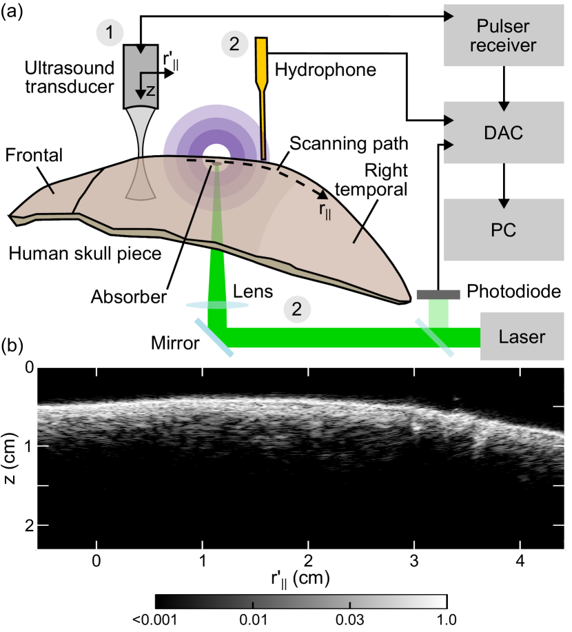
Fresh frozen frontotemporal bone sample derived from decompressive hemicraniectomy was collected according to protocols established by the ethical committee of the Department of Neurosurgery at the University Hospital Cologne. The skull sample was kept at -80∘C and was later degassed for 3 hours and immersed in 0.9 % saline solution during the imaging experiments that were performed in full compliance with the institutional guidelines of the Helmholtz Center Munich. To investigate on the exstence of GAW in a human skull, we adapted a similar approach to Estrada et al. (2017), where the exact shape of the skull surface was first obtained from a pulse-echo scan (Fig. 1(b)) performed by a focused transducer (20 mm focal distance, 15 MHz central frequency, Olympus) with a step size of 200 m. In order to excite broadband optoacoustic responses, a 200 m thick layer of a black burnish attached on the interior side of the skull sample was illuminated with a 1 mJ laser pulses of 10 ns duration and 532 nm wavelength (Spectra Physics, USA) focused down to a 1 mm spot. The generated responses were recorded by a needle hydrophone (0.5 mm diameter, Precision Acoustics, UK) that was scanned following the path in close proximity to the skull’s surface across the right temporal bone (see Fig. 1). The scanning step size (0.4 mm) was selected to allow the analysis in the spatial frequency domain up to 1.25 (mm-1). The laser pulse was fired at a repetition frequency of 15 Hz and the data acquisition was synchronized using a photodiode (DET10A, Thorlabs, USA), to avoid jitter in the laser trigger signal (Fig. 1). The laser energy fluctuations were further accounted for using pulse-to-pulse photodiode measurements. The data was digitised at 60 MSs by the data acquisition card (M3i.4142, Spectrum Systementwicklung Microelectronic, Germany) and stored for further analysis on a personal computer.
II.2 Simulations
As a first approximation, we modeled a flat multilayered viscoelastic solid embedded in a fluid Estrada et al. (2017) by means of the global matrix method Lowe (1995). For a given frequency and wavevector (incidence angle), a plane (inhomogeneous) wave is propagated from the input fluid, through the solid layers, to the output fluid. In the solid layers, longitudinal and transverse waves are considered in the propagation, as well as reflections at each interface between different media Estrada et al. (2017), forming a linear system of equations with 14 unknowns (complex transmitted and reflected amplitudes in each medium). First, the transmission problem was solved at a given region of the reciprocal space (frequency-wavevector) and then the transmission maxima at the output fluid were extracted. Second, a modal solution of the system was found by further refining the position of the extracted transmission maxima in reciprocal space using a golden-section search for singularity of the global matrix. The calculation of the linear system of equations Lowe (1995) was implemented in C++ and the analysis of the results was performed in Python. We assumed a total skull thickness of mm (manually measured average), and elastic constants close to what has been reported in the literature for cortical and trabecular bones Culjat et al. (2010) (see Table 1).
| Saline solution | Cortical bone | Diploë | |
|---|---|---|---|
| Thickness (mm) | (outer) 1.56 (inner) 1.44 | 3 | |
| Density (kg/m3) | 1000 | 1969 | 1055 |
|
Longitudinal wave
speed (m/s) |
1504 | 3476 | 1886 |
|
Transverse wave
speed (m/s) |
0 | 1520 | 830 |
|
Volumetric viscosity
(Pa s) |
0 | 0.1 | 1.5 |
|
Shear viscosity
(Pa s) |
0 | 1 | 3 |
III Results and discussion
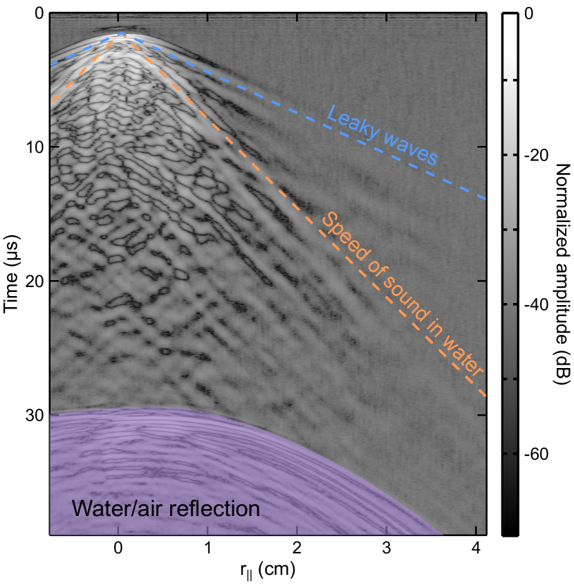
The amplitude of the detected signal is shown in Fig. 2 as a function of the distance to the optoacoustic source and the time of flight. The main wavefront is not registered at because the source is placed on the opposite side of the skull (see Fig. 1(a)) and requires a finite amount of time to propagate to the hydrophone. Waves propagating at a speed faster than the speed of sound in water () can be clearly identified, similarly to the previously reported murine skull measurements Estrada et al. (2017). The waves propagating with sonic speed are also distinguishable. However, the regime where waves propagate slower than is obscured by many oscillations that could be attributed to scattering events due to skull inhomogeneities or internal reverberations. To gain additional insight on the different simultaneously occurring ultrasound propagation regimes, one may further calculate the modes’ dispersion by means of a two-dimensional Fourier transform of the measured spatio-temporal data (Fig. 3). In the reciprocal space representation, the modes propagating with the speed of sound in water follow the green line. All the modes located above the sound line are therefore leaky while the ones below are non-leaky according to the Snell law. Several non-leaky modes are clearly distinguishable, in good agreement with the predictions of a flat multilayered solid model Estrada et al. (2017); Lowe (1995), which are labeled by solid white curves in Fig. 3. The modes follow an asymptotic trend propagating at 1343 (ms) for frequencies higher than 0.5 MHz (see Fig. 3(b)). We further evaluated the influence of first order corrections due to the skull’s curvature following methodology described in Biryukov et al. (1995). However, those turned negligible (-0.4 %) due to the large radius of curvature of the skull (10 cm) compared to the acoustic wavelength and the skull’s thickness ( 6 mm). The skull-guided waves in the case of murine skull were measured in the range, whereas, due to constrains of the experimental methodology and the sample geometry, the current results for the human skull are shown in a different regime, i.e. .
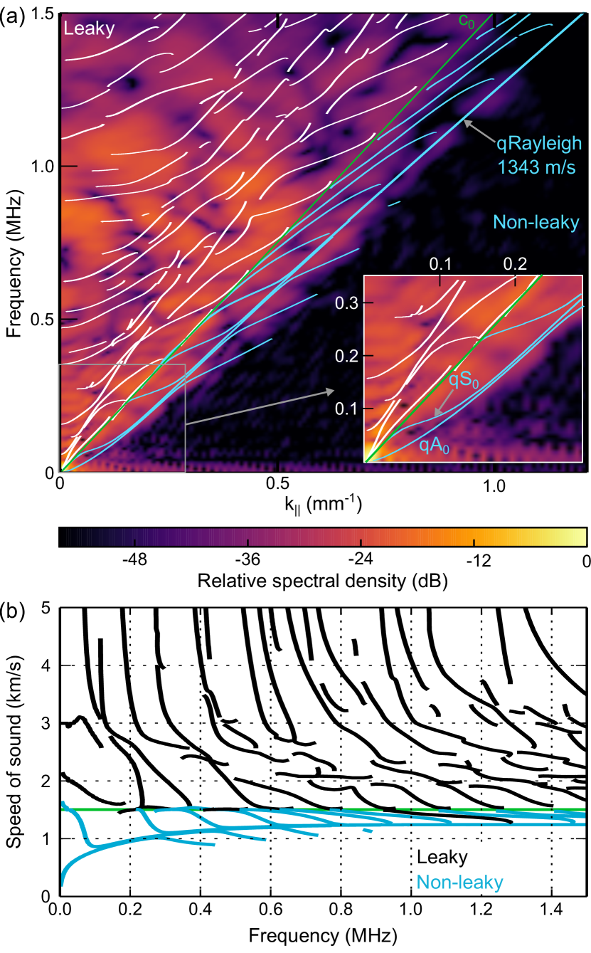
Note that windowing artifacts appear at frequencies below 0.2 MHz and high wave numbers, thus the effect of strong scattering due to the thick porous layer (diploë) in the cut-off free modes could not be reliably observed. Evidence exists of whole skull vibration modes in the audible frequency regime from experiments performed under airborne conditions McKnight et al. (2013). However, the intermediate regime where the diploë layer could play a major role is still to be explored.
The leaky skull-guided waves are further affected by scattering from inhomogeneities located outside the scanned region. The scattered waves could mimic a wave propagating faster than c0, when projected on the scanning path, thus interfering with real leaky skull-guided waves. This could explain the imperfect matching between experiments and numerical model for the leaky part of reciprocal space (Fig. 3(a)).
It is well known that leaky waves in a homogeneous plate can sustain full transmission at very specific frequencies and angles, even for large impedance mismatch between the plate and the surrounding fluid at normal incidence. Although no full transcranial transmission is expected due to the highly heterogeneous structure of the skull and the presence of the diploë, leaky skull-guided waves could be used to reach deep brain regions located at the skull’s far-field.
Current far-field array and single-element approaches are not optimized to target brain regions in close proximity to the skull. Transducer arrays and focusing algorithms optimized to work at angles close to normal incidence would suffer from increased aberrations if some of the array elements face the skull close to grazing incidence Pichardo and Hynynen (2007). On the other hand, single element focused transducers produce an elongated focal region in the order of centimeters at the low frequencies required to reach the brain transcranially at small-angle of incidence Legon et al. (2014). Thus, non-leaky skull-guided waves may present a more viable alternative to interrogate shallow brain cortex regions and the skull bone itself.
Proper design of strategies for direct-contact excitation of leaky and non-leaky skull guided waves requires prior knowledge on the skull’s near-field properties. Our work demonstrates the existence of guided wave phenomena, thus laying ground for further studies on skull-guided wave characterization and potential applications in transcranial ultrasound.
IV Conclusions
Ultrasonic wave propagation in the near-field region of a water-coupled human skull has been demonstrated theoretically and experimentally for the first time, thus deepening our current understanding of ultrasound transmission by the skull bone. In addition to leaky waves, we observed non-leaky skull-guided waves corresponding to Rayleigh-Lamb waves, as predicted by a multilayer flat plate model. Further experimental and theoretical work is neccesary to fully characterize skull-guided waves, as well as to identify their excitation strategies suitable for in vivo conditions. Observation and characterization of the skull-guided waves can be used for a more accurate interpretation of transcranial image data, such as optoacoustic images that are often afflicted by the complex wave propagation in the skull manifested via distortions in the location and shape of the vascular structures Kneipp et al. (2016). Our results may thus contribute to the development and optimization of non-invasive ultrasound-based techniques for diagnostic brain imaging Yao and Wang (2014); Meimani et al. (2017); Baykov et al. (2003); Lindsey et al. (2014); Shapoori et al. (2015), monitoring of neural activity Seo et al. (2016, 2013), guided surgery applications Bell et al. (2015), or cranial bone assesment without the use of ionizing radiation Bochud et al. (2017).
Acknowledgements.
Financial support is acknowledged from the European Research Council grant ERC-2015-CoG-682379 and the German research Foundation Grant RA1848/5-1.References
- Laugier and Haïat (2011) P. Laugier and G. Haïat, eds., Bone Quantitative Ultrasound (Springer, Dordrecht, 2011).
- Fry and Barger (1978) F. J. Fry and J. E. Barger, J. Acoust. Soc. Am. 63, 1576 (1978).
- Clement and Hynynen (2002a) G. Clement and K. Hynynen, Ultrasound Med. Biol. 28, 617 (2002a).
- Clement and Hynynen (2002b) G. T. Clement and K. Hynynen, Phys. Med. Biol. 47, 1219 (2002b).
- Clement et al. (2004) G. T. Clement, P. J. White, and K. Hynynen, J. Acoust. Soc. Am. 115, 1356 (2004).
- Marquet et al. (2009) F. Marquet, M. Pernot, J.-F. Aubry, G. Montaldo, L. Marsac, M. Tanter, and M. Fink, Phys. Med. Biol. 54, 2597 (2009).
- Pichardo et al. (2011) S. Pichardo, V. W. Sin, and K. Hynynen, Phys. Med. Biol. 56, 219 (2011).
- Pinton et al. (2012) G. Pinton, J.-F. Aubry, E. Bossy, M. Muller, M. Pernot, and M. Tanter, Med. Phys. 39, 299 (2012).
- Coluccia et al. (2014) D. Coluccia, J. Fandino, L. Schwyzer, R. O’Gorman, L. Remonda, J. Anon, E. Martin, and B. Werner, J. Ther. Ultrasound 2, 17 (2014).
- Yao and Wang (2014) J. Yao and L. V. Wang, Neurophotonics 1, 011003 (2014).
- Dean-Ben et al. (2017) X. L. Dean-Ben, S. Gottschalk, B. Mc Larney, S. Shoham, and D. Razansky, Chem. Soc. Rev. 46, 2158 (2017).
- Estrada et al. (2016) H. Estrada, J. Rebling, J. Turner, and D. Razansky, Phys. Med. Biol. 61, 1932 (2016).
- Kneipp et al. (2016) M. Kneipp, J. Turner, H. Estrada, J. Rebling, S. Shoham, and D. Razansky, Journal of Biophotonics 9, 117 (2016).
- Estrada et al. (2017) H. Estrada, J. Rebling, and D. Razansky, Phys. Med. Biol. 62, 4728 (2017).
- Moilanen (2008) P. Moilanen, IEEE Trans. Ultrason., Ferroelect., Freq. Control 55, 1277 (2008).
- Talmant et al. (2011) M. Talmant, J. Foiret, and J.-G. Minonzio, “Guided waves in cortical bone,” in Bone Quantitative Ultrasound, edited by P. Laugier and G. Haïat (Springer Netherlands, Dordrecht, 2011) pp. 147–179.
- Moilanen et al. (2014) P. Moilanen, Z. Zhao, P. Karppinen, T. Karppinen, V. Kilappa, J. Pirhonen, R. Myllylä, E. Hæggström, and J. Timonen, Ultrasound Med. Biol. 40, 521 (2014).
- Bochud et al. (2017) N. Bochud, Q. Vallet, J.-G. Minonzio, and P. Laugier, Sci. Rep. 7, 43628 (2017).
- Brookes and Revell (1998) M. Brookes and W. J. Revell, “Blood supply of flat bones,” in Blood Supply of Bone: Scientific Aspects (Springer London, London, 1998) pp. 64–74.
- Jilka (2013) R. L. Jilka, The Journals of Gerontology: Series A 68, 1209 (2013).
- Baykov et al. (2003) S. V. Baykov, L. V. Babin, A. M. Molotilov, S. I. Neiman, V. V. Riman, V. D. Svet, and A. I. Selyanin, Acoustical Physics 49, 389 (2003).
- Lindsey et al. (2014) B. D. Lindsey, H. A. Nicoletto, E. R. Bennett, D. T. Laskowitz, and S. W. Smith, Ultrasound in Medicine & Biology 40, 90 (2014).
- Shapoori et al. (2015) K. Shapoori, J. Sadler, A. Wydra, E. V. Malyarenko, A. N. Sinclair, and R. G. Maev, IEEE Transactions on Biomedical Engineering 62, 1253 (2015).
- Seo et al. (2016) D. Seo, R. M. Neely, K. Shen, U. Singhal, E. Alon, J. M. Rabaey, J. M. Carmena, and M. M. Maharbiz, Neuron 91, 529 (2016).
- Seo et al. (2013) D. Seo, J. M. Carmena, J. M. Rabaey, E. Alon, and M. M. Maharbiz, ArXiv e-prints (2013), arXiv:1307.2196 [q-bio.NC] .
- Lowe (1995) M. Lowe, IEEE Trans. Ultrason., Ferroelect., Freq. Control 42, 525 (1995).
- Culjat et al. (2010) M. O. Culjat, D. Goldenberg, P. Tewari, and R. S. Singh, Ultrasound Med. Biol. 36, 861 (2010).
- Biryukov et al. (1995) S. V. Biryukov, Y. V. Gulyaev, V. V. Krylov, and V. P. Plessky, Surface Acoustic Waves in Inhomogeneous Media, edited by L. B. L. F. H. Haus, Wave Phenomena (Springer-Verlag Berlin Heidelberg New York, 1995).
- McKnight et al. (2013) C. L. McKnight, D. A. Doman, J. A. Brown, M. Bance, and R. B. A. Adamson, J. Acoust. Soc. Am. 133, 136 (2013).
- Pichardo and Hynynen (2007) S. Pichardo and K. Hynynen, Physics in Medicine & Biology 52, 7313 (2007).
- Legon et al. (2014) W. Legon, T. F. Sato, A. Opitz, J. Mueller, A. Barbour, A. Williams, and W. J. Tyler, Nat Neurosci 17, 322 (2014).
- Meimani et al. (2017) N. Meimani, N. Abani, J. Gelovani, and M. R. Avanaki, Photoacoustics 7, 27 (2017).
- Bell et al. (2015) M. A. L. Bell, A. K. Ostrowski, K. Li, P. Kazanzides, and E. M. Boctor, Photoacoustics 3, 78 (2015).