Sex differences in network controllability as a predictor of executive function in youth
Abstract
Executive function is a quintessential human capacity that emerges late in development and displays different developmental trends in males and females. Sex differences in executive function in youth have been linked to vulnerability to psychopathology as well as to behaviors that impinge on health, wellbeing, and longevity. Yet, the neurobiological basis of these differences is not well understood, in part due to the spatiotemporal complexity inherent in patterns of brain network maturation supporting executive function. Here we test the hypothesis that sex differences in executive function in youth stem from sex differences in the controllability of structural brain networks as they rewire over development. Combining methods from network neuroscience and network control theory, we characterize the network control properties of structural brain networks estimated from diffusion imaging data acquired in males and females in a sample of 882 youth aged 8-22 years. We summarize the control properties of these networks by estimating average and modal controllability, two statistics that probe the ease with which brain areas can drive the network towards easy- versus difficult-to-reach states. We find that females have higher modal controllability in frontal, parietal, and subcortical regions while males have higher average controllability in frontal and subcortical regions. Furthermore, average controllability values in the medial frontal cortex and subcortex, both higher in males, are negatively related to executive function. Finally, we find that average controllability predicts sex-dependent individual differences in activation during an n-back working memory task. Taken together, our findings support the notion that sex differences in the controllability of structural brain networks can partially explain sex differences in executive function. Controllability of structural brain networks also predicts features of task-relevant activation, suggesting the potential for controllability to represent context-specific constraints on network state more generally.
Keywords:
network controllability, neurodevelopment, sex differences, executive function, working memory, fMRI BOLD, diffusion tensor imaging
Introduction
Executive function is necessary for regulation of goal-directed behavior, and encompasses cognitive processes including working memory, inhibition, task switching, and performance monitoring 1. Deficits in executive function are associated with increased risk taking and associated consequences 2; 3, and more generally hamper academic and occupational performance 4. Executive deficits frequently lead to personal, social, and professional consequences that accumulate throughout the course of a patient’s life 4. Importantly, the normative capacity for executive function rapidly increases during adolescence and differs by sex. Sex differences in the developmental trajectory of executive function have been linked to higher rates of impulsivity 5, ADHD diagnosis 6, criminality 7 and substance use 2 in males. Current interventions for disorders of executive function are relatively limited, usually do not consider sex, and rely primarily on psychotherapy and global manipulations via psychopharmacology 8.
A basic understanding of sex-related differences in executive function and their implications for the diagnosis and treatment of executive deficits requires an understanding of the normative maturation of underlying neural circuitry. Several recent studies highlight the fact that such maturation, and sex-differences in that maturation, span structure 9, anatomical connectivity 10, functional activity 11; 12; 13, and functional connectivity 14. Using structural MRI, a recent study 15 reported age-related, non-linear increases in gray matter density with concurrent decreases in cortical thickness. Interestingly, the maturation of these structural features was markedly different between the sexes, with females showing higher gray matter density globally and higher cortical thickness in frontal and insular regions 15. Several older structural studies 16 have also reported linear increases in frontoparietal white matter density throughout adolescence. Using diffusion-weighted MRI, another study found significantly greater within-hemisphere connectivity in males and greater between-hemisphere connectivity in females 17. Sex differences have also been identified in the clustered (or modular) structure in patterns of functional connectivity estimated from resting state fMRI data: males display higher between-module connectivity while females display higher within-module connectivity 18, and these differences were shown to predict individual differences in executive function 18. Although these descriptive studies have provided important insights, it remains difficult to specify in a mechanistic sense how executive function might arise from such complex, multimodal patterns of brain maturation in a sex-dependent manner.
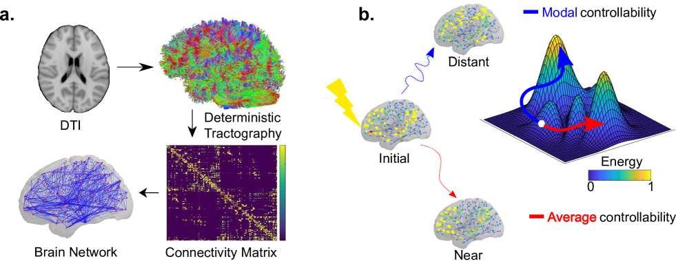
We address this challenge by positing that sex differences in executive function in youth stem from sex differences in the controllability of structural brain networks as they rewire over development. This notion intuitively bridges the control of behavior (executive function) with the control of brain dynamics (network controllability). Specifically, we capitalize on recent advances in network control theory 19; 20, an emerging branch of theoretical physics and systems engineering that builds on early efforts in control theory 21; 22 to offer a mechanistic model of how key nodes, or control points, can exert disproportionate influence over system function 19. Control points are identified with metrics that assess the ability of specific nodes to alter a system’s state, based on the underlying network topology 20 (Fig. 1). Specifically, the metric of average controllability reflects the average energy input required at a node to move the system from some initial state to all possible states. In contrast, the metric of modal controllability reflects the ease of transitioning the system from some initial state to a difficult-to-reach state. Prior work has demonstrated the utility of network control theory in understanding basic brain architecture and function 23; 24; 25 across spatial scales 26 and species 27, posited its relation to cognition 28; 29, and outlined its developmental course 29.
Here, we tested the hypothesis that developmental sex differences in network control underlie sex differences in executive functioning. Specifically, we predicted that (i) network controllability differs by sex, (ii) network controllability changes with age differently in males and females, (iii) sex differences in network controllability predict executive function, and (iv) network controllability predicts the activation of brain regions during a working memory task demanding executive function. To test these hypotheses, we constructed structural brain networks from diffusion tensor imaging data acquired in 882 healthy youth, ages 8-22 years, in the Philadelphia Neurodevelopmental Cohort (PNC) 30. Each brain network was comprised of 234 anatomically defined brain regions 31 connected by white matter tracts estimated from diffusion tractography. We show that regional controllability is a significant mediator of the relationship between sex and executive function, and that it predicts the magnitude of fMRI BOLD signal on an n-back working memory task. As described in detail below, our results suggest that sex differences in the controllability of structural brain networks predict executive function and the activity profiles that support that function.
Materials and Methods
Participants
Diffusion tensor imaging (DTI) data were obtained from youth who participated in a large community-based study of brain development, now known as the Philadelphia Neurodevelopmental Cohort (PNC) 30. Here we study 882 out of a total of 1601 subjects between the ages of 8 and 22 years (mean age = 15.06, SD = 3.15, 389 males, 493 females). Due to lack of complete diffusion scans () and incidental findings (), data from 244 participants was deemed unusable. The remaining 1357 participants underwent a rigorous manual and automated quality assurance protocol for DTI datasets 32, eliminating an additional 147 subjects with poor data quality. A subset of 93 of the remaining 1210 participants were excluded for low quality or incomplete FreeSurfer reconstruction of T1-weighted images. Further, 235 of the remaining 1117 participants were excluded for one or more of the following reasons: gross radiological abnormalities distorting brain anatomy, medical history that might impact brain function, history of inpatient psychiatric hospitalization, use of psychotropic medication at the time of imaging, or high levels of in-scanner head motion during the DTI scan, as defined by a mean relative displacement between non-weighted volumes of greater than 2 mm. These exclusions left us with a final sample of subjects 10; 29 between the ages of 8 and 22 years (mean age = 15.06, SD = 3.15, 389 males, 493 females).
Cognitive Phenotyping
Cognition was measured outside of the scanner using the Penn Computerized Neurocognitive Battery (CNB) 33; 34. Briefly, the 1-hour CNB was administered to all participants, and consisted of 14 tests that evaluated a broad range of cognitive functions. Twelve of the tests measure both accuracy and speed, while two of the tests (motor and sensorimotor) measure only speed.
Overall cognitive performance was summarized as the average -transformed accuracy and speed (as well as the difference between accuracy and median response time: efficiency) scores across all tests administered (as described in Moore et al.35 for complete details). Factor scores are described in Moore et al.35; here, we used the factor score for executive efficiency from a best-fitting four-factor solution comprising tests from the executive function domain (attention, abstraction and working memory). The tests contributing to the executive efficiency score include the Penn Continuous Performance Test, the Letter N-Back task, and (weakly) the Penn Verbal Reasoning Test 35; 36; 37 (analogical reasoning). In the present study, we use this factor score for executive efficiency as our primary measure of executive function, hereafter referred to as “executive function”.
Imaging Data Acquisition
MRI data were acquired on a 3 Tesla Siemens Tim Trio whole-body scanner and 32-channel head coil at the Hospital of the University of Pennsylvania. DTI scans were acquired via a twice-refocused spin-echo (TRSE) single-shot echo-planar imaging (EPI) sequence (TR = 8100ms, TE = 82ms, FOV = 240mm2/240mm2; Matrix = RL:128/AP:128/Slices:70, in-plane resolution (x and y) 1.875 mm2; slice thickness = 2mm, gap = 0; flip angle = 90/180/180 degrees, volumes = 71, GRAPPA factor = 3, bandwidth = 2170 Hz/pixel, PE direction = AP). This sequence utilizes a four-lobed diffusion encoding gradient scheme combined with a 90-180-180 spin-echo sequence designed to minimize eddy-current artifacts. The complete sequence consisted of 64 diffusion-weighted directions with = 1000s/mm2 and 7 interspersed scans where = 0s/mm2. Total scan time was approximately 11 min. The imaging volume was prescribed in axial orientation covering the entire cerebrum with the topmost slice just superior to the apex of the brain.
In addition to the DTI scan, a map of the main magnetic field (i.e., B0) was derived from a double-echo, gradient-recalled echo (GRE) sequence, allowing us to estimate field distortions in each dataset. Prior to DTI acquisition, a 5-minute magnetization-prepared, rapid acquisition gradient-echo T1-weighted (MPRAGE) image (TR 1810 ms, TE 3.51 ms, FOV 180 240 mm, matrix 256 192, effective voxel resolution of 1 1 1 mm) was acquired. This high-resolution structural image was used for tissue segmentation and parcellating gray matter into anatomically defined regions in native space. Rigorous manual and automated quality-assurance protocols for the T1-weighted structural imaging data were performed and cleared for the 882 subjects considered here 38. Subsequently, all structural images were processed using FreeSurfer (version 5.3) 39. FreeSurfer reconstructions underwent rigorous quality-assurance protocols 38; 40. The T1 image was parcellated into 234 regions by FreeSurfer according to the Lausanne Atlas 41. We define the subcortex of this 234-region parcellation to be comprised of the left and right hemispheric counterparts of the thalamus proper, caudate, putamen, pallidum, nucleus accumbens area, hippocampus and amygdala, while excluding the brainstem (14 regions). We define the cortex of this 234-region parcellation to be comprised of the remaining regions (219 regions).
fMRI BOLD data were acquired as subjects completed a version of the n-back task using fractal images 42. See the supplementary methods of ref. 43 for details regarding task presentation and structure. Functional images were obtained using a whole-brain, single-shot, multislice, gradient-echo echoplanar sequence (231 volumes; TR ms; TE ms; flip angle degrees; FOV = mm; matrix = ; slices ; slice thickness mm; slice gap mm; effective voxel resolution = mm).
Imaging Data Preprocessing
All DTI datasets were subject to a rigorous manual quality assessment procedure involving visual inspection of all 71 volumes 32. Each volume was evaluated for the presence of artifact, with the total number of volumes impacted summed over the series. This scoring was based on previous work describing the impact of removing image volumes when estimating the diffusion tensor 44; 45. Data was considered “Poor” if more than 14 (20%) volumes contained artifact, “Good” if it contained 1-14 volumes with artifact, and “Excellent” if no visible artifacts were detected in any volumes. All 882 subjects included in the present study had diffusion datasets identified as “Good” or “Excellent,” and had less than 2mm mean relative displacement between interspersed volumes. As described below, even after this rigorous quality assurance, motion was included as a covariate in all analyses.
The skull was removed for each subject by registering a binary mask of a standard fractional anisotropy (FA) map (FMRIB58 FA) to each subject’s DTI image using an affine transformation 46. Eddy currents and subject motion were estimated and corrected using the FSL eddy tool 47. Diffusion gradient vectors were then rotated to adjust for subject motion estimated by eddy. After the field map was estimated, distortion correction was applied to DTI data using FSL’s FUGUE 48.
BOLD time series were processed as described in 49; 43. Briefly, FSL 5 48 was used to analyze time series data from three condition blocks (0-back, 1-back and 2-back), with the primary contrast being 2-back 0-back. BOLD images were co-registered to the T1 image using boundary-based registration 50 with integrated distortion correction as implemented in FSL. Generalized linear model (GLM) beta weights were averaged across all voxels in each parcel of the 234-node Lausanne atlas. In our assessment of n-back performance-related activation, we use the difference in GLM beta weights between the 2-back and 0-back condition. For all analyses of fMRI data, we excluded 223 subjects with incomplete data or excessive head motion (mean relative displacement mm or maximum displacement mm), leaving remaining.
Structural Network Estimation
Structural connectivity was estimated from DTI data in order to generate the adjacency matrix representing the pattern of white matter tracts between large-scale brain areas. DSI Studio was used to estimate the diffusion tensor and perform deterministic whole-brain fiber tracking with a modified FACT algorithm that used exactly 1,000,000 streamlines per subject after removing all streamline with length mm 28. To extend regions into white matter, parcels defined using the Lausanne atlas were dilated by 4 mm 28; 29 and registered to the first non-weighted (b = 0) volume using an affine transform 28; 29. The number of streamlines connecting each node of the 234 region parcel end-to-end was used to define the edge weights of the adjacency matrix .
Network Controllability
We represent the streamline-weighted structural network estimated from diffusion tractography as the graph , where and are the vertex and edge sets, respectively. Let be the weight associated with the edge , and define the weighted adjacency matrix of as , where whenever . We associate a real value with each node to generate a vector describing the network state, and we define the map to describe the dynamics of the network state over time.
It is worth noting that this method assumes that the number of streamlines is proportional to the strength of structural connectivity with regards to propagation of activity between nodes according to a specified model of dynamics. Here we employ a simplified noise-free linear discrete-time and time-invariant model of such dynamics:
| (1) |
where describes the state (i.e. voltage, firing rate, BOLD signal) of brain regions over time. Thus, the state vector has length , where is the number of brain regions in the connectome parcellation, and the value of describes the brain activity state of that region. The matrix is symmetric, with the diagonal elements satisfying . Prior to calculating controllability values, we divide by , where is the largest eigenvalue of . The input matrix identifies the control point in the brain, where and
| (2) |
and denotes the -th canonical vector of dimension . The input denotes the control strategy.
To study the dynamics by which the activity of one brain region influences structurally connected regions, we apply the control theoretic notion of controllability to our dynamical model. Classic results in control theory ensure that controllability of the network, , from the set of network nodes is equivalent to the controllability Gramian being invertible, where
| (3) |
We calculate , or rather , with set equal to one canonical vector and repeat this process for all nodes 29; 28. Although it is well known that the activity of several brain regions and neuronal ensembles are related via non-linear dynamics, it has been shown that a linear approximation can explain features of the resting state fMRI BOLD signal 51; this suggests that a linear approximation can effectively capture the controllability properties of the original non-linear dynamics.
Controllability Metrics
Following ref.29; 28, controllability metrics for structural brain networks were calculated for two different control strategies which describe the ability to change in a particular fashion 20. Average controllability describes the ease of transition to energetically similar states, while modal controllability describes the ease of transition to difficult-to-reach states 20.
Average controllability of a network equals the average input energy applied to a set of control nodes required to reach all possible target states. It is known that average input energy is proportional to Trace(), the trace of the inverse of the controllability Gramian. Instead, as in ref. 29; 28, we use Trace() as a measure of average controllability because (i), Trace() and Trace() are related via inverse proportionality, and (ii), Trace() tends to be very ill-conditioned and cannot be accurately computed even at more coarse connectome parcellations. In addition to its relationship with Trace(), Trace() describes the energy of the network impulse response, or, equivalently, the network norm 21; 22. In summary, to compute the average controllability value for node in , we compute the Trace() when node is the only control node (i.e. ).
Modal controllability refers to the ability of a node to control the evolutionary modes of a dynamical network, and is most interpretable when used to identify states that are poorly controllable given . To calculate modal controllability, one must first obtain the eigenvector matrix of the adjacency matrix . If is small, then the -th evolutionary mode of the input-independent form of Eq. (1), , is poorly controllable from node . According to prior work 20, we define as a scaled measure of the modal controllability of each of the modes from node .
Finally, we calculate the mean of the regional controllability values either across the whole brain (Fig. A.1) or within the cortex and subcortex separately (Figs. 3, 4, 5, 6). For each subject, we define the mean modal controllability to be the sum of the values of for each node within , divided by the number of regions. Similarly, we define the mean average controllability to be the sum of the values of Trace() for each node within , divided by the number of regions.
Network Synchronizability
While the metrics of average and modal controllability provide complementary views on the diverse dynamics that a brain network can display, it is also useful to study contrasting metrics that probe the susceptibility of the network to be constrained within a narrower range of dynamics. Previous work 29 has demonstrated that a useful contrasting metric to controllability is synchronizability, which intuitively measures the susceptibility of a network to remain in a single synchronous state , i.e. . The master stability function (MSF) allows one to analyze the stability of this synchronous state without fully characterizing the properties of each feature of the system 52. Within this framework, linear stability depends on the positive eigenvalues of the Laplacian matrix defined by , where is the Kronecker delta function.
The condition for stability depends on the shape of the MSF, i.e. the stability of the synchronized state to linear perturbations holds when the MSF is negative for all non-zero eigenvalues of . This condition is more likely for a smaller range of eigenvalues, hence we can use the normalized spread of these eigenvalues to quantify network synchronizability as
| (4) |
and , the average coupling strength per node, which normalizes for the overall network strength. Using this definition, we calculated the synchronizability of for each subject.
Linear Regression of Network Control Metrics on Sex
For all analyses of network control metrics, we examined the effect of sex while controlling for age, total brain volume (segmented brain volume, as defined by FreeSurfer BrainSegVol metric), handedness, and motion during the diffusion scan. We used multiple ordinary least squares (OLS) linear regression with the command in R or the MATLAB function to fit the following general equation:
| (5) |
where is the controllability statistic (either for modal controllability or for average controllability), is age, is total intracranial volume, is the mean framewise displacement as a summary measure of in-scanner head motion during the diffusion imaging sequence, is handedness, and is sex. We then used the R package visreg to calculate 95% confidence intervals around fitted lines and generate partial residuals. Furthermore, when using the multiple OLS regression for the node-level analyses of controllability, we applied a false discovery rate (FDR) correction 53 () to control for Type I error due to multiple testing.
Non-Linear Fits of Executive Function and Network Metrics
Because age and executive function are nonlinearly related, we used a general additive model 54; 38 (GAM) to assess the relationship between executive efficiency and age using the package in R (Fig. 1b). A GAM is a generalized linear model in which the linear predictor is defined by unknown smooth functions of predictor variables. In this case, we use penalized regression splines for age, with conventional parametric components for the remaining predictors.
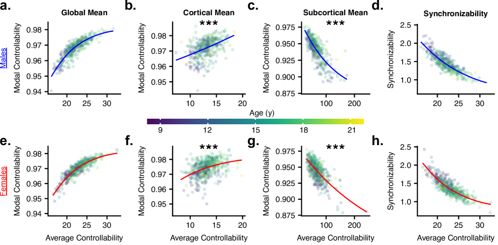
Following ref. 29, we used non-linear models to examine the relationships between synchronizability, average controllability, and modal controllability (Fig. 5). We generated estimates of parameters for models of the form via non-linear least squares using the function in R. To compare these fits between males and females, we performed an analysis of variance (ANOVA). For example, when considering sex differences in the nonlinear relation between modal controllability and average controllability, we examined the expression:
| (6) |
where is modal controllability, is average controllability, and is sex, coded as 0 for males and 1 for females, so that , and for males and the equation is reduced to the base form. The predicted values for males or females obtained from the full model were used to generate the curves in Fig. 5.
Mediation Testing
After identifying associations between sex, controllability, and executive function, we asked whether controllability is a statistically significant mediator of the relationship between sex and executive function. To test for a formal mediation, we performed non-parametric bootstrapping causal mediation analysis using the R function from the package mediation 55. It is worth noting that we cannot estimate causality in cross-sectional association data. The Average Causal Mediation Effect (ACME) quantifies the relationship between mediator and outcome independent of treatment, while the Average Direct Effect (ADE) quantifies the relationship between treatment and outcome, independent of the mediator 55.
Linear Regression of BOLD Data on Network Control Metrics
Finally, and in keeping with our linear systems framework, we sought to test whether controllability would predict brain state (), as defined by BOLD activation during a task demanding executive function. In these analyses (Fig. 8), we used the residuals from Eq. 5 for controllability. For activation, we used the residuals from the following equation:
| (7) |
where is 2-back minus 0-back activation (hereafter referred to as “activation”), is age, is handedness, is the mean framewise displacement during the n-back scan, and is sex. Because we had no prior knowledge about where controllability might predict activation, we fit linear models between controllability and activation at every possible pair of nodes, i.e. we performed regressions of controllability residuals at each node with activation residuals at each node. The matrices of -values for the slope of controllability on activation were FDR corrected () separately for average controllability and for modal controllability. To identify regions where activation was associated with executive function, we followed previous work 49; 43 by examining , a composite measure of n-back task performance which takes both correct responses and false positives into account to separate performance from response bias.
Results
Sex differences in executive function
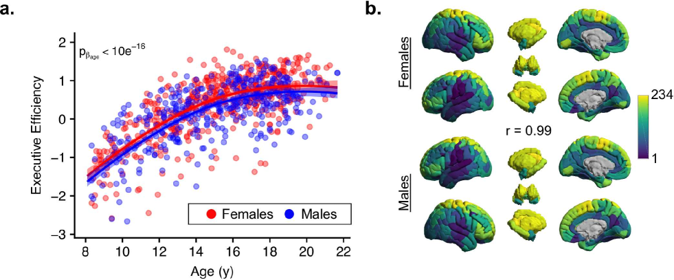
Our analysis of sex differences in the development of executive function and its neurobiological underpinnings began with a sex-stratified comparison of mean performance on executive efficiency, a cross-contextual summary measurement of executive function 35. Consistent with prior results from the PNC data 18; 30, the executive efficiency subscore of the PCNB was significantly higher in females (full model , , ; Fig. 2a) with no interaction between age and sex. Furthermore, executive efficiency increased with age in a non-linear fashion (, , ; Fig. 2a). This result suggests that development of executive function occurs via a similar course for males and females, but that females display higher scores in this cognitive domain for all ages studied. After performing this sex-stratified analysis of the developmental course of the executive efficiency score, we next turned to a consideration of its neurobiological underpinnings (Fig. 1a).
Sex differences in regional network controllability
Our general hypothesis was that sex differences in executive function in youth stem from sex differences in the controllability of structural brain networks as they rewire over development, a notion that bridges the control of behavior (executive function) with the control of brain dynamics (network controllability). To test this hypothesis, we examined the anatomical distribution of control points in the structural brain networks of males and females separately. We ranked the mean average controllability value at each region for males and females separately, which revealed that the distribution of controller strength is virtually identical between male and females (; Fig. 2b). This result suggests that there is no sex difference in the spatial distribution of controllers when classified by their relative magnitudes.
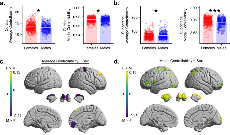
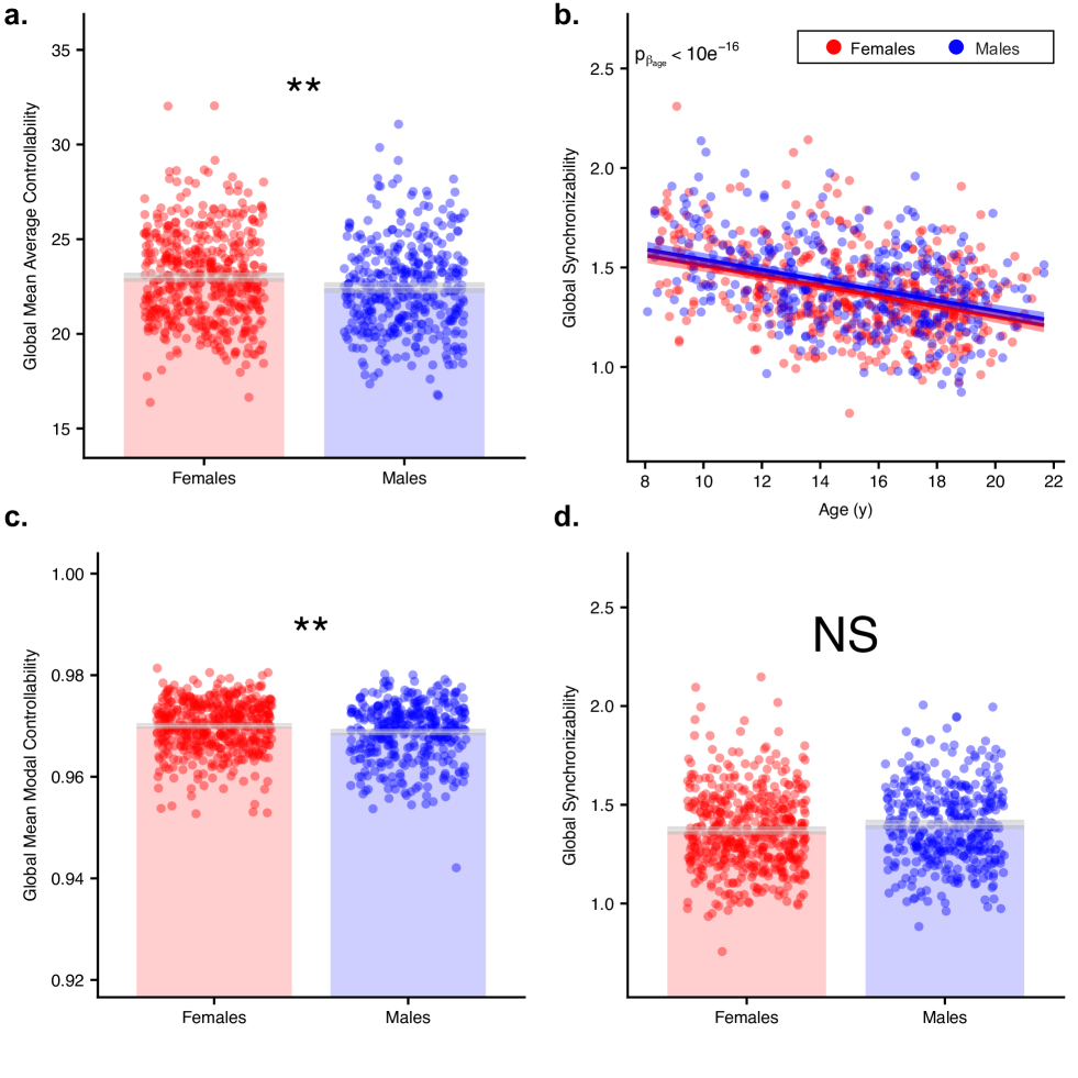
We next investigated whether the actual (rather than ranked) regional controllability estimates differed by sex, and we addressed this question first by considering the cortex and subcortex separately. Specifically, we computed mean controllability values for each subject across 219 cortical regions and 14 subcortical regions. In the cortex, mean average controllability (, , ; Fig. 3a) and mean modal controllability (, , ; Fig. 3a) are higher in females. Mean modal controllability in the subcortex is also higher in females (, , ; Fig. 3b), whereas mean average controllability in the subcortex is higher in males (, , ; Fig. 3b). Only with average controllability in the subcortex did males have higher controllability than females; this result suggests that the connectivity profile of subcortical regions may contribute to sex differences in functional brain dynamics.
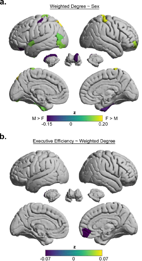
In a finer-grained analysis, we investigated whether the controllability of single regions differed by sex. Modal and average controllability were separately examined at each region, while accounting for age, total brain volume, handedness, and mean in-scanner head motion as model covariates. We found that average and modal controllability differed by sex at a subset of network nodes after FDR correction () for multiple comparisons. Nodes with average controllability values that differed by sex were almost all (4/5) higher in males and located in the frontal lobe or subcortex (Fig. 3c-d). Conversely, nodes with modal controllability values that differed by sex were all (18/18) higher in females and located in frontoparietal and subcortical systems. These results suggest that controller strength differs between males and females on a region-specific and control strategy-specific basis.
Development of network controllability across the sexes
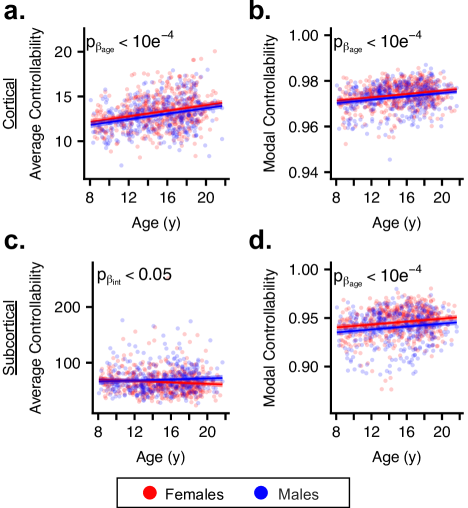
We next turned to assessing whether the developmental arc of network controllability differed by sex. We found that controllability in the cortex and subcortex tended to increase with age, with the exception of average controllability in the subcortex. In the cortex (, , ; Fig. 4b) and subcortex (, , ; Fig. 4d), mean modal controllability increases with age for males and females. Mean cortical average controllability also increases with age (, , ; Fig. 4b). Interestingly, there was a significant age-by-sex interaction only with subcortical average controllability, such that the slope was positive for males and negative for females (Fig. 4c). However, we also found that subcortical average controllability remains stable throughout development for both males (, , , ) and females (, , , ) 56. Taken together, these results suggest that controllability changes with age similarly for males and females, but that average controllability in the subcortex changes with age differently than modal controllability or than cortical average controllability.
With the knowledge that controllability has a sex-independent relationship with age, we were interested in testing the hypothesis that sex influences the relationship between different types of controllability in the developing brain. Following ref.29, we fit the relationships between average and modal controllability with the exponential function separately for each sex (Eq. 6). Our results showed that males and females did not have a statistically different relationship between average and modal controllability averaged across the whole brain (, ; Fig. 5a,e). In the cortex alone, average and modal controllability followed an increasing exponential form (Fig. 5b,f), similar to that of the whole brain. In contrast, in the subcortex alone average and modal controllability followed a decreasing exponential form (Fig. 5d,h). When sex was included in the model, fits improved significantly (cortex: , , Fig. 5b,f; subcortex: , , Fig. 5c,g), suggesting that these regions may contain sex-dependent differences in structural connections important for controlling brain network state.
Next, we performed a specificity analysis to determine whether our results could be further confirmed by sex differences in a contrasting metric – synchronizability – that probed the susceptibility of the network to be constrained within a narrow (rather than broad) range of dynamics. We found that synchronizability did not differ between males and females (A.1b) and decreased with age in a sex-independent fashion (A.1d). Moreover, the exponential relationship between synchronizability and average controllability did not differ by sex (, ; Fig. 5d,h), confirming the local (Fig. 3c-d) rather than global (Fig 2b) differences in metrics of network control and dynamics.
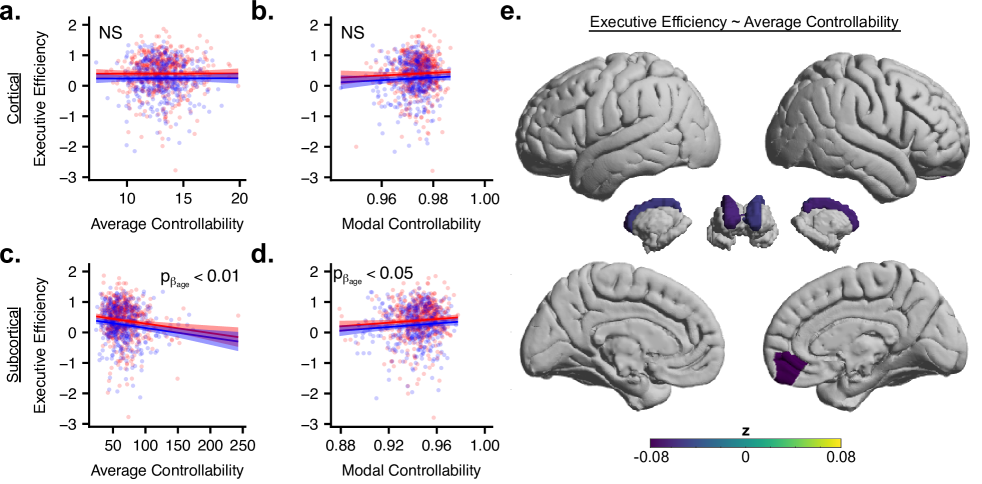
Sex differences in network controllability predict individual differences in executive function
While sex differences in network controllability are of interest in understanding the structural drivers of brain dynamics, their impact on behavior requires a link to cognition. Here we examine the relation between executive function and network controllability across cortex and subcortex separately, and then at individual brain regions. When considering mean cortical and subcortical controllability and executive function we found that neither average (, , ; Fig. 6a) nor modal controllability (, , ; Fig. 6b) in the cortex were associated with performance. A different trend was apparent in the subcortex: average controllability was negatively correlated with performance (, , ; Fig. 6c), while modal controllability was positively correlated with performance (, , ; Fig. 6d).
Next, we considered the 21 brain areas that we had previously found to display sex differences in controllability values (5 nodes for average controllability and 18 nodes for modal controllability, with 2 nodes overlapping). Nodes with significantly higher average controllability in males (right medial orbitofrontal cortex and bilateral caudate nuclei) had average controllability values that were negatively related to executive function (Fig. 6e; FDR corrected, ). Interestingly, modal controllability at any individual node was not significantly associated with executive function after FDR correction; however, two nodes with uncorrected (right caudate nucleus and left inferior parietal lobe) had modal controllability values that were higher in females and positively associated with executive function. This result suggests that modal controllability of one particular node is not as strong of a predictor of cognitive performance as mean modal controllability over the subcortex as a whole.
One parsimonious explanation for the results reported up to this point is that controllability mediates the relationship between sex and executive function: specifically, subcortical average controllability is higher in males, and increasing subcortical average controllability is associated with poorer performance on an assessment of executive function. We explicitly tested for such a mediation and found that, indeed, average controllability at the right medial orbitofrontal cortex, right superior frontal cortex, right postcentral gyrus and the bilateral caudate nuclei were statistically significant mediators of the relationship between sex and executive function (Fig. 7a). Similarly, subcortical modal controllability was higher in females, and increasing subcortical modal controllability was associated with better executive function. However, consistent with the fact that no individual node had modal controllability values associated with executive function, modal controllability was not found to be a mediator of the relationship between sex and executive function. Taken together, these results suggest that sex differences in average controllability may be a predictive biomarker for sex differences in executive function.
Sex-dependent relationships between n-back task activation magnitudes and controllability
Describing the relationship between sex, regional controllability, and cognitive performance provides us with a better understanding of the importance of structural brain networks in sex differences in executive function. The final aspect of our hypothesis pertains to whether network control theory can be used to explain how differences in brain network structure produce divergent patterns of brain activity that underpin executive function. We hypothesized that regional controllability values would predict regional n-back task activation magnitudes, and that associations between controllability and activation would differ by sex.
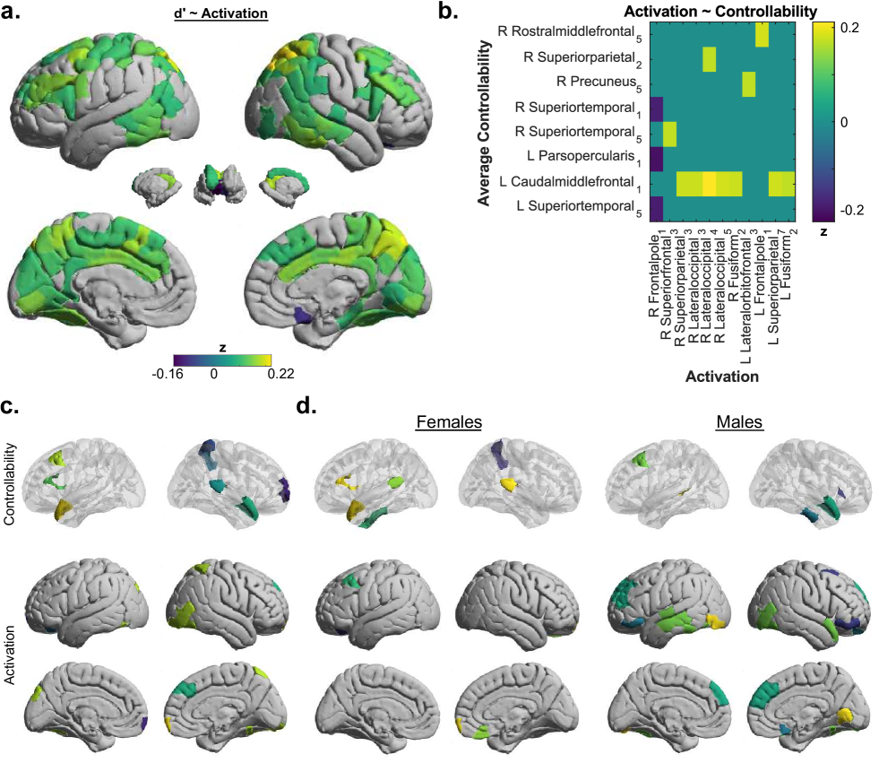
To obtain a reference point for activation profiles associated with strong executive function, we separately regressed activation at each node on 49. This analysis identified 113 nodes for which activation was associated with successful task performance (Fig. 8a). Note that in contrast to the use of a general factor score for executive efficiency in the previous analyses, here we considered the measure to summarize performance on the n-back task only 57 so that the performance measure is directly related to the context in which activation values are measured. Interestingly, sex was not associated with activation at any brain region (FDR corrected, , data not shown).
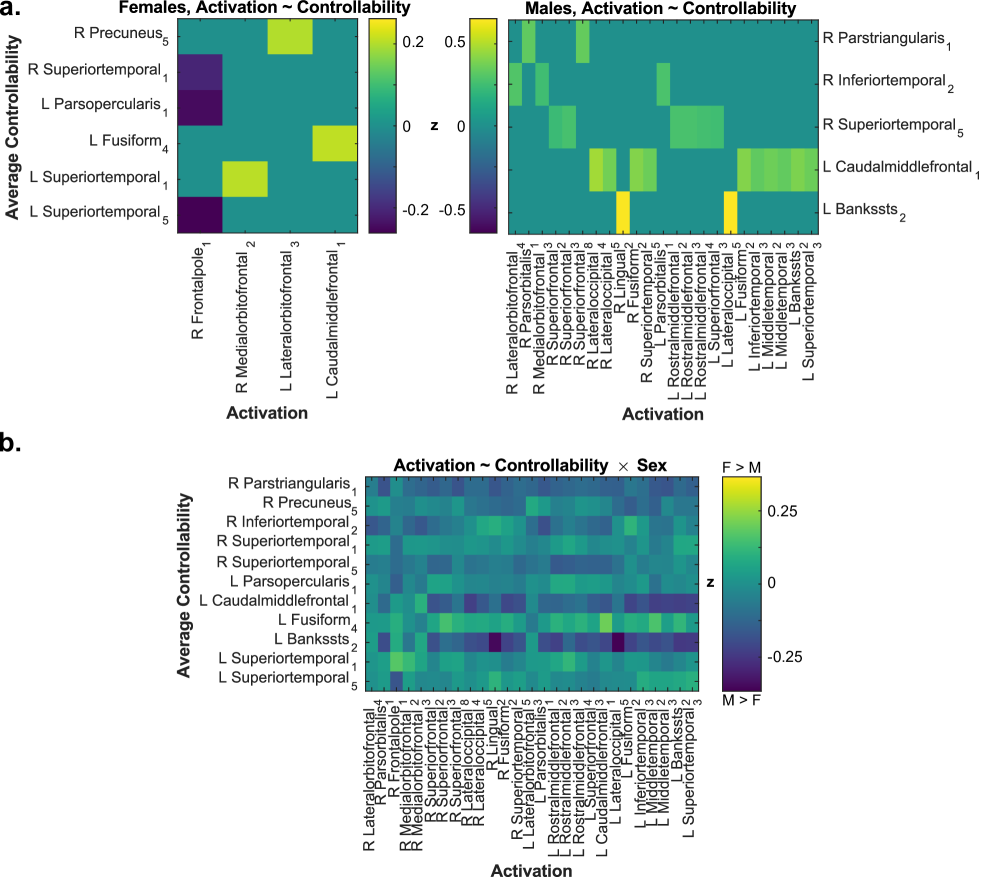
After identifying regions at which activation was associated with executive function, we sought to relate these activation values to controllability metrics of structural brain networks. We found a complex pattern of both positive and negative relationships between average controllability at 8 nodes with activation at 11 nodes (Fig. 8b). Average controllability at the left caudal middle frontal lobe was associated with activation at 7 different regions, which cluster together in a symmetric fashion in superior parietal and lateral occipital cortex (Fig. 8c). Modal controllability was not significantly associated with activation.
Next, we tested our hypothesis that the relationship between regional controllability and activation depends on sex. Specifically, we performed the same analysis on males and females, separately. Consistent with the pooled analysis, the profile of controllability-activation associations overlapped significantly between the sexes (Fig. 8d, Fig. A.4a). While there was no overlap in the precise regions involved in these sex-split analyses, there were gross anatomical similarities between males and females. In both sexes, controllability at regions near the temporal poles was associated with activation at medial frontal regions. We observed no significant interaction between controllability and sex in predicting activation (Fig. A.4b), although the directionality of the interaction terms was consistent with the observed sex differences (Fig. A.4a-b). These results support the notion that both similarities and differences in structure-function relationships exist across the sexes.
Weighted degree does not explain controllability trends
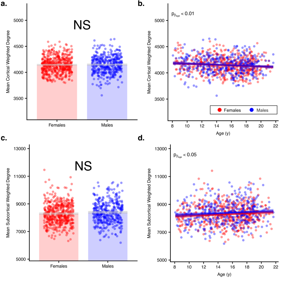
Weighted degree has previously been shown to correlate well with controllability metrics 28. We performed several specificity analyses to determine whether our results could be trivially explained by sex-differences in the weighted degree of nodes in the network. We identified 13 nodes where weighted degree was associated with sex (Fig. A.3a), and found that only 5/21 unique nodes with sex-associated controllability values also had a significant association between weighted degree and sex. Among those 5 unique nodes, only average controllability at the left caudate nucleus and the right post central gyrus had the same directionality of association with sex as the association with weighted degree (i.e. higher in males and females, respectively). When weighted degree was averaged across the anatomical cortex or subcortex, there were no significant associations between sex and cortical (, , ; Fig. A.2a) or subcortical (, , ; Fig. A.2c) weighted degree. These findings suggest that despite the known associations between controllability and weighted degree 28, weighted degree does not explain the observed sex differences in controllability.
Next, we tested whether developmental trends of weighted degree paralleled those of controllability metrics. We found that when averaged across the cortex or subcortex, weighted degree is positively associated with average controllability and negatively associated with modal controllability. However, mean weighted degree increased with age in the cortex (, , ; Fig. A.2b), but decreased with age in the subcortex (, , ; Fig. A.2d). Neither of these two trends (Fig. A.2b,d) were simple mirrors of the relationships between age and regional average or modal controllability (Fig. 4), suggesting that developmental trends of controllability reflect a more complex phenomenon than developmental trends of weighted degree. Furthermore, at nodes with sex-associated weighted degree or sex-associated controllability, weighted degree did not significantly mediate the relationship between sex and executive function (Fig. A.6). These findings suggest that weighted degree does not explain developmental trends of controllability, nor does it mediate the relationship between sex and executive function.
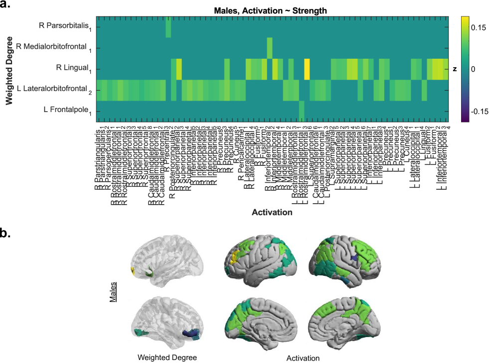
Finally, we assessed whether the associations between controllability and activation could be simply explained by the weighted degree of nodes in the network. We found no significant associations between activation and weighted degree in a pooled analysis. Moreover, including each subject’s global mean weighted degree in the model did not change the associations between average controllability and activation (data not shown). While there were significant associations between activation and weighted degree for males alone (Fig. A.5), weighted degree-activation profiles did not overlap well with average controllability-activation profiles. These results suggest that weighted degree does not explain the relation between controllability and activation.
Discussion
Our study demonstrates that network control theory can be used to explain how differences in brain network structure produce divergent patterns of brain activity that underpin executive function and its differences across males and females. We first showed that the relative locations of controllers by strength did not differ between males and females, suggesting that the overall structure of control points is similar across sex. Then, we showed that controllability covaried with age in a sex-independent fashion in both the cortex and subcortex. While global and developmental sex differences appear minimal, local sex differences in controllability exist and average controllability at those regions predicts executive function. Notably, average controllability significantly mediated the association between sex and executive function, such that subcortical average controllability was higher in males and is negatively associated with executive function. Crucially, consistent with predictions from network control theory, we also observed sex-dependent associations between nodal average controllability and n-back task BOLD activation magnitudes, demonstrating that differences in brain network structure produce divergent patterns of brain activity supporting executive function.
Implications for cognitive and clinical neuroscience
Executive function is generally lower in males, which is reflected in higher rates of impulsivity 5, ADHD diagnoses 6, criminality 7, and substance use 2. While it is known that sex differences exist in the prevalence of disorders of executive function and the putative neural circuitry involved 58; 59, it is unclear whether the pathophysiology of such disorders is sex-specific or sex-independent. Our study significantly extends the boundaries of knowledge in demonstrating neurophysiological markers of sex differences in executive function, and in couching such markers within a general network control theory of brain function.
The present work also provides important groundwork for clinical therapy. The delivery of psychiatric care is shifting towards personalized, targeted interventions. This shift can be supported by the methodology and computational tools of network neuroscience, where accurate models of brain structure and dynamics can be constructed for single individuals 60; 61. Such subject-, age-, and sex-specific models of brain structure and function can directly inform neuromodulatory therapies such as transcranial magnetic stimulation, by offering predictions about how stimulation will affect both the area being stimulated, and other areas connected to it, thereby producing a complex spatiotemporal influence on brain state. Specifically, assuming a model of brain dynamics allows us to determine which nodes could most easily drive transitions in the state of brain activity via some stimulatory input. The network control theory that we use here to study the internal modulation of brain state (via undertaking a task requiring executive function) also makes explicit predictions about the external modulation of brain state (via stimulation or neurofeedback) 62. Indeed, average controllability values were associated with sex-dependent, symmetric patterns of brain activity (Fig. 8c-d). Our results suggest that average controllability values contain useful information about brain state, beyond what can be predicted from a simple streamline-weighted adjacency matrix. These observations are the first steps towards a characterization of brain dynamics that would allow clinicians to predict the impact of stimulation given a subject-, age-, and sex-specific network architecture, thereby producing predictable changes in brain state.
An understanding of the relation between network controllability, sex, and executive function could provide important context for the study and diagnosis of neurological disease and psychiatric disorders whose prevalence may differ by sex and whose presentation includes alterations in executive function. In our study, the most robust associations between controllability, sex, and executive function are found in the subcortex and the prefrontal cortex. It is widely known that the prefrontal cortex is important for behavioral planning and working memory 63; 64, two key components of executive function. The subcortex alone is less commonly associated with executive function, but there is a strong precedent for the requirement of circuitry between dorsolateral prefrontal cortex (DLPFC) and subcortical structures for executive function 65; 66; 67; 68. Vascular 69 or neurodegenerative 68 (i.e. Huntington’s disease, Parkinson’s disease) lesions to the subcortex give rise to executive dysfunction, similar to what is observed with DLPFC lesions. Moreover, ADHD, a disorder characterized by impulsivity and executive dysfunction, is associated with decreased striatal dopamine transporter expression 58, aberrant striatal fMRI BOLD activation 70, and decreased striatal volume 71; 72. Sex differences in striatal volume 73 and striatal dopamine receptor binding 59 have also been reported. In light of these results, our study provides an important account of sex differences in subcortical connectivity from the dynamical perspective of network control theory. Our results suggest that the connectivity profile of subcortical structures is related to sex and is important for executive function, supporting the notion that disorders of executive function arise in part from altered structural connectivity.
Methodological Considerations
Several methodological considerations are pertinent to this work. First, we use a time-invariant, linear model of brain dynamics because its network control properties have been well characterized mathematically; however, it is known that the brain is highly non-linear 74. Yet, the associations between controllability and brain activity that we uncover here suggest that a simple linear model is sufficient to capture some aspect of the underlying brain dynamics. Second, axons transmit information in a unidirectional fashion, but diffusion imaging and associated tractography tools cannot elucidate the directionality of large axonal fiber bundles. Thus, we construct a symmetric adjacency matrix , assuming bidirectional influence between network nodes, and all interpretations of regional controllability depend on the realism of that assumption. Third, the eigenvalues of a symmetric matrix can only ever be real and thus the system will not oscillate, as neural systems are known to do. Finally, streamline counts and network density vary between scans 75 and with different tract reconstruction methods 76, which can complicate interpretation. Nevertheless, numerous DTI-based studies of neuropsychiatric illness have described differences in structural brain networks consistent with clinical and neuroscientific priors 77; 78, suggesting that DTI is capable of capturing subtle but meaningful variation in structural connectivity.
It is also important to acknowledge that our analysis of sex differences focuses on sex assigned at birth, which we refer to as “sex” throughout this paper. Our data does not include endocrinological measurements relevant to sex, nor does it include any psychosocial assessment of gender identity. Thus, we were not able to control for or assess gender-based differences in brain structure. Furthermore, while there exist two distinct classes of human genitalia, this fact does not imply that brains are also sexually dimorphic 79. Both “male” and “female” features exist in both male and female brains, though some features are more common in one sex than the other 79; 80. As a result, there may be more meaningful variance in neurologic phenotypes within each sex than between sexes. The divergent controllability-activation profiles in Fig. 8d may be in part due to this fact. Nevertheless, biological sex is an easily measured variable and identifying correlates of sex makes it a useful biomarker. In the future, identification of additional covariates might help uncover a more ubiquitous reason for sex-associated brain features 80.
Conclusion and Future Directions
First and foremost, our analysis of sex differences in structural brain networks showed that males and females are highly similar from the perspective of network controllability. The organization of relative controller strength was almost identical between males and females, and sex was not significantly associated with controllability values at most brain regions. However, the differences in average controllability between males and females predicted differences in cognitive performance and effects were most robust in subcortical and frontal regions. Given that BOLD signal is associated with network controllability, an interesting future study might use time-invariant, linear dynamics to predict changes in brain activity after stimulation of regions with high versus low controllability. Our results pave the way for a future study to use sex and age as features that may delineate the likely responses to brain stimulation a priori, obviating the need for pre-stimulation diffusion image acquisition and processing. Such an approach would allow the delivery of personalized psychiatric care at low cost with minimal requirements for imaging equipment and computational power.
Acknowledgments
This work was supported by an administrative supplement to NIH R21-M MH-106799 (Satterthwaite/Bassett MPI). DSB also acknowledges support from the John D. and Catherine T. MacArthur Foundation, the Alfred P. Sloan Foundation, the Army Research Office through contract number W911NF-14-1-0679, the Army Research Laboratory through contract number W911NF-10-2-0022, the National Institute of Health (2-R01-DC-009209-11, 1R01HD086888-01, R01-MH107235, R01-MH107703, R01MH109520, 1R01NS099348 and R21-M MH-106799), the Office of Naval Research, and the National Science Foundation (BCS-1441502, CAREER PHY-1554488, BCS-1631550, and CNS-1626008). TDS was supported by R01MH107703. DRR was supported by K01MH102609. FP acknowledges support from NSF-BCS-1631112 and NSF-BCS-1430279. The content is solely the responsibility of the authors and does not necessarily represent the official views of any of the funding agencies.
References
- Anderson et al. 2001 Vicki A Anderson, Peter Anderson, Elisabeth Northam, Rani Jacobs, and Cathy Catroppa. Development of executive functions through late childhood and adolescence in an australian sample. Developmental neuropsychology, 20(1):385–406, 2001.
- Romer et al. 2009 Daniel Romer, Laura Betancourt, Joan M. Giannetta, Nancy L. Brodsky, Martha Farah, and Hallam Hurt. Executive cognitive functions and impulsivity as correlates of risk taking and problem behavior in preadolescents. Neuropsychologia, 47(13):2916 – 2926, 2009. ISSN 0028-3932. doi: http://dx.doi.org/10.1016/j.neuropsychologia.2009.06.019. URL http://www.sciencedirect.com/science/article/pii/S002839320900270X.
- Barkley et al. 2002 Russell A Barkley, Kevin R Murphy, George J Dupaul, and Tracie Bush. Driving in young adults with attention deficit hyperactivity disorder: knowledge, performance, adverse outcomes, and the role of executive functioning. Journal of the International Neuropsychological Society, 8(5):655–672, 2002.
- Biederman et al. 2007 J. Biederman, C. R. Petty, R. Fried, A. E. Doyle, T. Spencer, L. J. Seidman, L. Gross, K. Poetzl, and S. V. Faraone. Stability of executive function deficits into young adult years: a prospective longitudinal follow-up study of grown up males with adhd. Acta Psychiatrica Scandinavica, 116(2):129–136, 2007. ISSN 1600-0447. doi: 10.1111/j.1600-0447.2007.01008.x. URL http://dx.doi.org/10.1111/j.1600-0447.2007.01008.x.
- Chapple and Johnson 2007 Constance L. Chapple and Katherine A. Johnson. Gender differences in impulsivity. Youth Violence and Juvenile Justice, 5(3):221–234, 2007. doi: 10.1177/1541204007301286. URL http://dx.doi.org/10.1177/1541204007301286.
- Willcutt 2012 Erik G Willcutt. The prevalence of dsm-iv attention-deficit/hyperactivity disorder: a meta-analytic review. Neurotherapeutics, 9(3):490–499, 2012.
- Cross et al. 2011 Catharine P Cross, Lee T Copping, and Anne Campbell. Sex differences in impulsivity: a meta-analysis. Psychological bulletin, 137(1):97, 2011.
- Hosenbocus and Chahal 2012 Sheik Hosenbocus and Raj Chahal. A review of executive function deficits and pharmacological management in children and adolescents. Journal of the Canadian Academy of Child and Adolescent Psychiatry, 21(3):223, 2012.
- Gogtay et al. 2004 N Gogtay, J N Giedd, L Lusk, K M Hayashi, D Greenstein, A C Vaituzis, T F 3rd Nugent, D H Herman, L S Clasen, A W Toga, J L Rapoport, and P M Thompson. Dynamic mapping of human cortical development during childhood through early adulthood. Proc Natl Acad Sci U S A, 101(21):8174–8179, 2004.
- Baum et al. 2017 G L Baum, R Ciric, D R Roalf, R F Betzel, T M Moore, R T Shinohara, A E Kahn, S N Vandekar, P E Rupert, M Quarmley, P A Cook, M A Elliott, K Ruparel, R E Gur, R C Gur, D S Bassett, and T D Satterthwaite. Modular segregation of structural brain networks supports the development of executive function in youth. Curr Biol, 27(11):1561–1572.e8, 2017.
- Schmithorst et al. 2015 V J Schmithorst, J Vannest, G Lee, L Hernandez-Garcia, E Plante, A Rajagopal, S K Holland, and CMIND Authorship Consortium. Evidence that neurovascular coupling underlying the BOLD effect increases with age during childhood. Hum Brain Mapp, 36(1):1–15, 2015.
- Nomi et al. 2017 J S Nomi, T S Bolt, C E C Ezie, L Q Uddin, and A S Heller. Moment-to-moment BOLD signal variability reflects regional changes in neural flexibility across the lifespan. J Neurosci, 37(22):5539–5548, 2017.
- Keulers et al. 2011 E H Keulers, P Stiers, and J Jolles. Developmental changes between ages 13 and 21 years in the extent and magnitude of the BOLD response during decision making. Neuroimage, 54(2):1442–1454, 2011.
- Fair et al. 2009 D A Fair, A L Cohen, J D Power, N U Dosenbach, J A Church, F M Miezin, B L Schlaggar, and S E Petersen. Functional brain networks develop from a “local to distributed” organization. PLoS Comput Biol, 5(5):e1000381, 2009.
- Gennatas et al. 2017 Efstathios D. Gennatas, Brian B. Avants, Daniel H. Wolf, Theodore D. Satterthwaite, Kosha Ruparel, Rastko Ciric, Hakon Hakonarson, Raquel E. Gur, and Ruben C. Gur. Age-related effects and sex differences in gray matter density, volume, mass, and cortical thickness from childhood to young adulthood. Journal of Neuroscience, 2017. ISSN 0270-6474. doi: 10.1523/JNEUROSCI.3550-16.2017. URL http://www.jneurosci.org/content/early/2017/04/21/JNEUROSCI.3550-16.2017.
- Blakemore and Choudhury 2006 Sarah-Jayne Blakemore and Suparna Choudhury. Development of the adolescent brain: implications for executive function and social cognition. Journal of Child Psychology and Psychiatry, 47(3-4):296–312, 2006. ISSN 1469-7610. doi: 10.1111/j.1469-7610.2006.01611.x. URL http://dx.doi.org/10.1111/j.1469-7610.2006.01611.x.
- Ingalhalikar et al. 2014 Madhura Ingalhalikar, Alex Smith, Drew Parker, Theodore D. Satterthwaite, Mark A. Elliott, Kosha Ruparel, Hakon Hakonarson, Raquel E. Gur, Ruben C. Gur, and Ragini Verma. Sex differences in the structural connectome of the human brain. Proceedings of the National Academy of Sciences, 111(2):823–828, 2014. doi: 10.1073/pnas.1316909110. URL http://www.pnas.org/content/111/2/823.abstract.
- Satterthwaite et al. 2014a Theodore D Satterthwaite, Daniel H Wolf, David R Roalf, Kosha Ruparel, Guray Erus, Simon Vandekar, Efstathios D Gennatas, Mark A Elliott, Alex Smith, Hakon Hakonarson, et al. Linked sex differences in cognition and functional connectivity in youth. Cerebral cortex, 25(9):2383–2394, 2014a.
- Liu et al. 2011 Y Y Liu, J J Slotine, and A L Barabasi. Controllability of complex networks. Nature, 473(7346):167–173, 2011.
- Pasqualetti et al. 2014 Fabio Pasqualetti, Sandro Zampieri, and Francesco Bullo. Controllability metrics, limitations and algorithms for complex networks. IEEE Transactions on Control of Network Systems, 1(1):40–52, 2014.
- Kailath 1980 T Kailath. Linear Systems. Prentice Hall, Englewood Cliffs, 1980.
- Kalman et al. 1963 R. E. Kalman, Y. C. Ho, and K. S. Narendra. Controllability of linear dynamical systems. Contributions to Differential Equations, 1:189–213, 1963.
- Muldoon et al. 2016 S F Muldoon, F Pasqualetti, S Gu, M Cieslak, S T Grafton, J M Vettel, and D S Bassett. Stimulation-based control of dynamic brain networks. PLoS Comput Biol, 12(9):e1005076, 2016.
- Betzel et al. 2016 R F Betzel, S Gu, J D Medaglia, F Pasqualetti, and D S Bassett. Optimally controlling the human connectome: the role of network topology. Sci Rep, 6:30770, 2016.
- Gu et al. 2017 S Gu, R F Betzel, M G Mattar, M Cieslak, P R Delio, S T Grafton, F Pasqualetti, and D S Bassett. Optimal trajectories of brain state transitions. Neuroimage, 148:305–317, 2017.
- Wiles et al. 2017 L Wiles, S Gu, F Pasqualetti, D S Bassett, and D F Meaney. Autaptic connections shift network excitability and bursting. Scientific Reports, In Press, 2017.
- Kim et al. 2017 J Kim, J M Soffer, A E Kahn, J M Vettel, F Pasqualetti, and D S Bassett. Role of graph architecture in controlling dynamical networks with applications to neural systems. Nature Physics, Epub Ahead of Print, 2017.
- Gu et al. 2015 Shi Gu, Fabio Pasqualetti, Matthew Cieslak, Qawi K Telesford, B Yu Alfred, Ari E Kahn, John D Medaglia, Jean M Vettel, Michael B Miller, Scott T Grafton, et al. Controllability of structural brain networks. Nature communications, 6, 2015.
- Tang et al. 2017 E Tang, C Giusti, G L Baum, S Gu, E Pollock, A E Kahn, D R Roalf, T M Moore, K Ruparel, R C Gur, R E Gur, T D Satterthwaite, and D S Bassett. Developmental increases in white matter network controllability support a growing diversity of brain dynamics. Nat Commun, 8(1):1252, 2017.
- Satterthwaite et al. 2014b T D Satterthwaite, M A Elliott, K Ruparel, J Loughead, K Prabhakaran, M E Calkins, R Hopson, C Jackson, J Keefe, M Riley, F D Mentch, P Sleiman, R Verma, C Davatzikos, H Hakonarson, R C Gur, and R E Gur. Neuroimaging of the philadelphia neurodevelopmental cohort. Neuroimage, 86:544–553, 2014b.
- Cammoun et al. 2012 L Cammoun, X Gigandet, D Meskaldji, J P Thiran, O Sporns, K Q Do, P Maeder, R Meuli, and P Hagmann. Mapping the human connectome at multiple scales with diffusion spectrum MRI. J Neurosci Methods, 203(2):386–397, 2012.
- Roalf et al. 2016 David R Roalf, Megan Quarmley, Mark A Elliott, Theodore D Satterthwaite, Simon N Vandekar, Kosha Ruparel, Efstathios D Gennatas, Monica E Calkins, Tyler M Moore, Ryan Hopson, et al. The impact of quality assurance assessment on diffusion tensor imaging outcomes in a large-scale population-based cohort. Neuroimage, 125:903–919, 2016.
- Gur et al. 2010 R C Gur, J Richard, P Hughett, M E Calkins, L Macy, W B Bilker, C Brensinger, and R E Gur. A cognitive neuroscience-based computerized battery for efficient measurement of individual differences: standardization and initial construct validation. J Neurosci Methods, 187(2):254–262, 2010.
- Gur et al. 2012 Ruben C Gur, Jan Richard, Monica E Calkins, Rosetta Chiavacci, John A Hansen, Warren B Bilker, James Loughead, John J Connolly, Haijun Qiu, Frank D Mentch, et al. Age group and sex differences in performance on a computerized neurocognitive battery in children age 8- 21. Neuropsychology, 26(2):251, 2012.
- Moore et al. 2015 Tyler M Moore, Steven P Reise, Raquel E Gur, Hakon Hakonarson, and Ruben C Gur. Psychometric properties of the penn computerized neurocognitive battery. Neuropsychology, 29(2):235, 2015.
- Moore et al. 2016 Tyler M Moore, Steven P Reise, David R Roalf, Theodore D Satterthwaite, Christos Davatzikos, Warren B Bilker, Allison M Port, Chad T Jackson, Kosha Ruparel, Adam P Savitt, et al. Development of an itemwise efficiency scoring method: Concurrent, convergent, discriminant, and neuroimaging-based predictive validity assessed in a large community sample. Psychol Assess., 28(12):1529–1542, 2016.
- Moore et al. 2017 Tyler M Moore, Ruben C Gur, Michael L Thomas, Gregory G Brown, Matthew K Nock, Adam P Savitt, John G Keilp, Steven Heeringa, Robert J Ursano, Murray B Stein, et al. Development, administration, and structural validity of a brief, computerized neurocognitive battery: Results from the army study to assess risk and resilience in service members. Assessment, Epub Ahead of Print, 2017.
- Vandekar et al. 2015 Simon N Vandekar, Russell T Shinohara, Armin Raznahan, David R Roalf, Michelle Ross, Nicholas DeLeo, Kosha Ruparel, Ragini Verma, Daniel H Wolf, Ruben C Gur, et al. Topologically dissociable patterns of development of the human cerebral cortex. Journal of Neuroscience, 35(2):599–609, 2015.
- Fischl 2012 B Fischl. Freesurfer. Neuroimage, 62:774–781, 2012.
- Rosen et al. 2017 Adon F.G. Rosen, David R. Roalf, Kosha Ruparel, Jason Blake, Kevin Seelaus, Lakshmi P. Villa, Rastko Ciric, Philip A. Cook, Christos Davatzikos, Mark A. Elliott, Angel Garcia de La Garza, Efstathios D. Gennatas, Megan Quarmley, J. Eric Schmitt, Russell T. Shinohara, M. Dylan Tisdall, R. Cameron Craddock, Raquel E. Gur, Ruben C. Gur, and Theodore D. Satterthwaite. Data-driven assessment of structural image quality. NeuroImage, pages –, 2017. ISSN 1053-8119. doi: https://doi.org/10.1016/j.neuroimage.2017.12.059. URL https://www.sciencedirect.com/science/article/pii/S1053811917310832.
- Cuadra et al. 2004 Meritxell Bach Cuadra, Claudio Pollo, Anton Bardera, Olivier Cuisenaire, J-G Villemure, and J-P Thiran. Atlas-based segmentation of pathological mr brain images using a model of lesion growth. IEEE transactions on medical imaging, 23(10):1301–1314, 2004.
- Ragland et al. 2002 J Daniel Ragland, Bruce I Turetsky, Ruben C Gur, Faith Gunning-Dixon, Travis Turner, Lee Schroeder, Robin Chan, and Raquel E Gur. Working memory for complex figures: an fmri comparison of letter and fractal n-back tasks. Neuropsychology, 16(3):370, 2002.
- Shanmugan et al. 2016 Sheila Shanmugan, Daniel H Wolf, Monica E Calkins, Tyler M Moore, Kosha Ruparel, Ryan D Hopson, Simon N Vandekar, David R Roalf, Mark A Elliott, Chad Jackson, et al. Common and dissociable mechanisms of executive system dysfunction across psychiatric disorders in youth. American journal of psychiatry, 173(5):517–526, 2016.
- Jones and Basser 2004 D K Jones and P J Basser. “squashing peanuts and smashing pumpkins”: how noise distorts diffusion-weighted MR data. Magn. Reson. Med., 52:979–993, 2004.
- Chen et al. 2015 Y Chen, O Tymofiyeva, C P Hess, and D Xu. Effects of rejecting diffusion directions on tensor-derived parameters. Neuroimage, 109:160–170, 2015.
- Jenkinson et al. 2002 M Jenkinson, P Bannister, M Brady, and S Smith. Improved optimization for the robust and accurate linear registration and motion correction of brain images. Neuroimage, 17:825–841, 2002.
- Andersson and Sotiropoulos 2016 J L R Andersson and S N Sotiropoulos. An integrated approach to correction for off-resonance effects and subject movement in diffusion MR imaging. Neuroimage, 125:1063–1078, 2016.
- Jenkinson et al. 2012 M Jenkinson, C F Beckmann, T E Behrens, M W Woolrich, and S M Smith. Fsl. Neuroimage, 62:782–790, 2012.
- Satterthwaite et al. 2013 Theodore D Satterthwaite, Daniel H Wolf, Guray Erus, Kosha Ruparel, Mark A Elliott, Efstathios D Gennatas, Ryan Hopson, Chad Jackson, Karthik Prabhakaran, Warren B Bilker, et al. Functional maturation of the executive system during adolescence. Journal of Neuroscience, 33(41):16249–16261, 2013.
- Greve and Fischl 2009 Douglas N Greve and Bruce Fischl. Accurate and robust brain image alignment using boundary-based registration. Neuroimage, 48(1):63–72, 2009.
- Honey et al. 2009 CJ Honey, O Sporns, Leila Cammoun, Xavier Gigandet, Jean-Philippe Thiran, Reto Meuli, and Patric Hagmann. Predicting human resting-state functional connectivity from structural connectivity. Proceedings of the National Academy of Sciences, 106(6):2035–2040, 2009.
- Pecora and Carroll 1998 Louis M Pecora and Thomas L Carroll. Master stability functions for synchronized coupled systems. Physical review letters, 80(10):2109, 1998.
- Benjamini and Hochberg 1995 Yoav Benjamini and Yosef Hochberg. Controlling the false discovery rate: a practical and powerful approach to multiple testing. Journal of the royal statistical society. Series B (Methodological), pages 289–300, 1995.
- Wood 2004 Simon N Wood. Stable and efficient multiple smoothing parameter estimation for generalized additive models. Journal of the American Statistical Association, 99(467):673–686, 2004.
- Tingley et al. 2014 Dustin Tingley, Teppei Yamamoto, Kentaro Hirose, Luke Keele, and Kosuke Imai. Mediation: R package for causal mediation analysis. 2014.
- Preacher et al. 2004 KJ Preacher, PJ Curran, and DJ Bauer. Simple intercepts, simple slopes, and regions of significance in mlr 2-way interactions. retrieved september 25, 2005, 2004.
- Snodgrass and Corwin 1988 Joan G Snodgrass and June Corwin. Pragmatics of measuring recognition memory: applications to dementia and amnesia. Journal of Experimental Psychology: General, 117(1):34, 1988.
- Castellanos and Tannock 2002 F Xavier Castellanos and Rosemary Tannock. Neuroscience of attention-deficit/hyperactivity disorder: the search for endophenotypes. Nature Reviews Neuroscience, 3(8):617–628, 2002.
- Pohjalainen et al. 1998 Tiina Pohjalainen, Juha O Rinne, Kjell Någren, Erkka Syvälahti, and Jarmo Hietala. Sex differences in the striatal dopamine d2 receptor binding characteristics in vivo. American Journal of Psychiatry, 155(6):768–773, 1998.
- Stephan et al. 2017 K.E. Stephan, F. Schlagenhauf, Q.J.M. Huys, S. Raman, E.A. Aponte, K.H. Brodersen, L. Rigoux, R.J. Moran, J. Daunizeau, R.J. Dolan, K.J. Friston, and A. Heinz. Computational neuroimaging strategies for single patient predictions. NeuroImage, 145(Part B):180 – 199, 2017. ISSN 1053-8119. doi: https://doi.org/10.1016/j.neuroimage.2016.06.038. URL http://www.sciencedirect.com/science/article/pii/S1053811916302877. Individual Subject Prediction.
- Izhikevich and Edelman 2008 Eugene M Izhikevich and Gerald M Edelman. Large-scale model of mammalian thalamocortical systems. Proceedings of the national academy of sciences, 105(9):3593–3598, 2008.
- Murphy and Bassett 2017 A C Murphy and D S Bassett. A network neuroscience of neurofeedback for clinical translation. Curr Opin Biomed Eng, 1:63–70, 2017.
- Tanji and Hoshi 2008 Jun Tanji and Eiji Hoshi. Role of the lateral prefrontal cortex in executive behavioral control. Physiological reviews, 88(1):37–57, 2008.
- Euston et al. 2012 David R Euston, Aaron J Gruber, and Bruce L McNaughton. The role of medial prefrontal cortex in memory and decision making. Neuron, 76(6):1057–1070, 2012.
- JL 1993 Cummings JL. Frontal-subcortical circuits and human behavior. Archives of Neurology, 50(8):873–880, 1993. doi: 10.1001/archneur.1993.00540080076020. URL +http://dx.doi.org/10.1001/archneur.1993.00540080076020.
- Duke and Kaszniak 2000 Lisa M Duke and Alfred W Kaszniak. Executive control functions in degenerative dementias: A comparative review. Neuropsychology review, 10(2):75–99, 2000.
- Bonelli and Cummings 2007 Raphael M Bonelli and Jeffrey L Cummings. Frontal-subcortical circuitry and behavior. Dialogues in clinical neuroscience, 9(2):141, 2007.
- Tekin and Cummings 2002 Sibel Tekin and Jeffrey L Cummings. Frontal–subcortical neuronal circuits and clinical neuropsychiatry: an update. Journal of psychosomatic research, 53(2):647–654, 2002.
- Bombois et al. 2007 Stéphanie Bombois, Stéphanie Debette, Xavier Delbeuck, Amélie Bruandet, Samuel Lepoittevin, Christine Delmaire, Didier Leys, and Florence Pasquier. Prevalence of subcortical vascular lesions and association with executive function in mild cognitive impairment subtypes. Stroke, 38(9):2595–2597, 2007. ISSN 0039-2499. doi: 10.1161/STROKEAHA.107.486407. URL http://stroke.ahajournals.org/content/38/9/2595.
- Plichta et al. 2009 Michael M. Plichta, Nenad Vasic, Robert Christian Wolf, Klaus-Peter Lesch, Dagmar Brummer, Christian Jacob, Andreas J. Fallgatter, and Georg Grön. Neural hyporesponsiveness and hyperresponsiveness during immediate and delayed reward processing in adult attention-deficit/hyperactivity disorder. Biological Psychiatry, 65(1):7 – 14, 2009. ISSN 0006-3223. doi: https://doi.org/10.1016/j.biopsych.2008.07.008. URL http://www.sciencedirect.com/science/article/pii/S0006322308008275. Perception, Empathy, and Reward in Attention-Deficit/Hyperactivity Disorder and Autism.
- Cubillo et al. 2012 Ana Cubillo, Rozmin Halari, Anna Smith, Eric Taylor, and Katya Rubia. A review of fronto-striatal and fronto-cortical brain abnormalities in children and adults with attention deficit hyperactivity disorder (adhd) and new evidence for dysfunction in adults with adhd during motivation and attention. Cortex, 48(2):194 – 215, 2012. ISSN 0010-9452. doi: https://doi.org/10.1016/j.cortex.2011.04.007. URL http://www.sciencedirect.com/science/article/pii/S001094521100102X. Frontal lobes.
- Vaidya 2011 Chandan J Vaidya. Neurodevelopmental abnormalities in adhd. In Behavioral neuroscience of attention deficit hyperactivity disorder and its treatment, pages 49–66. Springer, 2011.
- Raz et al. 1995 Naftali Raz, Ivan J Torres, and James D Acker. Age, gender, and hemispheric differences in human striatum: a quantitative review and new data from in vivo mri morphometry. Neurobiology of learning and memory, 63(2):133–142, 1995.
- Freeman and Skarda 1985 Walter J. Freeman and Christine A. Skarda. Spatial eeg patterns, non-linear dynamics and perception: the neo-sherringtonian view. Brain Research Reviews, 10(3):147 – 175, 1985. ISSN 0165-0173. doi: https://doi.org/10.1016/0165-0173(85)90022-0. URL http://www.sciencedirect.com/science/article/pii/0165017385900220.
- Zhu et al. 2011 Tong Zhu, Rui Hu, Xing Qiu, Michael Taylor, Yuen Tso, Constantin Yiannoutsos, Bradford Navia, Susumu Mori, Sven Ekholm, Giovanni Schifitto, et al. Quantification of accuracy and precision of multi-center dti measurements: a diffusion phantom and human brain study. Neuroimage, 56(3):1398–1411, 2011.
- Maier-Hein et al. 2017 Klaus H Maier-Hein, Peter F Neher, Jean-Christophe Houde, Marc-Alexandre Côté, Eleftherios Garyfallidis, Jidan Zhong, Maxime Chamberland, Fang-Cheng Yeh, Ying-Chia Lin, Qing Ji, et al. The challenge of mapping the human connectome based on diffusion tractography. Nature communications, 8:1349, 2017.
- Kubicki et al. 2007 Marek Kubicki, Robert McCarley, Carl-Fredrik Westin, Hae-Jeong Park, Stephan Maier, Ron Kikinis, Ferenc A Jolesz, and Martha E Shenton. A review of diffusion tensor imaging studies in schizophrenia. Journal of psychiatric research, 41(1):15–30, 2007.
- Pievani et al. 2011 Michela Pievani, Willem de Haan, Tao Wu, William W Seeley, and Giovanni B Frisoni. Functional network disruption in the degenerative dementias. The Lancet Neurology, 10(9):829–843, 2011.
- Joel et al. 2015 Daphna Joel, Zohar Berman, Ido Tavor, Nadav Wexler, Olga Gaber, Yaniv Stein, Nisan Shefi, Jared Pool, Sebastian Urchs, Daniel S Margulies, et al. Sex beyond the genitalia: The human brain mosaic. Proceedings of the National Academy of Sciences, 112(50):15468–15473, 2015.
- Joel and McCarthy 2017 Daphna Joel and Margaret M McCarthy. Incorporating sex as a biological variable in neuropsychiatric research: where are we now and where should we be? Neuropsychopharmacology, 42(2):379–385, 2017.
Figures

Average Controllability
| Control Node | ACME | ADE | ||
|---|---|---|---|---|
| R Medial orbito frontal, parcel 1 | 0.03 | 0.00 | 0.13 | 0.01 |
| R Superior frontal, parcel 4 | 0.01 | 0.43 | 0.15 | 0.01 |
| R Postcentral, parcel 5 | 0.01 | 0.10 | 0.15 | 0.00 |
| R Caudate | 0.03 | 0.01 | 0.13 | 0.01 |
| L Caudate | 0.02 | 0.03 | 0.14 | 0.01 |
Appendix A Supplementary Data
a. Weighted Degree
| ACME | p | ADE | p | Total Effect | p | Prop. Mediated | p | |
|---|---|---|---|---|---|---|---|---|
| R Medial orbito frontal1 | 0.01 | 0.54 | 0.15 | 0.00 | 0.16 | 0.00 | 0.06 | 0.55 |
| R Rostral middle frontal6 | 0.01 | 0.25 | 0.15 | 0.00 | 0.16 | 0.00 | 0.06 | 0.27 |
| R Superior frontal4 | 0.00 | 0.79 | 0.15 | 0.00 | 0.15 | 0.00 | 0.02 | 0.79 |
| R Postcentral5 | 0.01 | 0.77 | 0.15 | 0.00 | 0.15 | 0.00 | 0.05 | 0.77 |
| R Superior parietal1 | -0.00 | 0.91 | 0.16 | 0.00 | 0.15 | 0.00 | -0.00 | 0.91 |
| R Inferior parietal1 | -0.00 | 0.91 | 0.15 | 0.00 | 0.15 | 0.00 | -0.00 | 0.91 |
| R Inferior parietal3 | -0.00 | 0.91 | 0.16 | 0.00 | 0.15 | 0.00 | -0.01 | 0.91 |
| R Lateral occipital1 | -0.01 | 0.54 | 0.16 | 0.00 | 0.16 | 0.01 | -0.04 | 0.55 |
| R Fusiform4 | 0.00 | 0.85 | 0.15 | 0.00 | 0.16 | 0.00 | 0.03 | 0.86 |
| R Caudate | 0.01 | 0.06 | 0.15 | 0.00 | 0.16 | 0.00 | 0.08 | 0.12 |
| L Lateral orbitofrontal3 | -0.01 | 0.25 | 0.17 | 0.00 | 0.16 | 0.00 | -0.07 | 0.27 |
| L Pars triangularis1 | -0.00 | 0.91 | 0.16 | 0.00 | 0.16 | 0.00 | -0.00 | 0.91 |
| L Precentral4 | 0.00 | 0.91 | 0.16 | 0.00 | 0.16 | 0.00 | 0.01 | 0.91 |
| L Precentral8 | 0.00 | 0.91 | 0.16 | 0.00 | 0.16 | 0.00 | 0.01 | 0.91 |
| L Postcentral1 | -0.00 | 0.91 | 0.16 | 0.00 | 0.16 | 0.00 | -0.01 | 0.91 |
| L Postcentral3 | 0.00 | 0.91 | 0.15 | 0.00 | 0.16 | 0.00 | 0.00 | 0.91 |
| L Postcentral4 | -0.00 | 0.91 | 0.16 | 0.00 | 0.16 | 0.00 | -0.01 | 0.91 |
| L Superior parietal2 | -0.01 | 0.54 | 0.17 | 0.00 | 0.16 | 0.00 | -0.03 | 0.55 |
| L Superior parietal5 | -0.00 | 0.91 | 0.16 | 0.00 | 0.15 | 0.00 | -0.01 | 0.91 |
| L Inferior parietal3 | 0.00 | 0.86 | 0.16 | 0.00 | 0.16 | 0.00 | 0.01 | 0.86 |
| L Lateral occipital4 | 0.00 | 0.91 | 0.16 | 0.00 | 0.16 | 0.00 | 0.00 | 0.91 |
| L Lateral occipital5 | 0.00 | 0.86 | 0.15 | 0.00 | 0.16 | 0.00 | 0.02 | 0.86 |
| L Fusiform4 | 0.01 | 0.78 | 0.15 | 0.00 | 0.16 | 0.00 | 0.05 | 0.78 |
| L Superior temporal4 | -0.01 | 0.70 | 0.16 | 0.00 | 0.16 | 0.00 | -0.04 | 0.70 |
| L Insula3 | 0.00 | 0.86 | 0.15 | 0.00 | 0.16 | 0.00 | 0.01 | 0.86 |
| L Caudate | 0.01 | 0.54 | 0.15 | 0.00 | 0.16 | 0.00 | 0.06 | 0.55 |
| L Putamen | 0.00 | 0.70 | 0.15 | 0.00 | 0.16 | 0.00 | 0.02 | 0.70 |
| L Pallidum | 0.01 | 0.61 | 0.15 | 0.00 | 0.16 | 0.00 | 0.04 | 0.61 |
| L Amygdala | 0.01 | 0.54 | 0.15 | 0.00 | 0.16 | 0.00 | 0.04 | 0.55 |