Fluid flows shaping organism morphology
Abstract
A dynamic self-organized morphology is the hallmark of network-shaped organisms like slime moulds and fungi. Organisms continuously re-organize their flexible, undifferentiated body plans to forage for food. Among these organisms the slime mould P. polycephalum has emerged as a model to investigate how organism can self-organize their extensive networks and act as a coordinated whole. Cytoplasmic fluid flows flowing through the tubular networks have been identified as key driver of morphological dynamics. Inquiring how fluid flows can shape living matter from small to large scales opens up many new avenues for research.
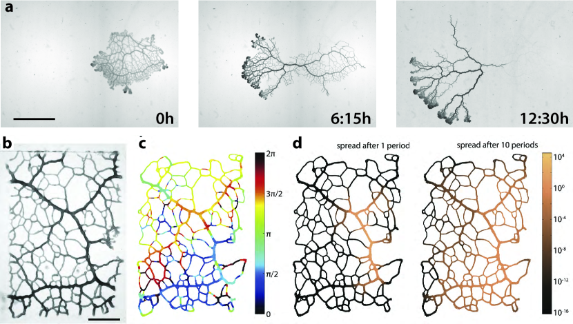
Many organisms, including a broad range of species of slime moulds and fungi, grow and forage as a single large network. Networks change their morphology over and over again during growth and migration to locate food in a patchy environment Boddy et al. (2009); Stephenson and Stempen (1994), see also Fig. 1. Moreover, network morphology adapts in response to newly acquired food sources, even connecting food sources in an efficient and robust manner Tero et al. (2010). The striking similarity of the morphological dynamics in foraging fungi and slime moulds is even more surprising if one takes into account that slime moulds and fungi are genetically distinct, with slime moulds being genetically even closer to animals than fungi. It is therefore likely that not biological make-up but the physics of fluid flows within the tubular networks are critical to the self-organization of network morphology across an individual. Both kinds of living, adaptive networks exhibit oscillatory, long-ranged fluid flows Tlalka et al. (2002, 2007); Kamiya (1950). Here, the syncytial plasmodia of the slime mould Physarum polycephalum emerged as a model system to understand the role of flows in coordinating morphology. Fluid flows in this organism are highly coordinated, driving intracellular transport on short time scales but also migration and likely morphological self-organization at long time scales.
I Cytoplasmic flows organized in peristaltic wave
In P. polycephalum the fluid cytoplasm within the tubular network streams forth and back in a shuttle flow Stewart and Stewart (1959); Kamiya (1950). Network sizes are macroscopic ranging from about to - experiments are typically conducted on specimen of up to few in size. Flows generally exhibit a Poiseuille profile Kamiya (1950); Bykov et al. (2009) with deviations likely in smaller tubes Romanovskii and Teplov (1995). Flow is dominated by small Reynolds number and small Womersley number , based on a representative tube radius of , a flow velocity reaching up to Kamiya (1950), a kinematic viscosity of cytoplasm Swaminathan et al. (1997) and an oscillation frequency of . The cytoplasm is enclosed by gel-like walls that are lined with an actin cortex Isenberg and sE Wohlfarth-Bottermann (1976); Naib-Majani et al. (1983). Actin organizes in circumferential fibrils that contract periodically Rieu et al. (2015) and drive the cytoplasms’ shuttle flow. Contrary to long-lasting speculations about localized pumps driving pressure difference, the common understanding now is that flows arise through network-wide, self-organized contractions of the actin cortex Alim et al. (2013); Teplov (2017).
The shuttelling cytoplasm itself is very rich in actin. In sized cytoplasm extract droplets, so called proto-plasmic droplets, the actin cortex and the contractions self-organize over time, showing first irregular contraction patterns and later highly coordinated spatio-temporal patterns including standing, travelling and spiral waves Matsumoto et al. (1988); Ueda (2005); Takagi and Ueda (2008, 2010). Similar dynamics and patterns can be reproduced in models by describing the proto-plasmic droplets by two phases of a viscoelastic solid phase representing the cytoskeleton, interpenetrated by a second fluid phase representing the cytosol and coupling both phases by a soluble molecule that activates tension in the solid phase Radszuweit et al. (2013); Alonso et al. (2016). Even on the much larger scale of an entire tubular network contractions are also highly coordinated, forming a peristaltic wave spanning specimen of at least in size Nakagaki et al. (2000); Alim et al. (2013), see Fig. 1. While the contraction period only increases moderately with network size Kuroda et al. (2015); Ueda and Kobatake (1982), the wavelength of the peristaltic wave matches organism size spanning two orders of magnitude Alim et al. (2013). It is fascinating to investigate how this scaling can arise. Given the success of mechanochemical models for proto-plasmic droplets it is likely that also the organism spanning peristaltic wave is a result of mechanochemical patterning, which could nicely complement our emerging understanding of this novel patterning mechanism widespread in biological systems Howard et al. (2011); Gross et al. (2017). From a fluid mechanics point a view a peristaltic wave matching organism size induces the highest flow velocities throughout a network. As flows change with organism morphology, they are likely not only important for organism homoeostasis but also for the coordination of morphological adaptation itself.
II Effective intracellular transport by oscillatory flow
Peristalsis is a common mechanism in biological systems, creating oscillatory flow and pumping fluid along a tube Shapiro et al. (1969); Shapiro and Jaffrin (1971); Selverov and Stone (2001); Li and Brasseur (1993). In P. polycephalum the tubular network can be considered to be of fixed volume on the time scales of tens of contraction periods. Therefore, net fluid transport is not relevant on these short time scales Iima and Nakagaki (2012); Alim et al. (2013). Yet, creating shuttle flows by a peristaltic wave of contractions is a simple but powerful mechanism to increase the spread of any particles, like metabolites or signalling molecules within this closed network. Rare measurements show organism-wide transport of particles within half a contraction period Nakagaki et al. (2000) out-competing diffusive spread that would have travelled only in that time frame. Because of the oscillatory nature of flow, particles flow mainly back to their initial site after a whole period. But the peristaltic flow also increases the effective diffusion according to Taylor dispersion in long slender tubes Aris (1956); Taylor (1953) also applicable to contractile tubes Mercer and Roberts (1990, 1994). Here, denotes the bare molecular diffusivity. Rapid diffusion across the tiny tube cross-sections allows particles to transition between fast and slow streamlines of the Poiseuille profile rapidly increasing their dispersion along the tube by an order of magnitude, see Fig. 2 c. The adjacency of big and small tubes and therefore different flow velocities in a slime mould network make it a hard task to theoretically map out how far particles can spread Heaton et al. (2012). One successful strategy is to map out an effective dispersion Saffman (1959); Marbach et al. (2016), which unveils for example that particle spread is increased by pruning/coarsening of the network when P. polycephalum is left to starve Marbach et al. (2016). This already provides a glimpse of the challenging question on how network morphology impacts network-wide transport Nakagaki et al. (2000). Is morphology geared to optimizing dispersion? If so, how does it evolve toward an optimized morphology? Transport by fluid flow seems to lie at the basis of network self-organization since advected signaling molecules may propagate information about the acquisition of a food source throughout the network by hijacking fluid flows Alim et al. (2017). Here, signaling molecules advected by fluid flow directly increase contraction activity. Further, flow is necessary to synchronize and coordinate contractions Yoshimoto and Kamiya (1978); Samans et al. (1984); Achenbach and Wohlfarth-Bottermann (1981). It is therefore likely that flows play a crucial role in coordinating contractions over space and time.
III Cell migration by pumping of peristaltic wave
Cytoplasmic flows form the basis of fast locomotion of very different kinds of amoeba Taylor et al. (1973); Lämmermann et al. (2008); Charras and Paluch (2008); Yoshida and Soldati (2006). Flows arise by local expansion of the actin cortex and subsequent myosin dependent contraction Lämmermann et al. (2008). Flows and contractility underlying cell migration are well accessible in the amoeboid plasmodia of P. polycephalum. Plasmodia of length adopt an amoeboid shape migrating rapidly Matsumoto et al. (2008); Zhang et al. (2017) by net fluid transport generated by a contractile peristaltic wave and cortex expansion at the front Lewis et al. (2015). Changes in contraction pattern affect locomotion velocity Rodiek et al. (2015). Plasmodia that exceed form a full network structure often including multiple migration fronts. Over the peristaltic cycle a front location advances and retracts asymmetrically leading to net advancement at long time scales Baumgarten and Hauser (2014). As networks grow they move faster Kuroda et al. (2015). When confined to lanes, migration velocity scales linearly with maximal plasmodium height, reaching locomotion speeds of up to Kuroda et al. (2015). Reorientation of migration direction for example toward a food source is associated with a redirection of the peristaltic wave direction to that site Alim et al. (2017) further substantiating that migration is governed by fluid flows on long time scales. Given that signaling molecules are advected by fluid flows affecting actin cortex dynamics Alim et al. (2017) it is seems possible that flows play an important role in the navigation of organisms, acting for example during chemotaxis.
IV Morphological changes triggered by cytoplasmic flows
Fluid flows not only transport particles and fluid mass but also exert forces themselves that may induce long-term changes to morphology. Forces may directly feed back onto biochemical reactions triggering complex spatiotemporal dynamics due to this mechanochemical coupling as currently more and more observed in morphogenetic processes Howard et al. (2011); Gross et al. (2017). Even without the ability to pin down a specific feedback on chemical reactions the influence of forces generated by flow can be investigated on a coarse-grained level. For example, in animal vasculature it is observed that tube diameters grow with increased flow rate regulating shear force to a balanced level Kamiya and Togawa (1980). This observation inspired the idea that fluid shear force induces morphological changes in vasculature, a concept also successfully used in models of P. polycephalum dynamics Nakagaki et al. (2000). Further support is the success of Murray’s law particularly in plant and animal vasculature Murray (1926) which predicts the ratio of tube diameters at a network node under the assumption of conserved shear force. Murray’s law is also consistent with minimizing dissipation inspiring theoretical work on optimal network architectures Corson (2010); Katifori et al. (2010); Durand (2007). Given that P. polycephalum grows its almost transparent tubes in a planar network, testing principles such as Murray’s law Akita et al. (2016) and in general relating morphological dynamics to flow properties is very feasible. In particular, the adaptability of the network morphology makes it a very suitable system to explore how well certain properties like dissipation, robustness Tero et al. (2010) or transport capabilities Meigel and Alim (2017); Chang et al. (2015) are optimized by living organisms. Equally, as flows are globally coupled throughout the network, there is considerable additional complexity in this system. Indeed, it might well be that precisely this added complexity due to the coupling is the key to have simple mechanisms based on fluid flow give rise to the complex dynamics of self-organization of morphology we observe.
Acknowledgement
This work has been supported by the Max Planck Society
References
- Boddy et al. (2009) L Boddy, J Hynes, D P Bebber, and M D Fricker. Saprotrophic cord systems: Dispersal mechanisms in space and time. Mycoscience, 50:9–19, 2009.
- Stephenson and Stempen (1994) S L Stephenson and H Stempen. Myxomycetes: a handbook of slime molds. Timber Press, Inc., Oregon, 1994.
- Tero et al. (2010) A Tero, S Takagi, T Saigusa, K Ito, D P Bebber, M D Fricker, K Yumiki, R Kobayashi, and T Nakagaki. Rules for biologically inspired adaptive network design. Science, 327:439–42, 2010.
- Tlalka et al. (2002) M Tlalka, S C Watkinson, P R Darrah, and M D Fricker. Continuous imaging of amino-acid translocation in intact mycelia of Phanerochaete velutina reveals rapid, pulsatile fluxes. New Phytol., 153:173–184, 2002.
- Tlalka et al. (2007) M Tlalka, D P Bebber, P R Darrah, S C Watkinson, and M D Fricker. Emergence of self-organised oscillatory domains in fungal mycelia. Fungal Genet. Biol., 44:1085–1095, 2007.
- Kamiya (1950) N Kamiya. The rate of the protoplasmic flow in the myxomycete plasmodium I. Cytologia, 15:183–193, 1950.
- Stewart and Stewart (1959) P A Stewart and B T Stewart. Protoplasmic movement in slime mold plasmodia: The diffusion drag force hypothesis. Exp. Cell Res., 17:44–58, 1959.
- Bykov et al. (2009) Alexander V Bykov, Alexander V Priezzhev, Janne Lauri, and Risto Myllylä. Doppler OCT imaging of cytoplasm shuttle flow in Physarum polycephalum. J. Biophoton., 2(8-9):540–547, 2009.
- Romanovskii and Teplov (1995) Yurii Romanovskii and VA Teplov. The physical bases of cell movement. The mechanisms of self-organisation of amoeboid motility. Phys.-Usp., 38(5):521–542, 1995.
- Swaminathan et al. (1997) R Swaminathan, C P Hoang, and A S Verkman. Photobleaching recovery and anisotropy decay of green fluorescent protein GFP-S65T in solution and cells: cytoplasmic viscosity probed by green fluorescent protein translational and rotational diffusion. Biophys. J., 72(4):1900–1907, 1997.
- Isenberg and sE Wohlfarth-Bottermann (1976) G Isenberg and K sE Wohlfarth-Bottermann. Transformation of cytoplasmic actin. Importance for the organization of the contractile gel reticulum and the contraction – Relaxation cycle of cytoplasmic actomyosin. Cell Tiss. Res., 173:495–528, 1976.
- Naib-Majani et al. (1983) W Naib-Majani, W Stockem, K Weber, J Wehland, and K E Wohlfarth-Bottermann. Cytoplasmic actin patterns in Physarum as revealed by NBD-phallacidin staining. Cell Biol. Int., 7(8):637–640, 1983.
- Rieu et al. (2015) J-P Rieu, H Delanoë-Ayari, S Takagi, Y Tanaka, and T Nakagaki. Periodic traction in migrating large amoeba of Physarum polycephalum. J. R. Soc. Interface, 12(106):20150099, 2015.
- Teplov (2017) Vladimir A Teplov. Role of mechanics in the appearance of oscillatory instability and standing waves of the mechanochemical activity in the Physarum polycephalum plasmodium. J. Phys. D: Appl. Phys., 50(21):213002–22, 2017.
- Matsumoto et al. (1988) K Matsumoto, T Ueda, and Y Kobatake. Reversal of thermotaxis with oscillatory stimulation in the plasmodium of Physarum polycephalum. J. Theor. Biol., 131(2):175–182, 1988.
- Ueda (2005) T Ueda. An intelligent slime mold: A self-organizing system of cell shape and information. World Scientific Lecture Notes in Complex Systems: Network of Interacting Machines. World Scientific Publishing Co., 2005.
- Takagi and Ueda (2008) S Takagi and T Ueda. Emergence and transitions of dynamic patterns of thickness oscillation of the plasmodium of the true slime mold Physarum polycephalum. Physica D, 237:420–427, 2008.
- Takagi and Ueda (2010) S Takagi and T Ueda. Annihilation and creation of rotating waves by a local light pulse in a protoplasmic droplet of the Physarum plasmodium. Physica D, 239(11):873–878, 2010.
- Radszuweit et al. (2013) Markus Radszuweit, Sergio Alonso, Harald Engel, and Markus Bär. Intracellular mechanochemical waves in an active poroelastic model. Phys. Rev. Lett., 110(13):138102, 2013.
- Alonso et al. (2016) Sergio Alonso, Ulrike Strachauer, Markus Radszuweit, Markus Bär, and Marcus J B Hauser. Oscillations and uniaxial mechanochemical waves in a model of an active poroelastic medium: Application to deformation patterns in protoplasmic droplets of Physarum polycephalum. Physica D, 318-319:58–69, 2016.
- Nakagaki et al. (2000) T Nakagaki, H Yamada, and T Ueda. Interaction between cell shape and contraction pattern in the Physarum plasmodium. Biophys. Chem., 84(3):195–204, May 2000.
- Alim et al. (2013) K Alim, G Amselem, F Peaudecerf, M P Brenner, and A Pringle. Random network peristalsis in Physarum polycephalum organizes fluid flows across an individual. Proc. Natl. Acad. Sci. U.S.A., 110(33):13306–13311, 2013.
- Fricker (2014) M D. Fricker. personal communication, 2014.
- Kuroda et al. (2015) S Kuroda, S Takagi, T Nakagaki, and T Ueda. Allometry in Physarum plasmodium during free locomotion: size versus shape, speed and rhythm. J. Exp. Biol., 218(23):3729–3738, 2015.
- Ueda and Kobatake (1982) T Ueda and Y Kobatake. Initiation, development and termination of contraction rhythm in plasmodia of myxomycete Physarum polycephalum. J. Theor. Biol., 97(1):87–93, 1982.
- Howard et al. (2011) Jonathon Howard, Stephan W Grill, and Justin S Bois. Turing’s next steps: the mechanochemical basis of morphogenesis. Nat. Rev. Mol. Cell Biol., 12(6):392–398, 2011.
- Gross et al. (2017) Peter Gross, K Vijay Kumar, and Stephan W Grill. How active mechanics and regulatory biochemistry combine to form patterns in development. Annu. Rev. Biophys., 46(1):337–356, 2017.
- Shapiro et al. (1969) AH Shapiro, MY Jaffrin, and SL Weinberg. Peristaltic pumping with long wavelengths at low Reynolds number. J. Fluid Mech., 37:799–825, 1969.
- Shapiro and Jaffrin (1971) A H Shapiro and M Y Jaffrin. Reflux in peristaltic pumping - is it determined by Eulerian or Lagrangian mean velocity. J. Appl. Mech., 38(4):1060–1062, 1971.
- Selverov and Stone (2001) K P Selverov and H A Stone. Peristaltically driven channel flows with applications toward micromixing. Phys. Fluids, 13(7):1837–1859, 2001.
- Li and Brasseur (1993) M Li and J G Brasseur. Non-steady peristaltic transport in finite-length tubes. J. Fluid Mech., 248:129–151, 1993.
- Iima and Nakagaki (2012) M Iima and T Nakagaki. Peristaltic transport and mixing of cytosol through the whole body of Physarum plasmodium. Math. Med. Biol., 29(3):263–281, 2012.
- Aris (1956) R Aris. On the dispersion of a solute in a fluid flowing through a tube. Proc. R. Soc. A, 235:67–77, 1956.
- Taylor (1953) G Taylor. Dispersion of soluble matter in solvent flowing slowly through a tube. Proc. R. Soc. A, 219:186–203, 1953.
- Mercer and Roberts (1990) G N Mercer and A J Roberts. A center manifold description of contaminant dispersion in channels with varying flow properties. SIAM J. Appl. Math., 50(6):1547–1565, 1990.
- Mercer and Roberts (1994) G N Mercer and A J Roberts. A complete model of shear dispersion in pipes. Japan J. Indust. Appl. Math., 11(3):499–521, 1994.
- Heaton et al. (2012) Luke L Heaton, Eduardo López, Philip K Maini, Mark D Fricker, and Nick S Jones. Advection, diffusion, and delivery over a network. Phys. Rev. E, 86(2):021905, 2012.
- Saffman (1959) PG Saffman. A theory of dispersion in a porous medium. J. Fluid Mech., 6:321–349, 1959.
- Marbach et al. (2016) S Marbach, K Alim, N Andrew, A Pringle, and M P Brenner. Pruning to increase Taylor dispersion in Physarum polycephalum networks. Phys. Rev. Lett., 117(17):178103, 2016.
- Alim et al. (2017) K Alim, N Andrew, A Pringle, and M P Brenner. Mechanism of signal propagation in Physarum polycephalum. Proc. Natl. Acad. Sci. U.S.A., 114(20):5136–5141, 2017.
- Yoshimoto and Kamiya (1978) Y Yoshimoto and N Kamiya. Studies on contraction rhythm of plasmodial strand 3. Role of endoplasmic streaming in synchronization of local rhythms. Protoplasma, 95(1-2):111–121, 1978.
- Samans et al. (1984) K.E. Samans, I. Hinz, Z. Hejnowicz, and K.E. Wohlfarth-Bottermann. Phase relation of oscillatory contraction cycles in Physarum plasmodia: I. A serial infrared registration device and its application to different plasmodial stages. J. Interdiscip. Cycle Res., 15(4):241–250, 1984.
- Achenbach and Wohlfarth-Bottermann (1981) U Achenbach and KE Wohlfarth-Bottermann. Synchronization and signal transmission in protoplasmic strands of Physarum. Planta, 151:574–583, 1981.
- Taylor et al. (1973) D L Taylor, J S Condeelis, P L Moore, and R D Allen. The contractile basis of amoeboid movement: I. The chemical control of motility in isolated cytoplasm. J. Cell Biol., 59(2):378–394, 1973.
- Lämmermann et al. (2008) T Lämmermann, B L Bader, S J Monkley, T Worbs, R Wedlich-Söldner, K Hirsch, M Keller, R Förster, D R Critchley, R Fässler, and M Sixt. Rapid leukocyte migration by integrin-independent flowing and squeezing. Nature, 453(7191):51–55, 2008.
- Charras and Paluch (2008) G Charras and E Paluch. Blebs lead the way: How to migrate without lamellipodia. Nat. Rev. Mol. Cell Biol., 9(9):730–736, 2008.
- Yoshida and Soldati (2006) K Yoshida and T Soldati. Dissection of amoeboid movement into two mechanically distinct modes. J. Cell Sci., 119:3833–3844, 2006.
- Matsumoto et al. (2008) K Matsumoto, S Takagi, and T Nakagaki. Locomotive mechanism of Physarum plasmodia based on spatiotemporal analysis of protoplasmic streaming. Biophys. J., 94:2492–504, 2008.
- Zhang et al. (2017) S Zhang, R D Guy, J C Lasheras, and J C del Álamo. Self-organized mechano-chemical dynamics in amoeboid locomotion of Physarum fragments. J. Phys. D: Appl. Phys., 50(20):204004, 2017.
- Lewis et al. (2015) O L Lewis, S Zhang, R D Guy, and J C del Alamo. Coordination of contractility, adhesion and flow in migrating Physarum amoebae. J. Roy. Soc. Interface, 12(106):20141359, 2015.
- Rodiek et al. (2015) B Rodiek, S Takagi, T Ueda, and M J B Hauser. Patterns of cell thickness oscillations during directional migration of Physarum polycephalum. Eur. Biophys. J., 44(5):349–358, 2015.
- Baumgarten and Hauser (2014) W Baumgarten and M J B Hauser. Dynamics of frontal extension of an amoeboid cell. Europhys. Lett., 108(5):50010, 2014.
- Kamiya and Togawa (1980) A Kamiya and T Togawa. Adaptive regulation of wall shear-stress to flow change in the canine carotid-artery. Am. J. Physiol., 239(1):H14–H21, 1980.
- Murray (1926) C D Murray. The physiological principle of minimum work: I. The vascular system and the cost of blood volume. Proc. Natl. Acad. Sci. U.S.A., 12(3):207–214, 1926.
- Corson (2010) F Corson. Fluctuations and redundancy in optimal transport networks. Phys. Rev. Lett., 104:048703, 2010.
- Katifori et al. (2010) E Katifori, G J Szöllősi, and M O Magnasco. Damage and fluctuations induce loops in optimal transport networks. Phys. Rev. Lett., 104:048704, 2010.
- Durand (2007) M Durand. Structure of optimal transport networks subject to a global constraint. Phys. Rev. Lett., 98(8):088701, 2007.
- Akita et al. (2016) D Akita, I Kunita, M D Fricker, S Kuroda, KA Sato, and T Nakagaki. Experimental models for Murray’s law. J. Phys. D: Appl. Phys., 50(2):024001, 2016.
- Meigel and Alim (2017) F. J. Meigel and K. Alim. Flow rate of transport network controls uniform metabolite supply to tissue. Roy. Soc. Interface, in press, 2018.
- Chang et al. (2015) S-S Chang, S Tu, Y-H Liu, V Savage, S-P L Hwang, and M Roper. Optimal occlusion uniformly partitions red blood cells fluxes within a microvascular network. PLos Comput. Biol., 13:e1005892, 2017.