Entropy of water and the temperature-induced stiffening of amyloid networks
Abstract
In water, networks of semi-flexible fibrils of the protein -synuclein stiffen significantly with increasing temperature. We make plausible that this reversible stiffening is a result of hydrophobic contacts between the fibrils that become more prominent with increasing temperature. The good agreement of our experimentally observed temperature dependence of the storage modulus of the network with a scaling theory linking network elasticity with reversible crosslinking enables us to quantify the endothermic binding enthalpy and an estimate the effective size of hydrophobic patches on the fibril surface.
I Introduction
Water and oil do not mix. Even after vigorous stirring, these two compounds spontaneously separate into distinct liquid phases. This is a macroscopic manifestation of the hydrophobic effect, a phenomenon driven by the microscopic behavior of water in the presence of nonpolar molecules. Ultimately, this is caused by hydrophobic molecules and assemblies thereof being incapable of forming hydrogen bonds. If introduced in an aqueous environment, hydrophobic molecular units perturb or even disrupt the dynamic hydrogen bonded network that is formed by the water molecules. As a result, water molecules self-organize into more strongly ordered structures in the vicinity of the hydrophobic molecular units. This response is entropically unfavorable, and, hence, hydrophobic solutes become sticky and tend to cluster together. The solvent-mediated interactions at play are known as hydrophobic interactions (HIs). A distinguishing hallmark of HIs is their non-trivial dependence on temperature. For small hydrophobic solutes as well as large ones characterized by small hydrophobic patches, below, say, 1nm2, HIs become stronger with increasing temperature Chandler (2005). This characteristic feature of HIs persists in liquid water and distinguishes HIs from all other types of non-covalent attractive interactions that become effectively weaker at higher temperature.
HIs play a central role in various phenomena in chemistry and biology, from the cleaning action of detergents and the production of micro-emulsions, to the in vivo assembly of biological macromolecules into complex structures. HIs often facilitate proteins in attaining their functional form by supporting their native fold or by binding to partners Dyson et al. (2006); Sturtevant (1977). HIs have been also implicated in promoting the self-assembly of proteins into oligomeric species and amyloid fibrils, a process accompanying many disease conditions Buell et al. (2012). Finally, HIs, in an intricate interplay with other types of non-covalent interactions, drive the self-assembly of virus coat proteins into virus capsids Kegel and van der Schoot (2004). This phenomenon has inspired the field of bionanotechnology to create novel self-assembled biosynthetic structures Comellas-Aragones et al. (2007); Hernandez-Garcia et al. (2014). Apart from being a characteristic feature of HIs, the unique temperature response makes this type of non-covalent interaction a suitable tool to manipulate the properties of materials that have exposed patches of hydrophobic surface. Nevertheless, controlling material properties through hydrophobic forces remains a challenge, arguably resulting from our limited understanding of HIs and the lack of design principles for the synthesis of tunable materials, the responsiveness of which is based on HIs.
Here, we make use of the neuronal protein alpha-synuclein (S) that under appropriate conditions self-assembles into amyloid fibrils, and take it as a model system in which material properties can be controlled by harnessing HIs. This protein exhibits a complex phase behavior and, depending on the physico-chemical conditions, organizes into hierarchical suprafibrillar aggregates with varying morphologies or into isotropic semi-flexible amyloid networks Semerdzhiev et al. (2014, 2017). We carefully choose the experimental conditions to steer the self-assembly into the region of the phase diagram where semi-flexible networks are formed. Fibril networks are a convenient platform i) to convincingly address the hydrophobic nature of the attractive interactions that drive the self-organization of S fibrils into larger scale structures, helping us to identify the role and formation mechanism of pathological fibril structures such as Lewy bodies that accompany the progression of Parkinson’s disease, and ii) to exploit hydrophobic interactions to tune the mechanical properties of a material and inspire design principles for the creation of novel temperature responsive materials. We use temperature as a ‘tuning knob’ to adjust the effective degree of crosslinking in the S fibril network and by doing so change the viscoelastic response of the material without forcing any permanent structural alterations in the material. Finally, we quantitatively connect the thermally induced enhancement of HIs to the observed stiffening in the viscoelastic response of the network by incorporating the effect of reversible crosslinking in established scaling theory for semi-flexible networks.
II Results and discussion
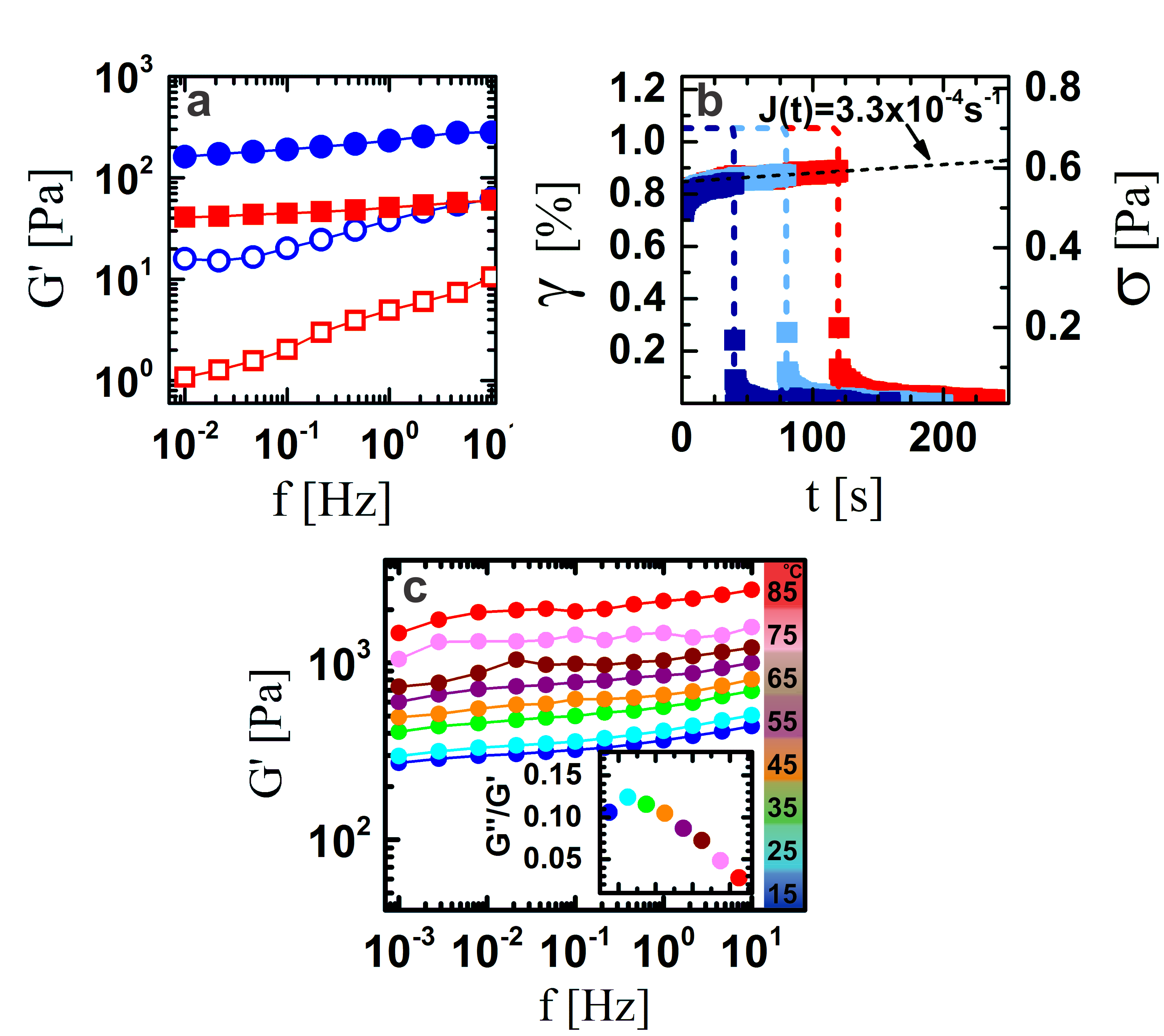
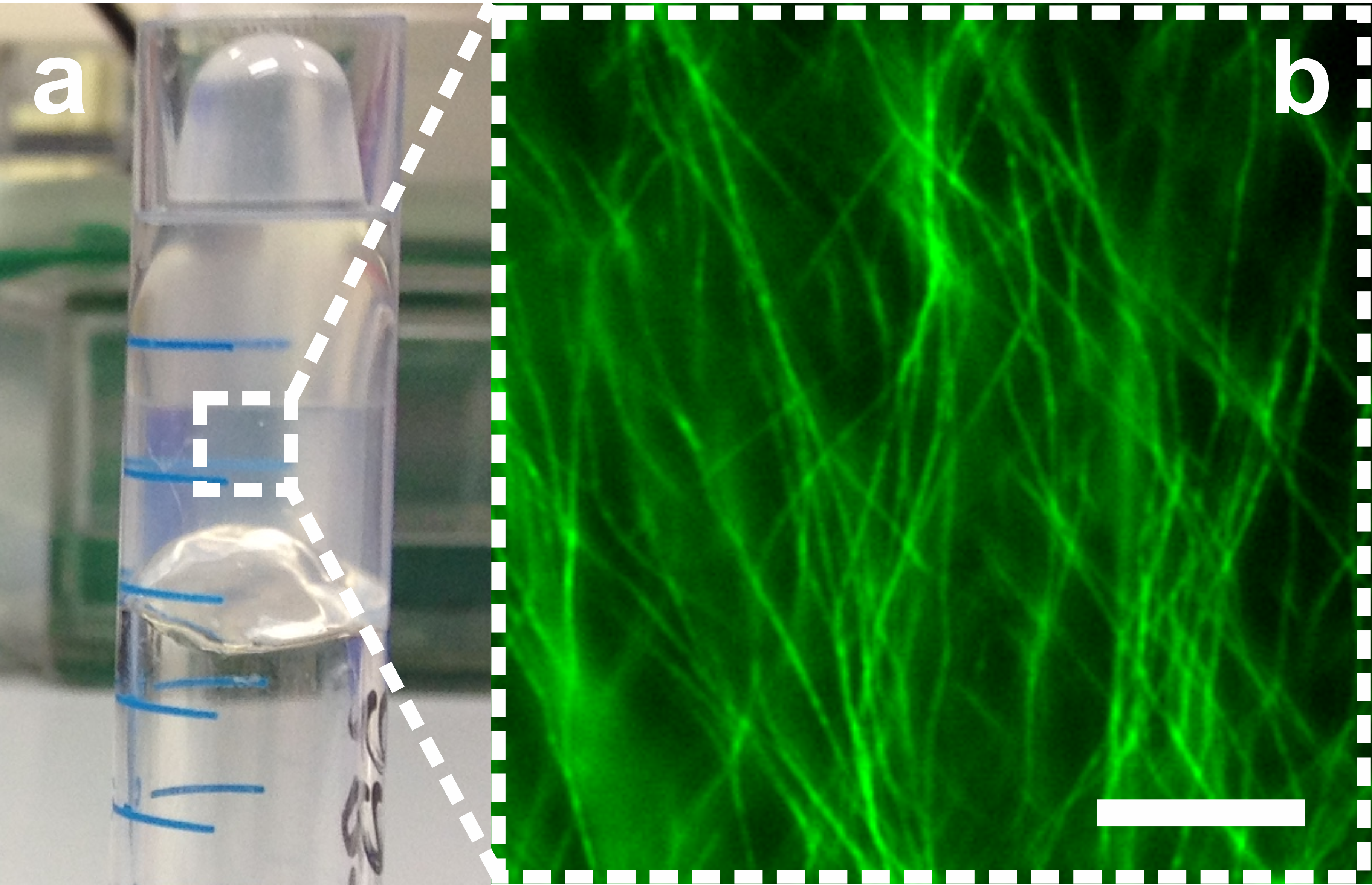
The polymerization of S into amyloid fibrils is a slow process. Within 7 days after the initiation of polymerization, a solution of monomeric S typically evolves into a gel (Fig. S1). It takes up to 37 days until all the protein has polymerized into fibrils (Fig. S2) and the network has equilibrated (Fig. 1a). Rheologically, the networks of semi-flexible amyloid fibrils behave as viscoelastic materials. Frequency sweeps of aged networks produce relatively featureless spectra. No cross-overs between the frequency dependent storage modulus and loss modulus are observed in the probed frequency range (Fig. 1a). The amyloid network properties are dominated by the storage modulus, as is characteristic for viscoelastic solids. Both and are weakly dependent on the frequency, a feature typical of cross-linked polymeric materials. However, since the S amyloid networks are not chemically cross-linked, this observation implies significant attractive inter-fibril interactions, physically cross-link the fibrils. The presence of associative inter-fibril interactions is further supported by creep-recovery experiments (Fig. 1b). Applying a constant stress on an S fibril network initially induces a time-dependent strain response of the material. Shortly after the stress is applied, the change in the strain reaches a steady state known as creep. The S fibril gel exhibits very low creep (Fig. 1b), which is again in line with the presence of (localized) attractive inter-fibril interactions. Once the stress is removed, the network shows very low levels of plastic deformation and recovers almost completely to its original state. The same behavior is observed after extending the period during which the sample is subjected to stress (Fig. 1b).
Given the triblock copolymer-like architecture of the S monomer, consisting of an amphiphilic domain, a hydrophobic domain and a net charged domain, it is not surprising that this protein exhibits multiple modes of intermolecular interactions. While the stability of the amyloid fibrils is provided by hydrogen bonding, the driving force for the self-assembly into amyloid fibrils is believed to be hydrophobic interactions. The latter most probably also play a role in inter-fibril interactions Buell et al. (2012). Considering that the S amyloid fold results from the subtle interplay between electrostatic interactions between the charged domains, hydrogen bonding and HIs, the minimum free energy conformation of the protein in the fibrils does not preclude some residual exposure of hydrophobic domains that can mediate HIs between fibrils Celej et al. (2008). The formation of S fibril clusters after a high-temperature treatment of fibril suspensions indicates that HIs are indeed also involved at the inter-fibril level Semerdzhiev et al. (2014). Plausibly, HIs are also responsible for the observed viscoelastic behavior of S networks (Fig. 1a).
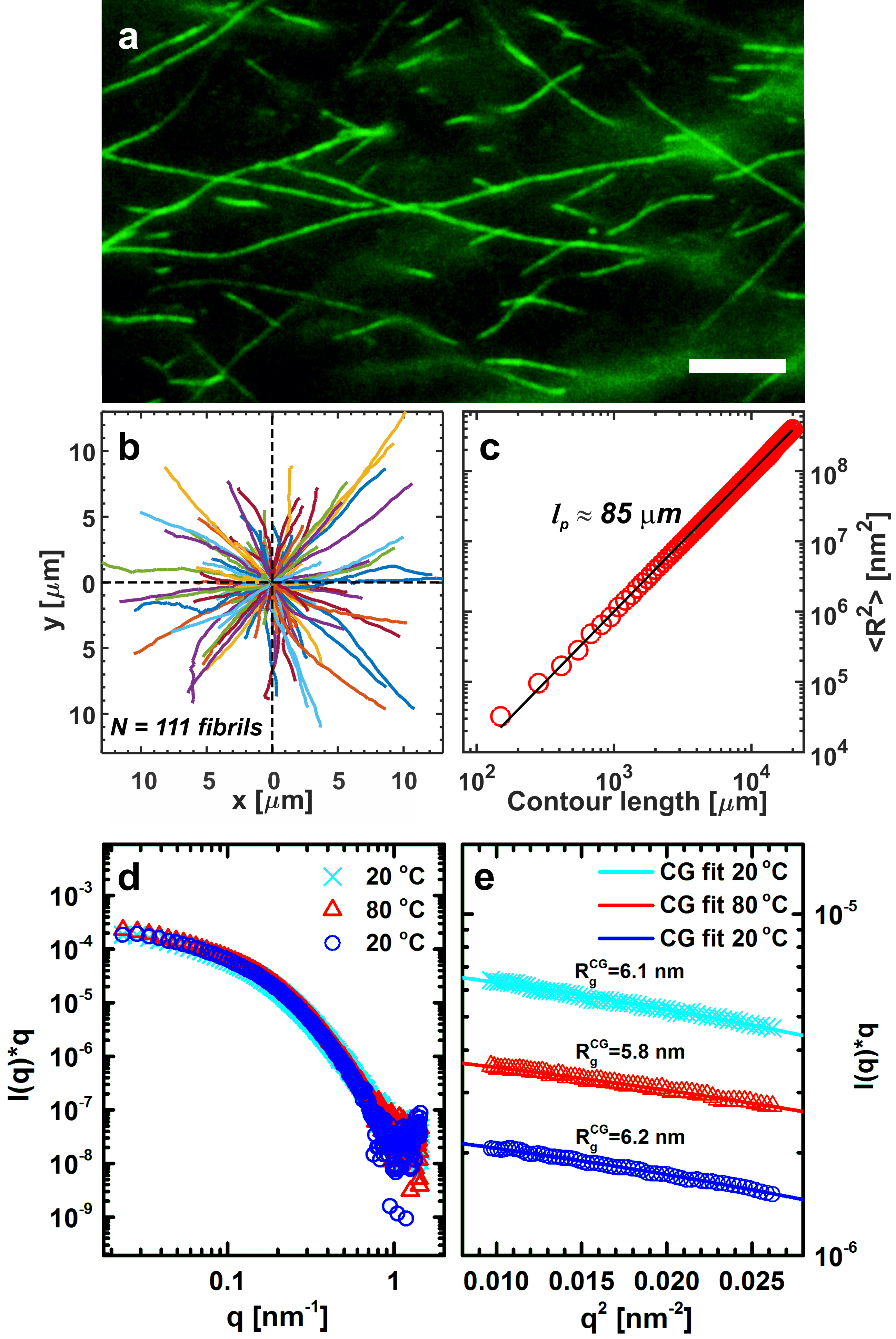
In the right settings, HIs can be made stronger by elevating the temperature of the system Chandler (2005). If HIs are indeed responsible for inter-fibril interactions in S amyloid networks, temperature should have a pronounced effect on the viscoelastic response of the network to applied deformations. Indeed, an S network significantly stiffens in the temperature range from 15 oC to 85 oC, which expresses itself in an order of magnitude increase of the storage modulus (Fig. 1c). This temperature-induced network stiffening is reversible. The value of tightly follows the changes in the temperature even if the network is repeatedly subjected to temperature cycles with different amplitudes (Fig. S3). This behavior is typically not observed in networks of semi-flexible polymers, yet does superficially resemble the elastic behavior of rubbers. In the latter, the free energy cost of stretching out the stored contour length of the crosslinked polymers becomes much larger at elevated temperatures, an effect associated with them being flexible rather than semi-flexible. However, S fibrils formed at the conditions used to prepare the amyloid gels appear to be very stiff as is evidenced by TIRF microscopy images (Fig. S4a). An end-to-end distance versus contour length analysis of the fibrils yields an estimate for the persistence length of 85 m, which is very much larger than the mesh size expected for a 300 M S fibril network (Fig. S4b and S4c). It is therefore unlikely that the increased free energy cost of reducing the conformational freedom of an individual fibril contributes significantly to the observed increase in with increasing temperature.
A huge experimental and theoretical research effort has been invested in the past few decades to better understand the viscoelastic properties of networks comprised of semi-flexible polymers Broedersz and MacKintosh (2014). These efforts have resulted in theoretical frameworks that describe scaling relations between quantities characterizing the properties of these networks, and which have been successfully applied to a wide range of biological and synthetic materials, all falling in the general class of semi-flexible polymer networks Gardel et al. (2004); Huisman et al. (2011); Kouwer et al. (2013). In view of the featureless frequency sweeps and the creep recovery experiments, the S fibril network seems to behave as a crosslinked network (Fig. 1a). With this in mind, we have adopted the scaling theory for cross-linked semi-flexible networks in order to interpret the temperature behavior of the S amyloid network. Because of the relatively large persistence length of S fibrils compared to the mesh size, we invoke a scaling theory based on the so-called floppy modes model, assuming a constant strain Heussinger and Frey (2006). According to this model, we have:
| (1) |
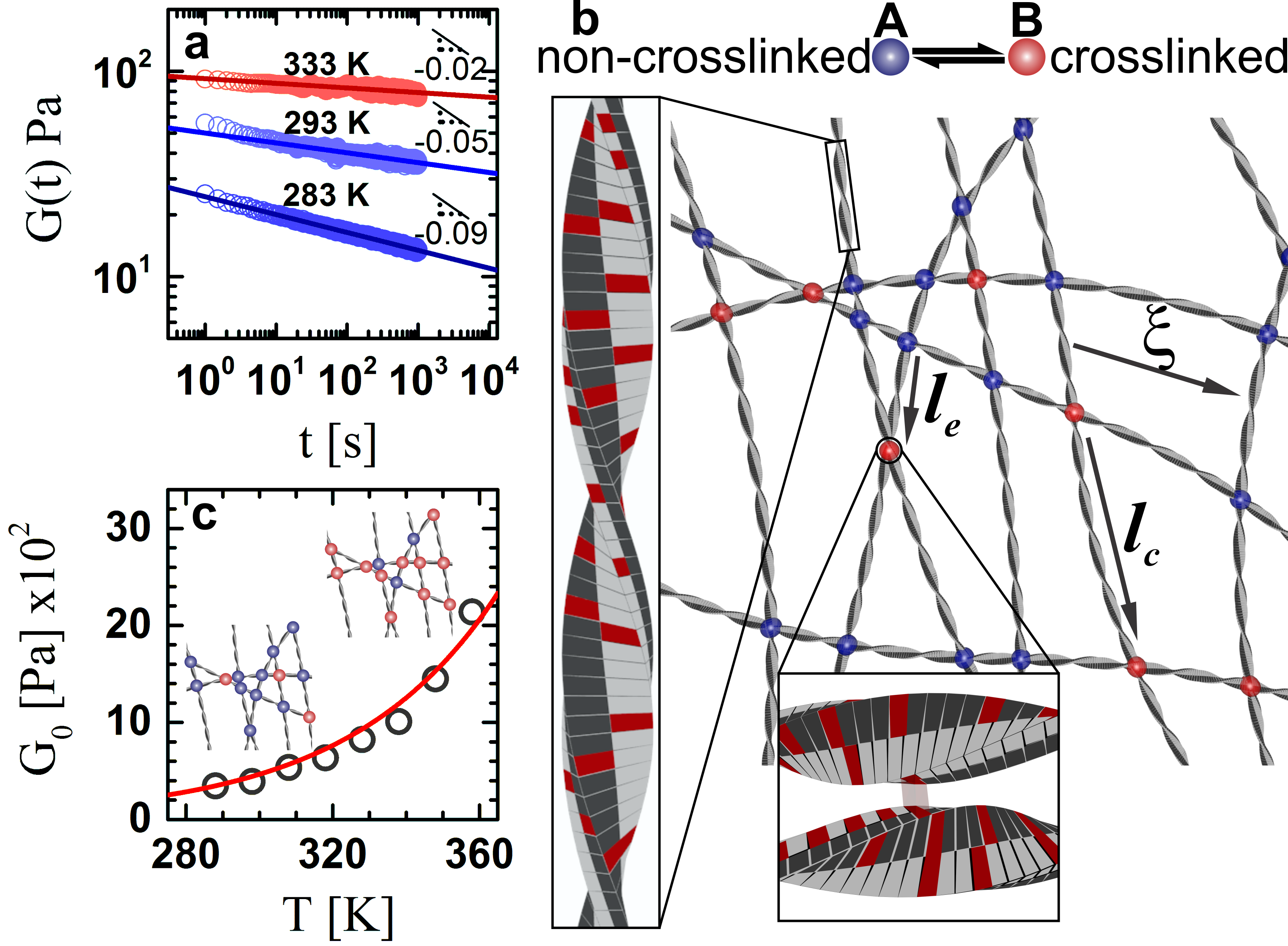
where is the plateau modulus of the network, is the bending stiffness of the semi-flexible chains, the average mesh size and the average distance between crosslinks Lieleg and Bausch (2007) (see also Fig. 2b). Note that the persistence length and bending stiffness are related according to . The strong dependence of the plateau modulus on the number of crosslinks (through ) is apparent. However, before focusing on this particular quantity, the possible contribution of the other two relevant parameters to the observed thermal stiffening in S networks, namely and , needs to be considered. Large temperature-induced stiffening of semi-flexible polymers has been observed in some synthetic systems. Driven by the enhanced hydrophobic interactions at higher temperatures, synthetic polymers may bundle into filaments with more than an order of magnitude larger rigidity Kouwer et al. (2013). The enhancement of can in that case be large enough to overcome the effect of the increased mesh size, which is expected to soften the network (eq. 1) and to produce an overall increase in the plateau modulus Tharmann et al. (2006). Even though hydrophobic interactions also seem to play an important role at the intra- and inter-fibril level in S networks, bundling is an unlikely mechanism to account for the experimental observations at higher temperatures. SAXS measurements do not provide any evidence for significant structural changes in the S network at these higher temperatures. The size of the fibril’s cross-section remains close to constant throughout the temperature ramps (Fig. S4d and S4e). Moreover, the SAXS curves remain identical at the different temperatures indicating that there are no sizable changes in the overall structure of the network and consequently in the mesh size .
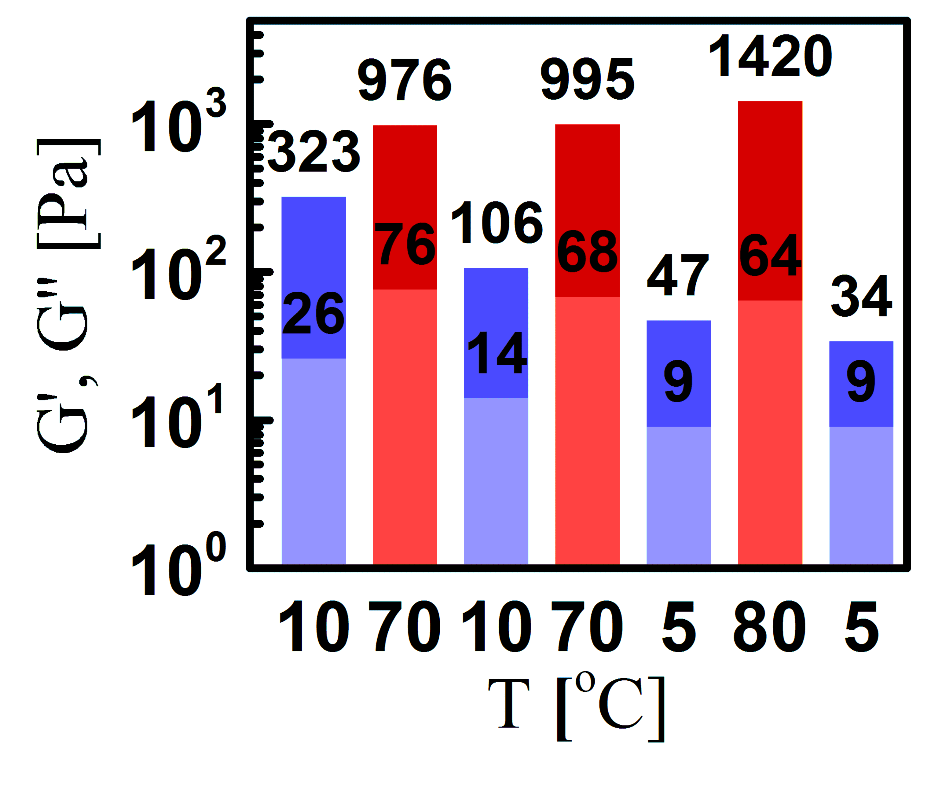
If bundling does not take place, then the strong temperature dependence of might result from a drastic change in the bending rigidity of the individual fibrils themselves with temperature. Since (eq. 1), the observed change in between 5 - 80 oC would imply a 13-fold increase over that temperature range. This estimate is based solely on changes in without taking into account the changes in the so-called entanglement length . If the sensitivity of on the temperature is also taken into account, using the known scaling relations and assuming crosslinks can only appear at entanglement points , then the increase of with temperature would need to be even larger (eq. 1) Isambert and Maggs (1996). The temperature dependence of is however generally very moderate for semi-flexible biopolymers. Additionally the sign of the change strongly depends on the biopolymer species. While, depending on GC content and salt concentration, double-stranded DNA seems to exhibit a 15-20 reduction in with increasing temperature by 30-45 oC, single-stranded DNA shows a 15 increase of over a comparable temperature range de Lorenzo et al. (2015); Geggier et al. (2011); Driessen et al. (2014). A drastic change in the mechanical properties of individual S fibrils also seems highly unlikely. As mentioned earlier, the protein monomers in the cross -sheets fibril backbone are held together by numerous intermolecular hydrogen bonds, which are the main determinant for the fibril stiffness Fitzpatrick et al. (2015). Driven by inter- and intra-molecular HIs, S monomers aggregate and attain a fold with optimized internalization of apolar residues in the fibril’s core. Keeping this in mind, higher temperatures stimulate two counteracting effects: on one hand, the breaking of hydrogen bonds should reduce the rigidity of the fibrils and, on the other, the enhancement of the hydrophobic interaction could increase the stiffness. However, it is unlikely that the strength of inter-monomer HIs increases sufficiently to compensate for the loss of hydrogen bonds, and produce the dramatic net increase in that would account for the unusual increase in the of the network. Indeed, other molecular assemblies that are held together by hydrophobic interactions also do not show signs of unusual stiffening, induced by an increase in temperature in the range comparable to the one used in this study. Lipid bilayers, for example, become easier to deform with increasing temperature Niggemann et al. (1995); Pan et al. (2008). The filamentous fd virus exhibits a non-monotonic change in the persistence length with temperature: at higher temperature, decreases, while an increase is found at the lower temperature range Tang et al. (1996). This variation in is however small, amounting to no more than 30.
With changes in being an unlikely cause for the large temperature-induced increment of , the only parameter left that could potentially account for this phenomenon is the mean distance between crosslinks . Heating up the system seems to strengthen the hydrophobic contacts between the fibrils, which ultimately results in a more densely crosslinked network. Results from stress-relaxation measurements on an S network at different temperatures are in line with this hypothesis. Instead of speeding up the relaxation processes, heating up the sample actually slows down the relaxation dynamics (Fig. 2a), probably due to the enhanced inter-fibrillar contacts (Fig. 2b).
To test the hypothesis that heating the amyloid network strengthens hydrophobic contacts between fibrils, we establish a quantitative relation between temperature and the storage modulus. For this purpose, the impact of temperature on the effective number of crosslinks in the system is evaluated at the level of a Boltzmann equilibrium and incorporated in the scaling relations for crosslinked semi-flexible networks (for the derivation see the Supplemental Material). This results in the following relation:
| (2) |
where is the temperature-dependent plateau modulus, and the plateau modulus at the reference temperature . The value of strongly depends on the architecture of S fibrils. Since there is no established model for this architecture, is left as a free parameter. The reference temperature , which is the lowest temperature at which the storage modulus was measured, is used to fit equation 2 to the experimental data. Equation 2 seems to describe the experimental observations very well indeed (Fig. 2c). The fit yields an endothermic value for . From the obtained value for we can estimate the apparent size of the hydrophobic patches using the expression for the enthalpy at the reference state of hydrophobic contacts: where is the energy cost per unit area of exposed hydrophobic surface and is the area Phillips et al. (2009). Taking into account that typically , the estimate for the size of the hydrophobic patches on the fibril surface is which is comparable to what has been found previously for virus coat proteins Kegel and van der Schoot (2004); Phillips et al. (2009); Kraft et al. (2012).
III Conclusion
In summary, S amyloid networks exhibit remarkable thermo-responsive properties. The fibrillar gel significantly stiffens at higher temperatures and completely recovers its original state once the temperature is lowered again. We propose that the thermo-stiffening of the S network is the consequence of enhanced inter-fibrillar hydrophobic contacts stimulated by the higher temperature. This is consistent with previously established qualitative findings, suggesting that the hydrophobic effect plays an essential role in the interaction between S fibrils Semerdzhiev et al. (2014). The presence of hydrophobic interactions between fibrils suggest that multiple hydrophobic domains in the fibril core remain solvent exposed. At higher temperatures these hydrophobic areas become “activated”, which effectively increases the number of contacts points between fibrils. An alternative explanation in which the exposure of the hydrophobic domains itself is a temperature-induced phenomenon could also be considered. At higher temperature segments of the S fibrils may unfold and reveal the hydrophobic stretches of the protein sequence to the solvent, which later become anchoring points between fibrils. Such a hypothetical scenario would be consistent with previous research, suggesting that at elevated temperatures the cross-beta sheet structure of the fibrils starts to fall apart Ikenoue et al. (2014). However, the experimental findings reported here do not give any clues supporting this interpretation. Numerous unfolding events in fibrils should have become apparent in the SAXS cross-sectional Guinier analysis, as it changes the effective cross-section of the fibril. We do not observe this in our SAXS data. Moreover, compromising the structural integrity of the fibrils should also have altered the mechanical response of the network towards softening rather than towards stiffening.Elucidating the cohesive forces between amyloid fibrils is crucial for obtaining a better understanding of the associated pathology and the physiological role of such structures. A correlation between the exposure of hydrophobic surface in amyloid aggregates and their toxicity has been suggested by numerous studies Bemporad and Chiti (2012); Mannini et al. (2014); Olzscha et al. (2011). Considering the nanoscale organization of the S fibrils it is unlikely that all hydrophobic patches on the fibril surface are protected from contact with the aqueous environment by inter-fibril interactions. The exposure of these hydrophobic fibril patches to the cytosol may induce interactions with other proteins. The accumulation of additional proteins in amyloid deposits may therefore not be a result of preserved functional interactions but rather be an effect of HIs. The accumulation of amyloid fibrils might increase the total hydrophobic surface present in the cell and thereby interfere with its normal functioning.
Understanding the inter-fibril interactions is also imperative for the successful utilization and manipulation of amyloid materials. Our data indicate that there are opportunities to harness these interactions and tune the mechanical properties of amyloid materials. Moreover, these findings indicate that it should be possible to design amyloid fibrils or other supramolecular assemblies with engineered hydrophobic patches and synthesize materials with imprinted temperature responsiveness. An important question remains, however. Are these interactions generic for amyloids or just specific for S? Exposure of amyloid networks comprised of the disease-unrelated protein -lactoglobulin does not seem to provoke the same response, indicating that the degree of thermo-stiffening observed for S gels cannot be expected to hold for all amyloid materials Gosal et al. (2004).
IV Acknowledgements
This work was supported by the “Nederlandse Organisatie voor Wetenschappelijk Onderzoek” (NWO) through NWO-CW Veni grant (722.013.013) to S.L., NWO-CW TOP program (700.58.302) to V.S., NWO-CW VIDI grant (700.59.423) to M.M.A.E.C., and through and Nanonext NL theme 8A. The authors thank Kirsten van Leijenhorst-Groener and Nathalie Schilderink for the expression and purification of α- synuclein. SAXS experiments were performed at ESRF, BM26B (DUBBLE) experiment number 26-02-664. We thank Dr. G. Portale (Beamline Scientist) for his help performing SAXS experiments.
V Supplemental Material
V.1 I. Materials and methods
V.1.1 A. S gel preparation
Expression of the human wild type S was performed in E. coli B121 (DE3) using the pT7-7 based expression system. Details on the purification procedure for S are described elsewhere Rooijen (2009). Gels of S amyloid fibrils were prepared in quiescent conditions. Fibril growth was seeded by 5 mol. preformed S seeds at a total protein concentration of 300 M S in 10 mM Tris, pH 7.4 and 10 mM NaCl (Sigma). The first 6 days samples were incubated at 37 oC and after that the gels were stored to mature at room temperature. The gels for SAXS measurement were directly grown in quartz capillaries (Hilgenberg GmbH, L=80, OD=1.5, Wall=0.01 mm) using the procedure described above. Samples for the TIRFM imaging were stained with the amyloid specific fluorescent dye Thioflavin T (ThT) and sealed in custom made glass chambers.
V.1.2 B. S gel preparation
A solution of fibrils was prepared by incubating 100 M of S in 10 mM Tris (Sigma), 2 mM NaCl (Sigma), pH=7.4, 37 oC and shaking at 900 RPM. Once the aggregation was completed, the fibril solution was sonicated (Branson, Sonifier 250) on ice. The solution was sonicated at the lowest power for 5 seconds and left at rest for another 55 second. The cycle was repeated five times. Subsequently, the sonicated samples were tested for seeding efficiency by incubation with S monomers.
V.1.3 C. SAXS
Experiments were performed at the BM26 DUBBLE (Dutch-Belgian Beamline, ESRF, Grenoble, France). Two dimensional images were collected using Pilatus 1M photon counting detector. The sample to detector distance was 6.6 m. The wavelength for the incident x-ray was 0.1 nm-1 and beam cross-section with dimensions 2.5 mm x 4.5 mm. The energy of the x-rays was 12 eV. The attained range was 0.03−1.5 nm-1. The ATSAS 2.6.0 software package was used for post-acquisition processing and analysis of the SAXS data Petoukhov et al. (2012).
V.1.4 D. Rheology
Rheology measurements were performed on an Anton Paar MCR 301 rheometer using a plate-plate geometry, with a plate diameter of 25 mm. Gel samples were carefully collected from the storage tubes using truncated pipette tips to minimize shearing and network disruption. The gel was then carefully deposited on the rheometer’s stage. Measurements were conducted at gap size of 0.16 mm and the samples were covered with mineral oil to avoid evaporation during the temperature runs. For each temperature step the sample was first left to equilibrated until the storage modulus measured at 0.5 Hz, 0.5, attained a stationary value (usually within 1 hour). Subsequently, a frequency spectrum was recorded at 1. Stress relaxation test were conducted by first equilibrating the sample at a given temperature and then subjecting it to a strain 3 which is within the linear viscoelastic response region of the sample. Subsequently the time relaxation modulus was recorded.
V.1.5 E. Total Internal Reflection Fluorescence Microscopy (TIRFM)
The TIRFM imaging was performed using a Nikon Ti-E microscopy setup coupled with an Argon laser (35-IMA-040, Melles Griot, USA). Images were acquired with a CFI Apo TIRF 100x objective (Nikon, Japan) and iXon 3 DU-897 EMCCD camera (Andor, UK) using the 457 nm line of the laser for the excitation of the ThT dye. The used filter cubes contained a 455 nm excitation filter with 10 nm bandpass, a 458 nm long pass dichroic mirror and a 485 nm emission filter with 30 nm bandpas
V.1.6 F. Determination of residual monomer concentration
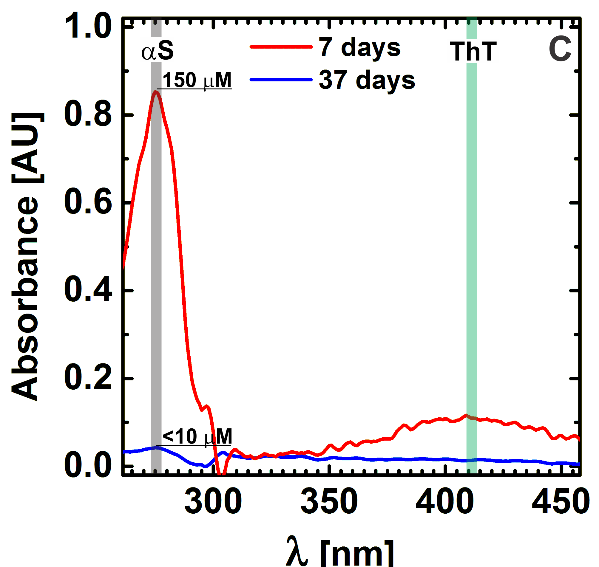
Determination of residual monomer concentration. Samples of the S amyloid gels were centrifuged using a Sorvall WX 80 ultracentrifuge (Thermo Scientific, USA) and Fiberlite F50L-24 x 1.5 fixed-angle rotor at 247 kG for 5 hours. Subsequently the concentration of free S monomers in the supernatant was determined using a UV-Vis spectrophotometer (UV 2401 PC, Shimadzu). Absorbance was measured at 276 nm and an extinction coefficient of 5600 M-1.cm-1 was used to calculate the protein concentration.
V.1.7 G. Persistence length analysis
Aliquots of a 300 M S gel, 10 mM NaCl and 10mM Tris were diluted in the same buffer keeping the ionic strength constant. Thioflavin T (ThT) was subsequently added to a final concentration of 1 M in order to stain the fibrils. A small volume of the diluted sample was sealed between a glass slide and a cover slip. The sample was left at rest for the fibrils to sediment on the cover slip and then imaged using Nikon Ti-E in TIRF mode (see section TIRFM). Images were analyzed using the MATLAB based package Easyworm Lamour et al. (2013). Briefly, the persistence length is determined via random resampling using bootstrap with replacement method. A bootstrap sample of n chains is randomly selected from the ensemble of all chains. For each bootstrap the average square of the end to end distance is binned at equal length intervals. Then all the data is fitted using the wormlike chain model:
| (1) |
where is the contour length.
V.1.8 H. Derivation of the equation linking hydrophobic interactions to the temperature induced stiffening of S fibril networks
Temperature seems to enhance the formation of physical crosslinks between fibrils in the S amyloid network. Identifying the impact of temperature on the effective number of crosslinks in the system, would enable us to incorporate the temperature effect in the scaling relations for the elastic response of crosslinked semi - flexible networks. To directly relate the enhanced hydrophobic interactions to the stiffening of the network, we introduce a two state model for the entanglements in the network , where and represent the ‘free’ and ‘crosslinked’ entanglements respectively (Fig 4B from the main article). Assuming that hydrophobic interactions drive the crosslinking of entanglements, the equilibrium dissociation constant can be written as Kegel and van der Schoot (2004); van der Schoot (2005):
| (2) |
where is the equilibrium dissociation constant at a reference temperature , and is the endothermic binding enthalpy at temperature for two hydrophobic surfaces coming into contact. Because , increases with as is typically observed for hydrophobic surfaces Kegel and van der Schoot (2004). We can calculate the fraction of entanglements that are in the crosslinked state by invoking Boltzmann statistics:
| (3) |
Since is presumably small, at low the hydrophobic interactions are known to be weak Dapoian et al. (1995) so is over the probed temperature range, we can approximate making use of a Taylor expansion:
| (4) |
In a semiflexible network, contacts between filaments appear at the entanglement points. Given that i) there are no significant structural rearrangements in the network during the crosslinking process, ii) the maximum number bonds equals the number of entanglement points, we conclude that the distance between crosslinks can be related to the entanglement length through the fraction :
| (5) |
Taking into account that , where represents the concentration of the filament forming protein, the entanglement length reads as Degennes et al. (1976):
| (6) |
where is the bending stiffness of the semi-flexible chains and the average mesh size. We know that for a network comprised of semi-flexible polymers that appear stiff between entanglement points, the storage modulus scales as (see eq. 1 and the discussion preceding it the main article):
| (7) |
where is the plateau modulus of the network. Substituting equations eq. S4 and eq. S6 in eq.S5, inserting the latter in eq. S7, and finally after some re-arrangements we arrive at:
| (8) |
V.2 II. Figures
VI References
References
- Chandler (2005) D. Chandler, Nature 437, 640 (2005).
- Dyson et al. (2006) H. J. Dyson, P. E. Wright, and H. A. Scheraga, Proceedings of the National Academy of Sciences of the United States of America 103, 13057 (2006).
- Sturtevant (1977) J. M. Sturtevant, Proceedings of the National Academy of Sciences of the United States of America 74, 2236 (1977).
- Buell et al. (2012) A. K. Buell, A. Dhulesia, D. A. White, T. P. J. Knowles, C. M. Dobson, and M. E. Welland, Angewandte Chemie-International Edition 51, 5247 (2012).
- Kegel and van der Schoot (2004) W. K. Kegel and P. van der Schoot, Biophysical Journal 86, 3905 (2004).
- Comellas-Aragones et al. (2007) M. Comellas-Aragones, H. Engelkamp, V. I. Claessen, N. A. J. M. Sommerdijk, A. E. Rowan, P. C. M. Christianen, J. C. Maan, B. J. M. Verduin, J. J. L. M. Cornelissen, and R. J. M. Nolte, Nature Nanotechnology 2, 635 (2007).
- Hernandez-Garcia et al. (2014) A. Hernandez-Garcia, D. J. Kraft, A. F. J. Janssen, P. H. H. Bomans, N. A. J. M. Sommerdijk, D. M. E. Thies-Weesie, M. E. Favretto, R. Brock, F. A. de Wolf, M. W. T. Werten, P. van der Schoot, M. C. Stuart, and R. de Vries, Nature Nanotechnology 9, 698 (2014).
- Semerdzhiev et al. (2014) S. A. Semerdzhiev, D. R. Dekker, V. Subramaniam, and M. M. Claessens, ACS Nano 8, 5543 (2014).
- Semerdzhiev et al. (2017) S. A. Semerdzhiev, V. V. Shvadchak, V. Subramaniam, and M. M. A. E. Claessens, Scientific Reports 7, 7699 (2017).
- Celej et al. (2008) M. S. Celej, E. A. Jares-Erijman, and T. M. Jovin, Biophysical Journal 94, 4867 (2008).
- Broedersz and MacKintosh (2014) C. P. Broedersz and F. C. MacKintosh, Reviews of Modern Physics 86, 995 (2014).
- Gardel et al. (2004) M. L. Gardel, J. H. Shin, F. C. MacKintosh, L. Mahadevan, P. Matsudaira, and D. A. Weitz, Science 304, 1301 (2004).
- Huisman et al. (2011) E. M. Huisman, Q. Wen, Y. H. Wang, K. Cruz, G. Kitenbergs, K. Erglis, A. Zeltins, A. Cebers, and P. A. Janmey, Soft Matter 7, 7257 (2011).
- Kouwer et al. (2013) P. H. J. Kouwer, M. Koepf, V. A. A. Le Sage, M. Jaspers, A. M. van Buul, Z. H. Eksteen-Akeroyd, T. Woltinge, E. Schwartz, H. J. Kitto, R. Hoogenboom, S. J. Picken, R. J. M. Nolte, E. Mendes, and A. E. Rowan, Nature 493, 651 (2013).
- Heussinger and Frey (2006) C. Heussinger and E. Frey, Physical Review Letters 97 (2006).
- Lieleg and Bausch (2007) O. Lieleg and A. R. Bausch, Physical Review Letters 99 (2007).
- Tharmann et al. (2006) R. Tharmann, M. M. A. E. Claessens, and A. R. Bausch, Biophysical Journal 90, 2622 (2006).
- Isambert and Maggs (1996) H. Isambert and A. C. Maggs, Macromolecules 29, 1036 (1996).
- de Lorenzo et al. (2015) S. de Lorenzo, M. Ribezzi-Crivellari, J. R. Arias-Gonzalez, S. B. Smith, and F. Ritort, Biophysical Journal 108, 2854 (2015).
- Geggier et al. (2011) S. Geggier, A. Kotlyar, and A. Vologodskii, Nucleic Acids Research 39, 1419 (2011).
- Driessen et al. (2014) R. P. C. Driessen, G. Sitters, N. Laurens, G. F. Moolenaar, G. J. L. Wuite, N. Goosen, and R. T. Dame, Biochemistry 53, 6430 (2014), pMID: 25291500, http://dx.doi.org/10.1021/bi500344j .
- Fitzpatrick et al. (2015) A. W. P. Fitzpatrick, G. M. Vanacore, and A. H. Zewail, Proceedings of the National Academy of Sciences of the United States of America 112, 3380 (2015).
- Niggemann et al. (1995) G. Niggemann, M. Kummrow, and W. Helfrich, Journal De Physique Ii 5, 413 (1995).
- Pan et al. (2008) J. Pan, S. Tristram-Nagle, N. Kucerka, and J. F. Nagle, Biophysical Journal 94, 117 (2008).
- Tang et al. (1996) J. X. Tang, S. Wong, P. T. Tran, and P. A. Janmey, Berichte der Bunsengesellschaft fГјr physikalische Chemie 100, 796 (1996).
- Phillips et al. (2009) R. Phillips, J. Kondev, and J. Theriot, Physical biology of the cell (Garland Science, New York, 2009) pp. xxiv, 807 p.
- Kraft et al. (2012) D. J. Kraft, W. K. Kegel, and P. van der Schoot, Biophysical Journal 102, 2845 (2012).
- Ikenoue et al. (2014) T. Ikenoue, Y. H. Lee, J. Kardos, M. Saiki, H. Yagi, Y. Kawata, and Y. Goto, Angewandte Chemie-International Edition 53, 7799 (2014).
- Bemporad and Chiti (2012) F. Bemporad and F. Chiti, Chemistry & Biology 19, 315 (2012).
- Mannini et al. (2014) B. Mannini, E. Mulvihill, C. Sgromo, R. Cascella, R. Khodarahmi, M. Ramazzotti, C. M. Dobson, C. Cecchi, and F. Chiti, ACS Chemical Biology 9, 2309 (2014).
- Olzscha et al. (2011) H. Olzscha, S. M. Schermann, A. C. Woerner, S. Pinkert, M. H. Hecht, G. G. Tartaglia, M. Vendruscolo, M. Hayer-Hartl, F. U. Hartl, and R. M. Vabulas, Cell 144, 67 (2011).
- Gosal et al. (2004) W. S. Gosal, A. H. Clark, and S. B. Ross-Murphy, Biomacromolecules 5, 2408 (2004).
- Rooijen (2009) B. D. v. Rooijen, Structural and functional insights into interactions of oligomeric alpha-synuclein with lipid membranes, Phd thesis, University of Twente, Enschede, The Netherlands (2009).
- Petoukhov et al. (2012) M. V. Petoukhov, D. Franke, A. V. Shkumatov, G. Tria, A. G. Kikhney, M. Gajda, C. Gorba, H. D. T. Mertens, P. V. Konarev, and D. I. Svergun, Journal of Applied Crystallography 45, 342 (2012).
- Lamour et al. (2013) G. Lamour, H. B. Li, and J. Gsponer, Biophysical Journal 104, 393a (2013).
- van der Schoot (2005) P. van der Schoot, “Theory of supramolecular polymerization,” in Supramolecular polymers, edited by A. Ciferri (Taylor & Francis, Boca Raton, 2005) pp. xiii, 761 p., 2nd ed.
- Dapoian et al. (1995) A. T. Dapoian, A. C. Oliveira, and J. L. Silva, Biochemistry 34, 2672 (1995).
- Degennes et al. (1976) P. G. Degennes, P. Pincus, R. M. Velasco, and F. Brochard, Journal De Physique 37, 1461 (1976).