Vibrational Energy Transfer from Photo-Excited Carbon Nanotubes to Proteins Observed by Coherent Phonon Spectroscopy
Abstract
Vibrational energy transfer from photo-excited single-wall carbon nanotubes (SWCNTs) to coupled proteins is a key to engineer thermally induced biological reactions such as photothermal therapy. Here, we explored vibrational energy transfer from the photo-excited SWCNTs to different adsorbed biological materials by means of a femtosecond pump-probe technique. We show that the vibrational relaxation time of the radial breathing modes (RBMs) in SWCNTs significantly depends on the structure of coupled materials, i.e. proteins or biopolymers, indicating the vibrational energy transfer is governed by overlap of phonon density of states between the SWCNTs and coupled materials.
The energy transfer from mechanical modes to organic layers or specimen has been examined extensively, because of broad interest in efficient energy conversion problems.Losego12 ; Hettich16 In particular, one-dimensional and two-dimensional structures made from carbon atoms have shown great advantages on their vivid applications in fundamental systems, such as biological and semiconductor devices,Modi03 ; Wang15 because of their extremely high carrier mobility, high thermal conductivity, and rigid structure.Zhang08 Carbon nanotubes (CNTs) are promising materials for fabricating nano-machines capable for medical treatments,Kong00 such as photothermal therapy. To realize this potential application, it is highly important to understand protein behaviors on CNTs. Although extensive efforts have been made to understand and control the attraction between CNTs and proteins, only limited knowledge has been obtained on their physical and chemical properties under static conditions.Horn12
Single-wall CNTs (SWCNTs) are one of the most popular and fundamental carbon materials, and they have metallic or semiconducting properties,Saito98 depending on the nanotube symmetry and diameter, which are specified by chiral vector.Rao97 Frequency-domain spectroscopy revealed diameter-selective Raman scattering from the radial breathing modes (RBMs) in SWCNTs,Jorio01 ; Dresselhaus05 where the frequency of the RBMs depends on the inverse of the diameter. Moreover, Hertel et al. investigated the effects of environment on the optical properties of SWCNTs. They found that interactions between the SWCNTs and their environment depended on their diameter and exciton energy.Hertel00 Very recently, reduction in antibacterial activity of protein with the help of SWCNTs has been demonstrated.Horn12 In addition, hen egg-white lysozyme (LYZ) has been demonstrated to disperse SWCNTs, depending on the pH values.Strano03 However, little is known about the vibrational energy transfer between SWCNTs and proteins, like LYZ, particularly at femtosecond to picosecond time scales, in which phonon-phonon coupling may play a significant role. Understanding and controlling the vibrational energy transfer between SWCNTs and proteins (or biopolymers) will be highly relevant to engineering thermally induced biological reactions.
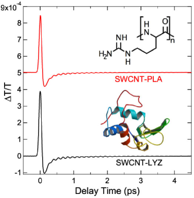
In this paper, coherent vibrational motion of the RBMs in SWCNTs coupled with proteins or biopolymers has been precisely measured in the femtosecond time scales. We show that relaxation dynamics of the coherent RBM depend on coupled species, from which it is possible to gain new insights into the vibrational energy transfer from photo-excited SWCNTs to coupled species. Here, we selected two kinds of cosolute, i.e. LYZ and poly-L-arginine (PLA), as materials interacting with SWCNTs to investigate their structural effects. Both of them have ability of dispersing SWCNTs, though these solutes have different structures from each other. LYZ consists of 129 amino acid residues (molecular weight: 14,307) with a basic isoelectric point (pI = 11). The tertiary structure of LYZ in aqueous solution is highly stable with secondary structures including -helix and -sheet (Fig 1 inset). On the other hand, PLA (molecule weight: 70,000) is made by polymerizing basic amino acid, arginine (pKa = 12.5) (Fig. 1 inset). As mentioned below, we could observe different dynamics in the coherent RBM phonon between these biomaterials, which is argued to be originated from their structures and thereby phonon density of states (PDOS).
The samples of SWCNTs were produced via the high-pressure catalytic CO decomposition process.Chiang01 Hen egg-white LYZ and poly-L-arginine (PLA) were obtained from Sigma-Aldrich. The protein solution was prepared as described previously.Hirano14 Briefly, a solution containing LYZ (0.2 mg/mL) or PLA (0.2 mg/mL) was mixed with the SWCNT powder. Then the SWCNT solution was dispersed by ultrasonication for 60 minutes at 18∘C using ultrasonic processor. Finally, highly dispersed SWCNT solution was obtained from supernatant of the sample solution after ultracentrifugation (210,000 g for 30 min at 25∘C) using an ultracentrifuge. On the other hand, the bundled sample was also obtained from supernatant of the dispersed solution after gentle centrifugation (16,100 g for 1 minute at 25∘C).
The transient transmission () of the samples, i.e. SWCNT dispersed by LYZ (SWCNT-LYZ) or by PLA (SWCNT-PLA), was measured in a 5-mm-thick quartz cell by employing the fast scanning delay method.Hase03 The pump and probe pulses (20 fs pulse duration; 830 nm wavelength; 80 MHz repetition rate) were focused to a 70-m-diameter spot in the sample with a mutual angle of 10∘. The pump pulse with a constant average power (40 mW) generated a carrier density of 4 1019 cm-3; the probe power was less than 2 mW. Under the constant pump power and the focused spot size, the pump fluence should be constant through the measurements, and therefore the effect of excitation density on the vibrational relaxation dynamics, i.e. carrier-density-dependent carrier-phonon scattering, was negligible in the present study. Polarization of the two beams was linear with a mutual angle of 55∘ (magic angle).Makino09
The signals were recorded as a function of the pump-probe time delay as shown in Fig. 1. The transient electronic response is observed just at the zero time delay, which is followed by the coherent phonon oscillations due to the RBMs, whose frequency () inversely depends on the diameter of SWCNTs (),Makino09
| (1) |
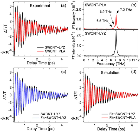
The transient electronic response, appearing just at the zero time delay, corresponds to the coherent arrangement of the SWCNTs molecules induced by the electric field of the femtosecond laser pulse, and is governed by the third-order nonlinear susceptibility [ref. Shen84 ]. To subtract the transient electronic response, we fitted the time domain data (Fig. 1) with a double-exponential-decay function. Subsequently, we subtracted the double-exponential-decay function from the time-domain data. Thus, we obtained only oscillatory parts of the time-domain signals (Fig. 2). Comparing the signal obtained in the different systems, i.e., SWCNT-LYZ and SWCNT-PLA, the coherent phonon spectra are indeed different from each other, indicating the interaction between SWCNTs and biomaterials depends on their structures [Fig. 2(a)]. By using the Kataura’s plot, it is possible to derive the electronic states of SWCNTs and hence to identify whether they are semiconducting or metallic. In our samples, the diameter of SWCNTs has a range of 0.9-1.3 nm. Considering the photon energy (830 nm, i.e., 1.49 eV) and the broad bandwidth (50 nm in FWHM) of the laser used, the excitonic transitions around the critical point of S22 (the second-order semiconducting optical transition) are available at 800-900 nm [ref. Kataura99 ]. We can thus conclude that the semiconducting SWCNTs are resonantly excited through the impulsive stimulated Raman scattering.Lim06 ; Gambetta06 Actually, the peaks at 6.5, 6.9, and 7.2 THz observed in the Fourier transformed (FT) spectra [Fig. 2(b)] are assigned to the 1.14-, 1.08-, and 1.03-nm-diameter tubes, respectively, based on eq. (1). Note that chirality (n, m) of these modes can be (9, 7), (8, 7), and (12, 1) for the 1.14-, 1.08-, and 1.03-nm-diameter tubes, respectively.Goupalov06 The time-domain data were fit to a linear combination of damped harmonic oscillators,
| (2) |
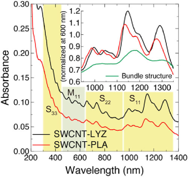
where (, , ), (, , ), (, , ), and (, , ) are the amplitude, the relaxation time, the frequency, and the initial phase of the three coherent RBMs of the first ( = 6.5 THz), the second ( = 6.9 THz), and the third ( = 7.2 THz) modes, respectively. The time-domain data are well fit to eq. (Vibrational Energy Transfer from Photo-Excited Carbon Nanotubes to Proteins Observed by Coherent Phonon Spectroscopy) as shown in Fig. 2(c), and the fitting parameters obtained are listed in Table 1. By comparing the amplitude of the dominant RBM (7.2 THz) for two biomaterials, we found that the initial vibrational amplitude for the SWCNT-LYZ system is 1.6 times larger than that for the SWCNT-PLA system (see Table 1 and Fig. 2(d)), which is dominantly ascribable to the difference in the concentration of SWCNTs between these samplesTanaka09 ; note that the absorbance of the SWCNT-LYZ at a 810 nm peak after the background subtraction was 2.1-fold higher than that of the SWCNT-PLA (Fig. 3).
Here, we focus on the strongest RBM at 7.2 THz by extracting the vibrational energy transfer dynamics from the fitting results. Importantly, the vibrational relaxation time for the SWCNT-LYZ (ca. 1.41 ps) at 7.2 THz was shorter than that for the SWCNT-PLA (ca. 1.66 ps), suggesting that the vibrational energy transfer from the photo-excited SWCNT to LYZ is faster than that from SWCNT to PLA for the 7.2 THz mode. These vibrational relaxation time constants are significantly shorter than those obtained for the SWCNTs dispersed by sodium dodecyl sulfate (SWCNT-SDS) (ca. 2.0 ps), which was observed by the same pump-probe technique,Makino09 indicating that the vibrational relaxation time of the coherent RBM in the present complex systems was indeed changed by the adsorbed biomaterials. Although decay of the coherent RBM is generally accounted for by anharmonic phonon-phonon energy relaxation into underlying acoustic phonons,Makino09 this decay path depends only on the temperature, so that the change in the relaxation time observed in Fig. 2 at the constant room temperature cannot be accounted for by the anharmonic phonon-phonon energy relaxation. Note that the total relaxation rate 1/ is the sum of the new path and the existing path (anharmonic phonon-phonon energy relaxation), 1/ = 1/+1/, where we will discuss the origin of the new path () in details below.
It should be also noted that LYZ is adsorbed onto the SWCNTs through hydrophobic and interactionsCalvaresi12 whereas PLA is adsorbed onto the SWCNTs through hydrophobic and van der Waals interactions and partly through interaction between the guanidium group of arginine and the SWCNT surfaces.Hirano14 Because of the different adsorption mechanisms, the coverage of the SWCNTs will not be the same for the two types of cosolute. The difference in the coverage of the SWCNTs can be reflected in difference in carrier doping of SWCNTs.Hirano16 However, in terms of the carrier doping, the differences in the coverage will not affect the vibrational relaxation time of the SWCNTs, since we have already demonstrated that the carrier doping did not affect the relaxation time of the RBMs.Makino09
Since SWCNTs dispersed in aqueous solution generally exist as debundled or bundled forms,Hirano14 change in the relaxation time might be associated with the intertube interactions. In fact, the bundling effects on the phonon relaxation have been examined using a SWCNT film and SWCNT solution for the -modeLee10 as well as for the RBM,Honda13 indicating that the bundling makes phonon relaxation time shorter than that of individually dispersed SWCNTs. However, our samples used here were individually dispersed since they were collected from the supernatants after the ultracentrifugation. To test this expectation, we measured the absorption spectra of the SWCNTs dispersed solutions as well as the bundled form (Fig. 3). Although the absorption intensity of the S11 band was found to be slightly smaller and broader for the SWCNT-PLA than that for the SWCNT-LYZ, the intensity of the S11 band was explicitly stronger than that of the bundled form (see the inset of Fig. 3).Oconnell02 These results indicate that both the SWCNT-LYZ and SWCNT-PLA were well dispersed. Thus, the decay-time shortening of the RBMs seen with LYZ can be explained mainly by the energy transfer from the SWCNTs to LYZ and not by the bundling effect.
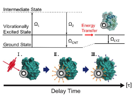
To further discuss the difference in the vibrational energy transfer observed in Fig. 2, the phonon-phonon coupling between the SWCNTs and biopolymers should be taken into account.Leitner01 ; Hase02 Recently Li et al. observed anisotropic energy flow from proteins to ligands on albumin molecules using ultrafast infrared vibrational spectroscopy.Magana14 The efficient vibrational energy transfer was thought to occur through the connecting helix structures. In SWCNT-protein systems, similarly, favorable coupling would occur between -helix of proteins and SWCNT.Hu16 Therefore, it is expected that the vibrational energy transfer from SWCNTs to coupled LYZ might be more efficient than that from the SWCNT to PLA, since only LYZ has -helix structure. We thus examine the vibrational energy transfer from SWCNTs to coupled biomaterials in terms of phonon-phonon coupling based on the PDOS.Leitner01 The main peak in the PDOS at 240 cm-1 (= 7.2 THz) for LYZSvanidze07 is well overlapped with the energy of the RBM (7.2 THz), while the overlap of the main peak (250 cm-1 = 7.5 THz) in the PDOS for PLA with the RBM (7.2 THz) is poor.Sharma02 Therefore, there can exist resonant coupling between the RBM (7.2 THz) in the SWCNTs and the optical phonons in LYZ through -helix of proteins, leading to efficient vibrational energy transfer. A possible scenario for the vibrational relaxation dynamics in the SWCNT-LYZ complex at 7.2 THz is schematically presented in Fig. 4, which exhibits that the vibrational amplitude of the SWCNT readily decays by the vibrational energy transfer to LYZ.
To further support our interpretation, we then focused on the vibrational relaxation time of the 6.9 THz mode. As shown in Table 1, the relaxation time for SWCNT-PLA (ca. 2.00 ps) was significantly shorter than that for SWCNT-LYZ (ca. 3.76 ps) , both of which are shorter than that of the 6.9 THz RBM in SWCNT-SDS (5.50 0.17 ps)(data not shown). This result suggests that the vibrational energy transfer from the photo-excited SWCNT to PLA is faster than that from SWCNT to LYZ, which is concordant with the fact that the peak in the PDOS for PLA (230 cm-1 = 6.9 THz) well overlaps with the energy of the RBM (6.9 THz).Sharma02 Importantly, the difference in the relaxation time of the 7.2 THz mode between LYZ and PLA is very small (i.e. 1.41 and 1.66 ps, respectively), while that of the 6.9 THz mode is relatively large (i.e. 3.76 and 2.00 ps, respectively). A plausible reason for this lies in the natural linewidth of the PDOS inside a protein or biopolymer; specifically, the linewidth of the 240 cm-1 (7.2 THz) mode for LYZ ( 10 cm-1)Svanidze07 is much broader than that of the 230 cm-1 (6.9 THz) mode for PLA ( 10 cm-1).Sharma02 . Therefore, the resonant energy transfer will be more pronounced for PLA than for LYZ at the respective peak position in PDOS. This could explain why the difference in the relaxation time is larger in the case of the 6.9 THz mode. According to the above results and discussions, we concluded that the rate of the phonon energy flow significantly depends on the PDOS of the biomaterials.
| Sample | Mode | (THz) | 106 | (ps) | (deg.) |
|---|---|---|---|---|---|
| SWCNT | 1st | 6.5 | 0.59 0.05 | 2.16 0.20 | -109.1 2.5 |
| -LYZ | 2nd | 6.9 | 0.39 0.04 | 3.76 0.50 | 74.8 3.2 |
| 3rd | 7.2 | 6.38 0.06 | 1.41 0.01 | 23.4 0.3 | |
| SWCNT | 1st | 6.5 | 0.54 0.06 | 1.94 0.22 | -130.4 3.4 |
| -PLA | 2nd | 6.9 | 0.78 0.08 | 2.00 0.17 | 58.6 2.9 |
| 3rd | 7.2 | 4.03 0.06 | 1.66 0.03 | 18.5 0.4 |
In summary, we explored the vibrational energy transfer between the two different biomaterials and SWCNTs by means of the femtosecond transient transmission technique. The vibrational relaxation time of the RBMs dramatically changes, depending on the PDOS of the coupled biomaterials. Our findings are particularly useful for designing a highly efficient phonon energy flow system from photo-excited SWCNT to biomaterials, and such vibrational energy transfer can be controlled by the PDOS originated from the structure of coupled biomaterials.
The authors thank S. Hasegawa and H. Tadokoro for the assistance at the early stage of the experiments. This work was supported in part by KAKENHI-23104502, from MEXT, Japan.
References
- (1) M. D. Losego, M. E. Grady, N. R. Sottos, D. G. Cahill, and P. V. Braun, Nat. Mater. 11, 502 (2012).
- (2) M. Hettich, K. Jacob, O. Ristow, M. Schubert, A. Bruchhausen, V. Gusev, and T. Dekorsy, Sci. Rep. 6, 33471 (2016).
- (3) A. Modi, N. Koratkar, E. Lass, B. Wei, and P. M. Ajayan, Nature 424, 171 (2003).
- (4) F. J. Wang, D. Kozawa, Y. Miyauchi, K. Hiraoka, S. Mouri, Y. Ohno, and K. Matsuda, Nat. Commun. 6, 6305 (2015).
- (5) M. Zhang, T. Murakami, K. Ajima, K. Tsuchida, A. S. Sandanayaka, O. Ito, S. Iijima, and M. Yudasaka, PNAS 105, 14773 (2008).
- (6) J. Kong, N. R. Franklin, C. Zhou, M. G. Chapline, S. Peng, K. Cho, and H. Dai, Science 287, 622 (2000).
- (7) D. W. Horn, K. Tracy, C. J. Easley, and V. A. Davis, J. Phys. Chem. C 116, 10341 (2012).
- (8) R. Saito, G. Dresselhaus, and M. S. Dresselhaus: Physical Properties of Carbon Nanotubes (Imperial College Press, London, 1998).
- (9) A. M. Rao, E. Richter, S. Bandow, B. Chase, P. C. Eklund, K. A. Williams, S. Fang, K. R. Subbaswamy, M. Menon, A. Thess, R. E. Smalley, G. Dresselhaus, and M. S. Dresselhaus, Science 275, 187 (1997).
- (10) A. Jorio, R. Saito, J. H. Hafner, C. M. Lieber, M. Hunter, T. McClure, G. Dresselhaus, and M. S. Dresselhaus, Phys. Rev. Lett. 86, 1118 (2001).
- (11) M. S. Dresselhaus, G. Dresselhaus, R. Saito, and A. Jorio, Phys. Rep. 409, 47 (2005).
- (12) T. Hertel and G. Moos, Phys. Rev. Lett. 84, 5002 (2000).
- (13) M. S. Strano, C. B. Huffman, V. C. Moore, M. J. O’Connell, E. H. Haroz, J. Hubbard, M. Miller, K. Rialon, C. Kittrell, S. Ramesh, R. H. Hauge, and R. E. Smalley, J. Phys. Chem. B 107, 6979 (2003).
- (14) I. W. Chiang, B. E. Brinson, A. Y. Huang, P. A. Willis, M. J. Bronikowski, J. L. Margrave, R. E. Smalley, and R. H. Hauge, J. Phys. Chem. B 105, 8297 (2001).
- (15) A. Hirano, T. Tanaka, H. Kataura, and T. Kameda, Chem. Eur. J. 20, 4922 (2014).
- (16) M. Hase, M. Kitajima, A. M. Constantinescu, and H. Petek, Nature 426, 51 (2003).
- (17) K. Makino, A. Hirano, K. Shiraki, Y. Maeda, and M. Hase, Phys. Rev. B 80, 245428 (2009).
- (18) Y. -R. Shen, The Principle of Nonlinear Optics (John Wiley & Sons, New York, 1984).
- (19) H. Kataura, Y. Kumazawa, Y. Maniwa, I. Umezu, S. Suzuki, Y. Ohtsuka, and Y. Achiba, Synthetic Metals 103, 2555 (1999).
- (20) Y. -S. Lim, K. -J. Yee, J. -H. Kim, E. H. Hároz, J. Shaver, J. Kono, S. K. Doorn, R. H. Hauge, and R. E. Smalley, Nano Lett. 6, 2696 (2006).
- (21) A. Gambetta, C. Manzoni, E. Menna, M. Meneghetti, G. Cerullo, G. Lanzani, S. Tretiak, A. Piryatinski, A. Saxena, R. L. Martin, and A. R. Bishop, Nat. Phys. 2, 515 (2006).
- (22) S. V. Goupalov, B. C. Satishkumar, and S. K. Doorn, Phys. Rev. B 73, 115401 (2006).
- (23) T. Tanaka, Y. Urabe, D. Nishide, and H. Kataura, Appl. Phys. Exp. 2, 125002 (2009).
- (24) M. Calvaresi, S. Hoefinger, and F. Zerbetto, Chem. Eur. J. 18, 4308 (2012).
- (25) A. Hirano, T. Kameda, Y. Yomogida, M. Wada, T. Tanaka, and H. Kataura, ChemNanoMat 2, 911 (2016).
- (26) Y. J. Lee, S. H. Parekh, J. A. Fagan, and M. T. Cicerone, Phys. Rev. B 82, 165432 (2010).
- (27) Y. Honda, E. Maret, A. Hirano, T. Tanaka, K. Makino, and M. Hase, Appl. Phys. Lett. 102, 222109 (2013).
- (28) M. J. O’connell, S. M. Bachilo, C. B. Huffman, V. C. Moore, M. S. Strano, E. H. Haroz, K. L. Rialon, P. J. Boul, W. H. Noon, C. Kittrell, J. Ma, R. H. Hauge, R. B. Weisman, and R. E. Smalley, Science 297, 593 (2002).
- (29) D. M. Leitner, Phys. Rev. Lett. 87, 188102 (2001).
- (30) M. Hase, M. Kitajima, S. Nakashima, and K. Mizoguchi, Phys. Rev. Lett. 88, 067401 (2002).
- (31) G. Li, D. Magana, and R. B. Dyer, Nat. Commun. 5, 3100 (2014).
- (32) X. Hu, L. Hong, M. D. Smith, T. Neusius, X. Cheng, and J. C. Smith, Nat. Phys. 12, 171 (2016).
- (33) A. V. Svanidze, I. L. Sashin, S. G. Lushnikov, S. N. Gvasaliya, K. K. Turoverov, I. M. Kuznetsova, and S. Kojima, Ferroelectrics 348, 154 (2007).
- (34) P. Sharma, N. K. Misra, P. Tandon, and V. D. Gupta, J. Macromol. Sci. B 41, 319 (2002).