Spectroscopy of the transition in Yb II: Isotope shifts, hyperfine splitting and branching ratios
Abstract
We report on spectroscopic results on the transition in single trapped Yb+ ions. We measure the isotope shifts for all stable Yb+ isotopes except 173Yb+, as well as the hyperfine splitting of the state in 171Yb+. Our results are in agreement with previous measurements but are a factor of 5-9 more precise. For the hyperfine constant MHz our results also agree with previous measurements but deviate significantly from theoretical predictions. We present experimental results on the branching ratios for the decay of the state. We find branching fractions for the decay to the state and state of 0.17(1)% and 1.08(5)%, respectively, in rough agreement with theoretical predictions. Furthermore, we measured the isotope shifts of the transition and determine the hyperfine structure constant for the state in 171Yb+ to be MHz.
I Introduction
Laser-cooled ions in Paul traps form one of the most mature laboratory systems for performing optical metrology, precision measurements as well as quantum-computation and quantum-simulation Diddams et al. (2001); Fortson (1993); Häffner et al. (2000); Cirac and Zoller (1995); Blatt and Roos (2012). The ion species Yb+ is a particularly versatile system for many of these applications owing to its rich electronic structure with multiple meta-stable states Tamm et al. (2009); Godun et al. (2014). Furthermore, the hyperfine structure of 171Yb+ provides a first-order magnetic field-insensitive qubit in the electronic ground state Blatt et al. (1983); Fisk et al. (1997); Olmschenk et al. (2007) that may be used in quantum information applications Islam et al. (2013); Debnath et al. (2016).
While many transitions between low-lying electronic states in Yb+ have been studied with great precision Taylor et al. (1997, 1999); Yu and Maleki (2000); Olmschenk et al. (2007); Tamm et al. (2007), there has been only one measurement of the isotope shifts in the (D2) transition as well as of the hyperfine splitting of the state Berends and Maleki (1992) so far, which was performed in a hollow-cathode discharge lamp. Remarkably, the experimental result for the hyperfine splitting disagrees with theoretical predictions Martensson-Pendrill et al. (1994); Safronova and Safronova (2009); Porsev et al. (2012); Sahoo and Das (2011); Dzuba and Flambaum (2011). Although there has been a lot of theoretical work on transition amplitudes for the decay of the state Safronova and Safronova (2009); Biemont et al. (1998); Porsev et al. (2012); Sahoo and Das (2011); Roberts et al. (2014), there seems to be no experimental data available for the branching ratios of the decay of the state up until now.
Here, we present experimental results on the isotope shifts in the D2-transition, the hyperfine splitting of the state as well as on the branching ratios of its decay obtained from a single trapped and laser-cooled ion. Furthermore, we present measurements of the isotope shift in the transition, as well as the hyperfine splitting in the state in 171Yb+. Single trapped ions are very well suited to perform such precision measurements, as both state preparation and detection can be performed with great accuracy while at the same time errors due to back-ground gas collisions are negligible. Using isotope-selective photo-ionization to load the Paul trap, we are able to conduct the experiments even with the rare isotope 168Yb+ (0.13% abundance,NIS ) for which no previous data seems to exist.
II Experimental setup
The experiments have been performed in a linear Paul trap described in Ref. Joger et al. (2017). We load a single Yb+ ion into the trap by two-step photo-ionization with lasers at 399 nm wavelength for the resonant excitation of the transition in neutral Yb and 369 nm wavelength for the excitation into the ionization continuum. Tuning the wavelength of the first step to the resonance of a specific isotope allows for isotope selective loading of Yb+ ions. Due to overlapping resonances Kleinert et al. (2016), 170Yb+ and 172Yb+ cannot be loaded deterministically, but only in combination with 171Yb+ and 173Yb+, respectively. However, by temporarily lowering the trap drive amplitude we can expel the heavier isotopes from the trap and keep only the isotope 170Yb+ or 172Yb+, respectively.
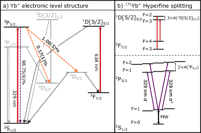
Lasers near wavelengths of 369 nm and 935 nm are used to Doppler cool the ion on the transition and pump population trapped in the metastable state back into the cooling cycle via excitation to the state, as shown in Fig. 1a. We image the ion’s fluorescence at 369 nm wavelength to a photo-multiplier-tube (PMT) for detection.
For cooling of the isotope 171Yb+, which has a nuclear spin of and accordingly hyperfine splittings of the electronic states, we use the closed transition . However, due to off-resonant excitation of the state, the ion occasionally decays to the state. Microwave radiation at 12.6 GHz couples the and ground states to ensure continuous cooling. In order to prepare the ion in the state, we excite the transition resonantly while the microwave radiation is switched off.
| Yb+ Isotope | – this work | – Ref. Berends and Maleki (1992) | – this work |
| 168 | 3007.8(30) MHz | – | 6.04(2) GHz |
| 170 | 1457.9(30) MHz | 1459(21) MHz | 2.93(2) GHz |
| 171 | 922.5(25) MHz | 920(15) MHz | 1.87(2) GHz |
| 172 | 0 | 0 | 0 |
| 174 | -1152.3(15) MHz | -1154(11) MHz | -2.26(2) GHz |
| 176 | -2254.8(15) MHz | -2259(13) MHz | -4.41(2) GHz |
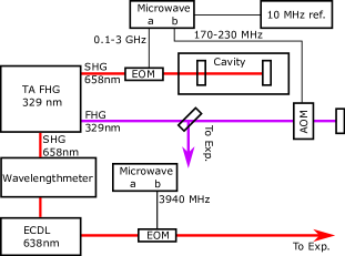
In addition to the lasers required for cooling and detection of the ion, we use light near the wavelengths of 329 nm and 638 nm to drive the transitions and , respectively. We generate light at 329 nm wavelength with a frequency quadrupled, amplified diode laser. After the first doubling cavity, light at 658 nm wavelength is coupled into a high bandwidth fiber-electro-optical modulator (EOM). Sidebands at frequencies of 0.1-3 GHz are modulated onto the light and used to stabilize the laser to an external reference cavity. Thus, the laser is stabilized to the fixed reference cavity with a variable frequency offset which is given by the modulation frequency of the EOM. The reference cavity consists of two mirrors with a reflectivity of glued to a 10 cm long Zerodur spacer in a temperature stabilized vacuum housing. The laser is frequency stabilized by the Pound-Drever-Hall technique Drever et al. (1983).
For further frequency scanning and pulse shaping we use an acousto-optical modulator (AOM) in double-pass configuration with a center frequency of 200 MHz and a bandwidth of 100 MHz. The signals for the AOM and the fiber EOM are generated by a two channel microwave generator, which is stabilized to a 10 MHz reference signal from a signal generator. A mechanical shutter prevents any light from reaching the ion if switched off. Light from the first doubling cavity is coupled to a commercial wavelength meter, allowing for a course absolute frequency determination of the frequency-quadrupled light with an accuracy of 60 MHz according to specification.
We generate light at 638 nm wavelength with a home-built ECDL. This laser is stabilized to the wavelength meter to compensate for frequency drifts and has a short-time frequency stability of better than 10 MHz. We switch the light with a mechanical shutter. The light is coupled to a fiber EOM which allows for modulating sidebands in order to drive transitions between multiple hyperfine states in the case of 171Yb+, and guided to the experiment. The part of the setup relevant for the spectroscopy is shown in Fig.2.
III Results
III.1 Isotope shifts and hyperfine splitting
In case of the isotopes without nuclear spin, we measure the resonance frequency of the D2-transition by applying laser pulses with a width of 5 s and a saturation parameter to the ion. From the state, there is a probability of about 1% for the ion to decay to the metastable state ( ms) from where it decays with 83% probability Taylor et al. (1997) to the long-lived state. An ion in either of these states does not scatter light during Doppler cooling. On resonance, about 50% of the population decays to the dark state in 5 s. To detect whether the ion is in one of the dark states, we image the ion’s fluorescence to a PMT for 4 ms during Doppler cooling, allowing for almost perfect state detection. We scan the laser over the atomic resonance in steps of 2 MHz by tuning the drive frequency of the AOM (). We compensate for the frequency-dependence of the diffraction efficiency in the AOM by supplying appropriate radio-frequency power at each frequency. After the detection, we pump the ion back into the cooling cycle by exciting the transition. A post-selection measurement is performed before each spectroscopy pulse in order to check if the ion was successfully pumped out of the state. The measurement data for a single scan of the transition in 174Yb+ is plotted in Fig. 3a.
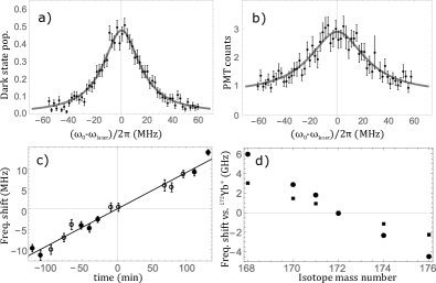
We repeat the experiment for the isotopes 168Yb+, 170Yb+, 172Yb+, 174Yb+ and 176Yb+. For each measurement, we frequency-stabilize the laser to the same cavity mode, but with different offset frequencies given by the modulation frequency of the fiber EOM. The relative frequency of the spectroscopy light compared to the fixed cavity resonance is determined by . We estimate the drift of the cavity by measuring the same resonance at different times as shown in Fig. 3c.
The uncertainties in the energy shifts are given by the statistical error of the fitted resonance and the uncertainty in the cavity drift during the measurement time. In principle the error should not depend on the abundance of the isotope. However, the measurements with the rare isotopes 168Yb+ (0.13% abundance) and 170Yb+ (3.05% abundance, can only be loaded in combination with 172Yb+) NIS take significantly longer due to low ion loading rates. This leads to larger uncertainties for the cavity drift. We use -polarized light and a small magnetic field of mT to avoid errors due to Zeeman-shifts of the D2-transition. The results are shown in Fig. 3d and Tab. 1.
| Martensson-Pendrill et al. (1994) | Safronova and Safronova (2009) | Porsev et al. (2012)a | Porsev et al. (2012)b | Sahoo and Das (2011) | Dzuba and Flambaum (2011) | Berends and Maleki (1992) | This work | |
|---|---|---|---|---|---|---|---|---|
| 391 | 311.5 | 330 | 765 | 322 | 388 | 877(16) | 875.4(10) | |
| - | - | - | - | - | - | - | -107(6) |
In case of 171Yb+ we make use of the hyperfine structure (see Fig. 1b) in order to detect the excitation to the state. We prepare the ion in the ground state before transferring it to the state via rapid adiabatic passage (RAP) using microwave radiation. We apply a laser pulse with a width of 200 ns to excite the or states, followed by a second RAP pulse on the transition. At the end of this sequence, we perform state-selective fluorescence detection, based on Doppler cooling without microwave coupling of the and ground states. During the detection, an ion in the state appears dark while an ion in the state scatters light, which allows for detection of the induced population transfer out of the initial state.
Experiments with 171Yb+ are conducted in a magnetic field of 0.18-0.23 mT in order to allow for efficient Doppler cooling on the transition Olmschenk et al. (2007). We use linear polarized light () at 329 nm wavelength to avoid the dipole forbidden transition. Due to the symmetric excitation of the transition (see Fig. 1b) the magnetic field should not lead to a frequency shift of the transition. Measurements at different fields corroborate this assumption.
We repeat the experiment for both hyperfine states and determine the hyperfine energy splitting given in Tab.2. Together with the well-known energy splitting of the ground state of 12642.812 MHz Fisk et al. (1997) we calculate the isotope shift of the state given in Tab. 1.
During the spectroscopy of the transition, the state with a lifetime of yr Roberts et al. (1997) is populated via decay of the state. After the state-detection, we pump population out of the state by excitation of the transition. Wavelength meter readings during the measurement give us the isotope shifts of this transition with an accuracy of 20 MHz. To the best of our knowledge, this is the most complete (in terms of measured isotopes) and precise measurement of the isotope shifts of the transition. In we drive the transitions and (see Fig. 1b). We use sidebands at 3940 MHz to excite both transitions efficiently. With a hyperfine splitting of the state of 3620(2) MHz Taylor et al. (1999), we determine an energy splitting of the upper state of 320(20) MHz and a hyperfine constant MHz. There seems to be no previous experimental data available for the hyperfine splitting, only a theoretical estimation of MHz Petrasiunas et al. (2012), which deviates significantly from the value we find in the experiment.
III.2 Branching fractions
| Branching from to: | Biemont et al. (1998) | Porsev et al. (2012)a | Porsev et al. (2012)b | Sahoo and Das (2011) | Roberts et al. (2014) | This work |
|---|---|---|---|---|---|---|
| 98.77% | 98.86% | 99.09% | 98.86% | 98.83% | 98.75(6)% | |
| 0.21% | 0.18% | 0.15% | 0.18% | 0.19% | 0.17(1)% | |
| 1.02% | 0.96% | 0.76% | 0.96% | 0.98% | 1.08(5)% |
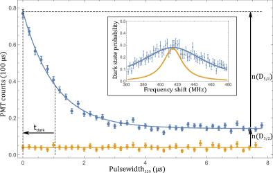
To measure the branching fractions for decay out of the state, we excite the D2-transition with a short pulse of resonant light, followed by fluorescence detection of 100 s duration. During the detection, an ion initially in the or state scatters light, while an ion in the state appears dark. Scanning the pulse width, we obtain the time constant s for the exponential decay of the PMT counts versus pulse width plotted in Fig. 4. From this time constant, the lifetime of the state of ns Pinnington et al. (1997) and the probability to be in the excited state during the laser-pulse , we determine the combined decay probability to both states as follows:
| (1) |
The saturation parameter and thus the excited state population probability is determined by a frequency scan over the resonance. We normalize the power in the 329 nm beam during the scan by appropriate power settings of the AOM as described above. We choose a short pulse width of ns to avoid saturation of the decay to the state. We measure a saturation parameter of , the data is shown in the inset of Fig. 4. For the branching ratio experiment we tune the laser to resonance and operate the AOM at the frequency of maximum diffraction efficiency which allows us to increase the power in the laser pulse by a factor of 2.2 compared to the scan. Accordingly we calculate a saturation parameter of for the branching ratio experiment, corresponding to a probability of being in the excited state of . This scan is repeated frequently during the measurement to monitor intensity and frequency drifts.
The ratio between the decay to the state and state is given by the ratio between the fluorescence for a pulse width , all population in the state (), compared to the fluorescence when all population is pumped to the manifold (), as indicated in Fig. 4.
We determine branching fractions to the state and state of 1.08(5)% and 0.17(1)% respectively which is in agreement with theoretical predictions from Ref. Biemont et al. (1998). The errors include the statistical uncertainties in , , as well as uncertainties in the excited state population and the lifetime of the state.
The cumulative duration of the excitation pulse (s) and the detection bin (s) is small compared to the lifetimes of the ( ms Yu and Maleki (2000)) and ( ms Taylor et al. (1997)) states. The probability for a decay back to the ground state during that time is less than 0.3% and thus negligible compared to other experimental errors.
IV Conclusions
The isotope shifts in the transition in Yb+ and hyperfine splitting (in 171Yb+) of the state have been determined. Our results on both agree with previous results from Ref. Berends and Maleki (1992) but are more precise by a factor of 5-9. Both experiments contradict theoretical predictions obtained from single-electron approaches for the hyperfine splitting by a factor 2-3. The results from a many-electron approach Porsev et al. (2012) agree much better with the experimental results. This corroborates their hypothesis that mixing with the energetically close state has a strong influence on the state, which can not be described by a single-electron approach. We find the branching fractions for decay of the state to the state and states to be 1.08% and 0.17%, respectively, in rough agreement with theoretical predictions.
Acknowledgements.
We thank M. Safronova for helpful comments on the manuscript. This work was supported by the European Union via the European Research Council (Starting Grant 337638) and the Netherlands Organization for Scientific Research (Vidi Grant 680-47-538) (R.G.).References
References
- Diddams et al. (2001) S. A. Diddams, T. Udem, J. C. Bergquist, E. A. Curtis, R. E. Drullinger, L. Hollberg, W. M. Itano, W. D. Lee, C. W. Oates, K. R. Vogel, and D. J. Wineland, Science 293, 825 (2001).
- Fortson (1993) N. Fortson, Phys. Rev. Lett. 70, 2383 (1993).
- Häffner et al. (2000) H. Häffner, T. Beier, N. Hermanspahn, H. J. Kluge, W. Quint, S. Stahl, J. Verdu, and G. Werth, Phys. Rev. Lett. 85, 5308 (2000).
- Cirac and Zoller (1995) J. I. Cirac and P. Zoller, Phys. Rev. Lett. 74, 4091 (1995).
- Blatt and Roos (2012) R. Blatt and C. F. Roos, Nat. Phys. 8, 277 (2012).
- Tamm et al. (2009) C. Tamm, S. Weyers, B. Lipphardt, and E. Peik, Phys. Rev. A 80, 043403 (2009).
- Godun et al. (2014) R. M. Godun, P. B. R. Nisbet-Jones, J. M. Jones, S. A. King, L. A. M. Johnson, H. S. Margolis, K. Szymaniec, S. N. Lea, K. Bongs, and P. Gill, Phys. Rev. Lett. 113, 210801 (2014).
- Blatt et al. (1983) R. Blatt, H. Schnatz, and G. Werth, Z. Phys. A 312, 143 (1983).
- Fisk et al. (1997) P. T. H. Fisk, M. J. Sellars, M. A. Lawn, and C. Coles, IEEE Trans. Ultrasonics Ferroelectrics and Frequency Control 44, 344 (1997).
- Olmschenk et al. (2007) S. Olmschenk, K. C. Younge, D. L. Mochring, D. N. Matsukevich, P. Maunz, and C. Monroe, Phys. Rev. A 76, 052314 (2007).
- Islam et al. (2013) R. Islam, C. Senko, W. C. Campbell, S. Korenblit, J. Smith, A. Lee, E. E. Edwards, C.-C. J. Wang, J. K. Freericks, and C. Monroe, Science 340, 583 (2013).
- Debnath et al. (2016) S. Debnath, N. M. Linke, C. Figgatt, K. A. Landsman, K. Wright, and C. Monroe, Nature 536, 63–66 (2016).
- Taylor et al. (1997) P. Taylor, M. R. ans S. V. Gateva-Kostova, R. B. M. Clarke, G. P. Barwood, W. R. C. Rowley, and P. Gill, Phys. Rev. A 56, 2699 (1997).
- Taylor et al. (1999) P. Taylor, M. Roberts, G. M. Macfarlane, G. P. Barwood, W. R. C. Rowley, and P. Gill, Phys. Rev. A 60, 2829 (1999).
- Yu and Maleki (2000) N. Yu and L. Maleki, Phys. Rev. A 61, 022507 (2000).
- Tamm et al. (2007) C. Tamm, B. Lipphardt, H. Schnatz, R. Wynands, S. Weyers, T. Schneider, and E. Peik, IEEE Trans. Instrum. Meas. 56, 601 (2007).
- Berends and Maleki (1992) R. W. Berends and L. Maleki, J. Opt. Soc. Am. B 9, 332 (1992).
- Martensson-Pendrill et al. (1994) A.-M. Martensson-Pendrill, D. S. Gough, and P. Hannaford, Phys. Rev. A 49, 3351 (1994).
- Safronova and Safronova (2009) U. I. Safronova and M. S. Safronova, Phys. Rev. A 79, 022512 (2009).
- Porsev et al. (2012) S. G. Porsev, M. S. Safronova, and M. G. Kozlov, Phys. Rev. A 86, 022504 (2012).
- Sahoo and Das (2011) B. K. Sahoo and B. P. Das, Phys. Rev. A 84, 010502(R) (2011).
- Dzuba and Flambaum (2011) V. A. Dzuba and V. V. Flambaum, Phys. Rev. A 83, 052513 (2011).
- Biemont et al. (1998) E. Biemont, J. F. Dutrieux, I. Martin, and P. Quinet, J. Phys. B: At. Mol. Opt. Phys. 31, 3321 (1998).
- Roberts et al. (2014) B. M. Roberts, V. A. Dzuba, and V. V. Flambaum, Phys. Rev. A 89, 012502 (2014).
- (25) NIST Atomic Spectra Database https://physics.nist.gov/.
- Joger et al. (2017) J. Joger, H. Fürst, N. Ewald, T. Feldker, M. Tomza, and R. Gerritsma, Phys. Rev. A 96, 030703(R) (2017).
- Kleinert et al. (2016) M. Kleinert, M. E. G. Dahl, and S. Bergeson, Phys. Rev. A 94, 052511 (2016).
- Pinnington et al. (1997) E. H. Pinnington, G. Rieger, and J. A. Kernahan, Phys. Rev. A 56, 2421 (1997).
- Roberts et al. (1997) M. Roberts, P. Taylor, G. P. Barwood, P.Gill, H. A. Klein, and W. R. C. Rowley, Phys. Rev. Lett. 78, 1876 (1997).
- Drever et al. (1983) R. W. P. Drever, J. L. Hall, F. V. Kowalski, J. Hough, G. M. Ford, A. J. Munley, and H. Ward, Appl. Phys. B 31, 97 (1983).
- Petrasiunas et al. (2012) M. J. Petrasiunas, E. W. Streed, T. J. Weinhold, B. G. Norton, and D. Kielpinski, Appl. Phys. B 107, 1053 (2012).