Time calibration of the J-PET detector
Abstract
The Jagiellonian Positron Emission Tomograph (J-PET) project carried out in the Institute of Physics of the Jagiellonian University is focused on construction and tests of the first prototype of PET scanner for medical diagnostic which allows for the simultaneous 3D imaging of the whole human body using organic scintillators. The J-PET prototype consists of 192 scintillator strips forming three cylindrical layers which are optimized for the detection of photons from the electron-positron annihilation with high time- and high angular-resolutions. In this article we present time calibration and synchronization of the whole J-PET detection system by irradiating each single detection module with a source and a small detector providing common reference time for synchronization of all the modules.
PACS: 06.20.fb, 36.10.Dr, 11.30.Er, 24.80.+y
1 Introduction
Positron emission tomography (PET) imaging is a very important tool in medical diagnostics, in particular in oncology, cardiology, neurology, gastrology and psychiatry. Currently, all commercial PET devices are built with scintillation crystals [1, 2, 3].
There are few known methods for PET scanners using time calibration. Time-of-flight (TOF)-PET synchronisation is carried out with radioactive isotopes like sodium or germanium, placed inside the PET device, typically in
its geometric center. The gamma quanta from radioactive source are scattered (due to applied shield) allowing synchronisation of all PET components [4, 5]. There are also methods for time synchronization using several radioactive sources simultaneously [6] as well as using a rotating source along the scintillation chamber [7].
J-PET is the first positron emission tomography scanner built from plastic scintillators which,
as organic detectors, are relatively cheap and easy to shape as well as are characterized by very
good time measurement resolution. In the J-PET detection system, information about the place of
quanta interaction is extracted solely from timing measurement instead of energy deposition
measurement [8, 9, 10, 11, 12, 13, 14, 15, 16]. Therefore, it is crucial to perform precise time calibration of the detection setup.
The time calibration for the J-PET scanner is carried out based on measurements performed with
a reference detector and radioactive sodium source. Collected data allowed us to perform a time calibration
for each of the 192 scintillator strips (i. e. the time difference calibration of a single detection module),
synchronize between the strips in a single cylindrical layer, as well as synchronize between
the three scintillator layers. The time calibration method is briefly described in this report.
2 J-PET calibration with a reference detector
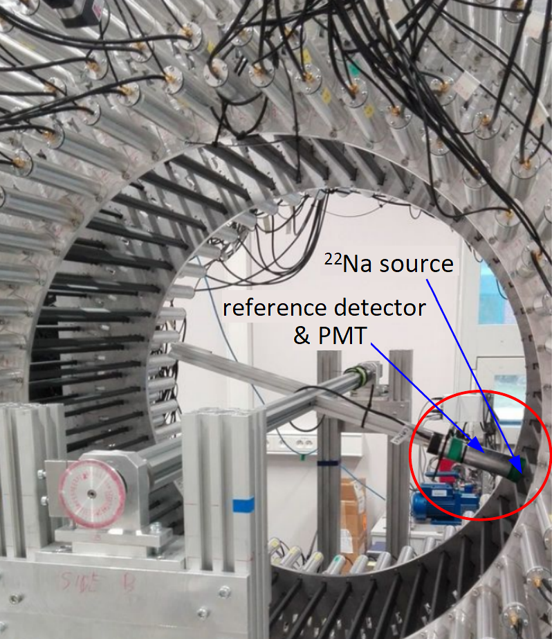
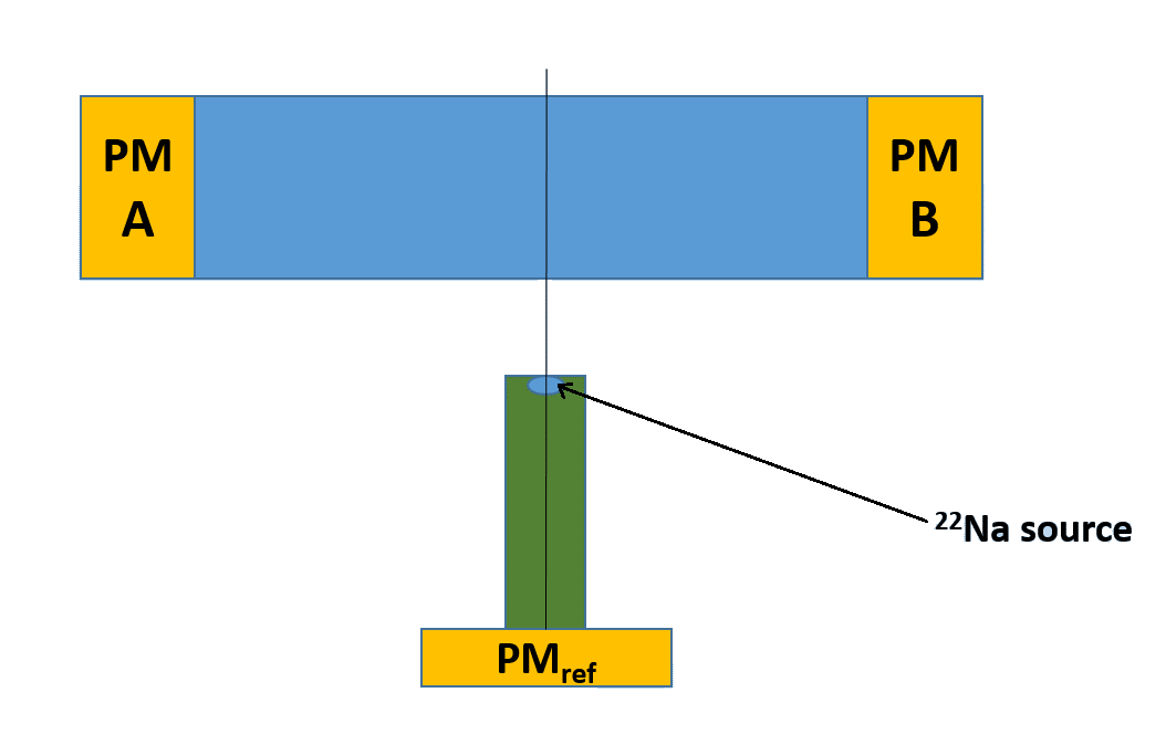
Measurements used for the calibration and synchronization of the J-PET detector modules was performed using a 5 5 19 mm3 BC-420 plastic reference detector coupled to a single photomultiplier and with a source placed on it [17]. The whole system was mounted on a metal arm inside the J-PET detector as shown in Fig. 1. A single measurement is carried out with the reference detector pointing at the center of the scintillator strip to be measured, schematically presented in Fig. 2. The data for each detection module were taken in coincidence with signals from the reference detector which, due to small size of the reference scintillator, selects a well defined beam of gamma quanta annihilation for calibration.
The measurement procedure was repeated for each of the 192 J-PET scintillator strips arranged
in three cylindrical layers. Since the J-PET front-end electronics are able to probe signals
at four different thresholds, and on both the leading and trailing edges [19, 20]
the calibration was done for times measured on each threshold of both signal edges.
Collected data were analysed using the J-PET Framework software [18] w. r. t.
the time calibration of each separate module (so called ”A-B” synchronization) and w. r. t. the TOF between scintillators.
The calibration was performed taking advantage of the fact that the beam of selected annihilation
quanta hits each scintillator in the center. Thus, in an ideal case the difference between the
times of the signals registered at both sides of a single scintillator
must be equal zero. But in reality the measured times are shifted with respect to the true values
by some constant factors accounting for the delays in the photomultipliers and electronic components. Thus, and , and the
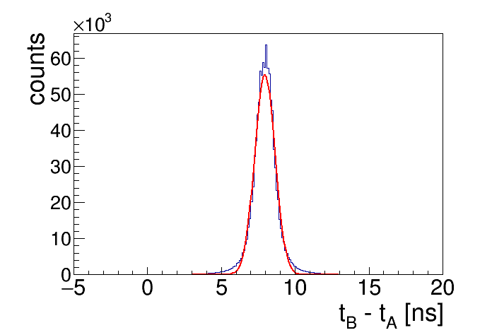
time difference will be non-zero. For the ”A-B” synchronization ()
we can determine the distribution for each detection module. By performing a gaussian fit to each of these distributions one can extract the effective time offsets ():
| (1) |
An example of a spectrum for one of the scintillator strips in the first layer of the J-PET detector is presented in Fig. 3.
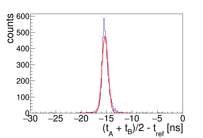
In order to perform simultaneous synchronization of all modules in a single detector layer, a difference between the time of gamma quanta hit in the module and the time measured with the reference detector was determined . Again, a fit to the distributions gives the common reference time for all the modules, i.e. time synchronization, and gives the calibration constant () related to the time offsets on both sides of a strip in the following way:
| (2) |
An example of a raw (without any selection conditions) spectrum for a strip in the first J-PET layer is presented in Fig. 4.
Solving the set of equations 1 and 2 gives finally the following time offsets:
| (3) |
| (4) |
Synchronization between layers was carried out with respect to the first internal layer. The constant (in Eq. 3 and 4) was then corrected for strips in the other layers () with time constants and corresponding to the time elapsed for gamma quanta traveling from layer 1 to layer 2, or to layer 3, respectively. Time differences between layers were calculated based on known distances between layers and were found to be equal to [ns] and [ns].
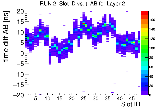
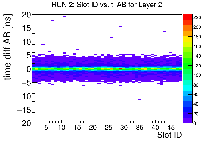
The time calibration method was validated with independent measurements performed using a collimated 22Na radioactive source installed in the geometrical center of the J-PET barrel [21]. As an example, in Fig. 5 we show the time difference for each strip of layer 2 before (upper panel) and after applying calibration constants (lower panel). As one can see, the is distributed around zero as expected for a properly calibrated detector.
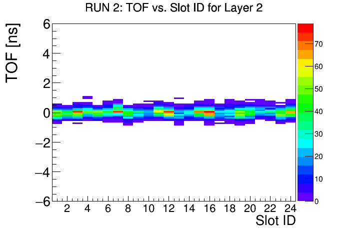
The synchronization of modules can be checked by studying the TOF of annihilation gamma quanta for two modules located opposite to each other. TOF is defined as the difference between the measured times of two back-to-back gamma quanta hits. Fig. 6 shows the TOF spectrum for pairs of modules in layer 2 after the synchronization. As in the previous case we expect the distribution to be peaked around zero for all the modules since the source was placed in the geometrical center of the detector.
3 Summary
We have presented the method used for time calibration of the J-PET detector. It is based on data taken by irradiating each detector module with a radioactive sodium source in coincidence with reference detector. This data was used to calibrate the time difference measurement within each single module and for time synchronization of modules in all detector layers. The method was validated with independent measurements using a collimated 22Na source placed in the center of the detector, demonstrating that the developed procedure gives satisfying results. There are other methods which may be used for the J-PET calibration and monitoring. For example, a measurement referenced to cosmic radiation [22]; the performance and limitations of this and other methods is now under investigation.
4 Acknowledgements
The authors acknowledge the technical support by A. Heczko, W. Migdał, the financial support from the Polish National Center for Development and Research through grant INNOTECH-K1/IN1/64/159174/NCBR/12, the EU and MSHE Grant no. POIG.02.03.00-161 00-013/09, and the National Science Center Poland based on decision number DEC-2013/09/N/ST2/02180 and UMO-2016/21/B/ST2/01222. B. C. Hiesmayr acknowledges gratefully the Austrian Science Fund FWF-P26783.
References
- [1] P. J. Slomka, T. Pan, G. Germano, Semin. Nucl. Med. 46, 5 (2016). DOI:10.1053/j.semnuclmed.2015.09.006
- [2] S. Vandenberghe, E. Mikhaylova, E. D’Hoe, P. Mollet and J. S. Karp, EJNMMI Phys. 3, 3 (2016). DOI:10.1186/s40658-016-0138-3
- [3] J. S. Karp, S. Surti, M. E. Daube-Witherspoon, G. Muehllehner, J. Nucl. Med. 49, 462 (2008). DOI:10.2967/jnumed.107.044834
- [4] J. J. Griesmer, T. L. Laurence, Patent No. US7414246 (2008).
- [5] T. Laurence, J. J. Griesmer, Patent Application No. US78209075 (2010).
- [6] G. Muehllehner, J. S. Karp, Patent No. US7557350 (2009).
- [7] C. W. Stearns, Patent No. US5272343 (1993).
- [8] P. Moskal, Sz. Niedźwiecki, T. Bednarski, E. Czerwiński, Ł. Kapłon, E. Kubicz, I. Moskal, M. Pawlik-Niedźwiecka, N.G. Sharma, M. Silarski, M. Zieliński, N. Zoń, P. Białas, A. Gajos, A. Kochanowski, G. Korcyl, J. Kowal, P. Kowalski, T. Kozik, W. Krzemień, M. Molenda, M. Pałka, L. Raczyński, Z. Rudy, P. Salabura, A. Słomski, J. Smyrski, A. Strzelecki, A. Wieczorek, W. Wiślicki, Nucl. Instrum. Meth. A764, 317 (2014). DOI: 10.1016/j.nima.2014.07.052
- [9] A. Gajos, D. Kamińska, E. Czerwiński, D. Alfs, T. Bednarski, P. Białas, B. Głowacz, M. Gorgol, B. Jasińska, Ł. Kapłon, G. Korcyl, P. Kowalski, T. Kozik, W. Krzemień, E. Kubicz, M. Mohammed, Sz. Niedźwiecki, M. Pałka, M. Pawlik-Niedźwiecka, L. Raczyński, Z. Rudy, O. Rundel, N.G. Sharma, M. Silarski, A. Słomski, A. Strzelecki, A. Wieczorek, W. Wiślicki, B. Zgardzińska, M. Zieliński, P. Moskal, Nucl. Instrum. Meth. A819, 54 (2016). DOI: 10.1016/j.nima.2016.02.069
- [10] D. Kamińska, A. Gajos, E. Czerwiński, D. Alfs, T. Bednarski, P. Białas, C. Curceanu, K. Dulski, B. Głowacz, N. Gupta-Sharma, M. Gorgol, B. C. Hiesmayr, B. Jasińska, G. Korcyl, P. Kowalski, W. Krzemień, N. Krawczyk, E. Kubicz, M. Mohammed, Sz. Niedźwiecki, M. Pawlik-Niedźwiecka, L. Raczyński, Z. Rudy, M. Silarski, A. Wieczorek, W. Wiślicki, B. Zgardzińska, M. Zieliński, P. Moskal, Eur. Phys. J. C76, 445 (2016). DOI: 10.1140/epjc/s10052-016-4294-3
- [11] P. Moskal, D. Alfs, T. Bednarski, P. Białas, E. Czerwiński, C. Curceanu, A. Gajos, B. Głowacz, M. Gorgol, B.C. Hiesmayr B. Jasińska, D. Kamińska, G. Korcyl, P. Kowalski, T. Kozik, W. Krzemień, N. Krawczyk, E. Kubicz, M. Mohammed, Sz. Niedźwiecki, M. Pawlik-Niedźwiecka, L. Raczyński, Z. Rudy, M. Silarski, A. Wieczorek, W. Wiślicki, M. Zieliński, Acta Phys. Polon. BB47, 509 (2016). DOI:10.5506/APhysPolB.47.509
- [12] P. Moskal, O. Rundel, D. Alfs, T. Bednarski, P. Białas, E. Czerwiński, A. Gajos, K. Giergiel, M. Gorgol, B. Jasińska, D. Kamińska, Ł. Kapłon, G. Korcyl, P. Kowalski, T. Kozik, W. Krzemień, E. Kubicz, Sz. Niedźwiecki, M. Pałka, L. Raczyński, Z. Rudy, N. G. Sharma, A. Słomski, M. Silarski, A. Strzelecki, A. Wieczorek, W. Wiślicki, P. Witkowski, M. Zieliński, N. Zoń, Phys. Med. Biol. 61, 2025 (2016). DOI:10.1088/0031-9155/61/5/2025
- [13] P. Moskal, N. Zoń, T. Bednarski, P. Białas, E. Czerwiński, A. Gajos, D. Kamińska, Ł. Kapłon, A. Kochanowski, G. Korcyl, J. Kowal, P. Kowalski, T. Kozik, W. Krzemień, E. Kubicz, Sz. Niedźwiecki, M. Pałka, L. Raczyński, Z. Rudy, O. Rundel, P. Salabura, N.G. Sharma, M. Silarski, A. Słomski, J. Smyrski, A. Strzelecki, A. Wieczorek, W. Wiślicki, M. Zieliński, Nuclear Inst. and Methods in Physics Research A775, 54 (2015). DOI:10.1016/j.nima.2014.12.005
- [14] L. Raczynski, P. Moskal, P. Kowalski, W. Wiślicki, T. Bednarski, P.Białas, E. Czerwiński, Ł. Kapłon, A. Kochanowski, G. Korcyl, J. Kowal, T. Kozik, W. Krzemień, E. Kubicz, M. Molenda, Sz. Niedźwiecki, M. Pałka, M Pawlik., Z. Rudy, P. Salabura, N. G. Sharma, M. Silarski, A. Słomski, J. Smyrski, A. Strzelecki, A. Wieczorek, M. Zieliński, N. Zoń, Nuclear Inst. and Methods in Physics Research A764, 186 (2014). DOI:10.1016/j.nima.2014.07.032
- [15] L. Raczynski, P. Moskal, P. Kowalski, W. Wiślicki, T. Bednarski, P. Białas, E. Czerwiński, A. Gajos, Ł. Kapłon, A. Kochanowski, G. Korcyl, J. Kowal, T. Kozik, W. Krzemień, E. Kubicz, Sz. Niedźwiecki, M. Pałka, Z. Rudy, O. Rundel, P. Salabura, N.G. Sharma, M. Silarski, A. Słomski, J. Smyrski, A. Strzelecki, A. Wieczorek, M. Zieliński, N. Zoń, Nuclear Inst. and Methods in Physics Research A786, 105 (2015). DOI:10.1016/j.nima.2015.03.032
- [16] L. Raczynski, W. Wiślicki, W. Krzemień, P. Kowalski, D. Alfs, T. Bednarski, P. Białas, C. Curceanu, E. Czerwiński, K. Dulski, A. Gajos, B. Głowacz, M. Gorgol, B. Hiesmayr, B. Jasińska, D. Kamińska, G. Korcyl, T. Kozik, N. Krawczyk, E. Kubicz, M. Mohammed, M. Pawlik-Niedźwiecka, S. Niedźwiecki, M. Pałka, Z. Rudy, O. Rundel, N. Gupta-Sharma, M. Silarski, J. Smyrski, A. Strzelecki, A. Wieczorek, B. Zgardzińska, M. Zieliński and P. Moskal, Phys. Med. Biol. 62, 5076 (2017). DOI:10.1088/1361-6560/aa7005
- [17] T. Bednarski, PhD thesis (2016).
- [18] W. Krzemień, A. Gajos, A. Gruntowski, K. Stola, D. Trybek, T. Bednarski, P. Białas, E. Czerwiński, D. Kamińska, L. Kapłon, A. Kochanowski, G. Korcyl, J. Kowal, P. Kowalski, T. Kozik, E. Kubicz, P. Moskal, Sz. Niedźwiecki, M. Pałka, L. Raczyński, Z. Rudy, P. Salabura, N. G. Sharma, M. Silarski, A. Słomski, J. Smyrski, A. Strzelecki, A. Wieczorek, W. Wiślicki, M. Zieliński, N. Zoń, et al., Acta Phys. Polon., A127 1491 (2015). DOI:10.12693/APhysPolA.127.1491
- [19] G. Korcyl, D. Alfs, T. Bednarski, P. Białas, E. Czerwiński, K. Dulski, A. Gajos, B. Głowacz, B. Jasińska, D. Kamińska Ł. Kapłon, P. Kowalski, T. Kozik, W. Krzemień, E. Kubicz, M. Mohammed, Sz. Niedźwiecki, M. Pałka, M. Pawlik-Niedźwiecka, L. Raczyński, Z. Rudy, O. Rundel, N.G. Sharma, M. Silarski, A. Słomski, K. Stoła, A. Strzelecki, A. Wieczorek, W. Wiślicki, B.K. Zgardzińska, M. Zieliński, P. Moskal, Acta Phys. Polon. B47, 491 (2016). DOI:10.5506/APhysPolB.47.491
- [20] M. Pałka, P. Strzempek, G. Korcyl, T. Bednarski, Sz. Niedźwiecki, P. Białas, E. Czerwiński, K. Dulski, A. Gajos, B. Głowacz M. Gorgol, B. Jasińska, D. Kamińska, M. Kajetanowicz, P. Kowalski, T. Kozik, W. Krzemień, E. Kubicz, M. Mohhamed, L. Raczyński, Z. Rudy, O. Rundel, P. Salabura, N.G. Sharma, M. Silarski, J. Smyrski, A. Strzelecki, A. Wieczorek, W. Wiślicki, M. Zieliński, B. Zgardzińska, P. Moskal, JINST 12, P08001 (2017). DOI:10.1088/1748-0221/12/08/P08001
- [21] E. Kubicz, M. Silarski, A. Wieczorek, D. Alfs, T. Bednarski, P. Białas, E. Czerwiński, A. Gajos, B. Głowacz, B. Jasińska D. Kamińska, G. Korcyl, P. Kowalski, T. Kozik, W. Krzemień, M. Mohammed, I. Moskal, S. Niedźwiecki, M. Pawlik-Niedźwiecka, L. Raczyński, Z. Rudy, A. Strzelecki, W. Wiślicki, M. Zieliński, B. Zgardzińska, P. Moskal, Acta Phys. Polon. B47, 537 (2016). DOI:10.5506/APhysPolB.47.537
- [22] M. Silarski, E. Czerwiński, T. Bednarski, P. Moskal, P. Białas, Ł. Kapłon, A. Kochanowski, G. Korcyl, J. Kowal, P. Kowalski, T. Kozik, W. Krzemień, M. Molenda, Sz. Niedźwiecki, M. Pałka, M. Pawlik, L. Raczyński, Z. Rudy, P. Salabura, N.G. Sharma, A. Słomski, J. Smyrski, A. Strzelecki, W. Wiślicki, M. Zieliński, N. Zoń, Bio-Alg. Med-Syst. 10, 19 (2014). doi:10.1515/bams-2013-0105