Nearest-neighbor Kitaev exchange blocked by charge order in electron doped -RuCl3
Abstract
A quantum spin-liquid might be realized in -RuCl3, a honeycomb-lattice magnetic material with substantial spin-orbit coupling. Moreover, -RuCl3 is a Mott insulator, which implies the possibility that novel exotic phases occur upon doping. Here, we study the electronic structure of this material when intercalated with potassium by photoemission spectroscopy, electron energy loss spectroscopy, and density functional theory calculations. We obtain a stable stoichiometry at K0.5RuCl3. This gives rise to a peculiar charge disproportionation into formally Ru2+ (4) and Ru3+ (4). Every Ru 4 site with one hole in the shell is surrounded by nearest neighbors of 4 character, where the level is full and magnetically inert. Thus, each type of Ru sites forms a triangular lattice and nearest-neighbor interactions of the original honeycomb are blocked.
The quantum spin liquid (QSL) is an exotic state of matter which carries fractionalized excitations, completely different from the standard spin waves found for conventional magnetic order Balents (2010). The long search for a realization of this state has recently led to -RuCl3, a layered honeycomb Mott-insulator with a 4 configuration and substantial spin-orbit coupling Plumb et al. (2014); Banerjee et al. (2016). From measurements of the magnetic excitation spectrum Banerjee et al. (2016); Sandilands et al. (2015); Banerjee et al. (2017), but also from other experiments Majumder et al. (2015); Sears et al. (2015); Johnson et al. (2015); Cao et al. (2016); Sandilands et al. (2016a); Koitzsch et al. (2016), and theory Jackeli and Khaliullin (2009); Kim et al. (2015); Chaloupka and Khaliullin (2016); Yadav et al. (2016); Nasu et al. (2016), evidence is mounting that -RuCl3 is close to a Kitaev QSL, that is, a realization of the exactly solvable Kitaev model Kitaev (2006), with some modifications due to Heisenberg interactions Chaloupka et al. (2010, 2013); Majumder et al. (2015); Kubota et al. (2015). In particular, there are indications of a QSL state in an external magnetic field Yadav et al. (2016); Baek et al. (2017); Wolter et al. (2017); Hentrich et al. (2017); Leahy et al. (2017); Zheng et al. (2017). This offers the opportunity to study the fractionalized excitations, most prominently Majorana Fermions and, possibly, to exploit the fact that they are protected from decoherence for quantum information processing schemes Kitaev (2003).
The magnetic state is usually described within the framework of Heisenberg–Kitaev Hamiltonians. However, these attempts face the difficulty that the exchange parameters are not exactly known and higher order interactions (i.e. beyond nearest neighbor) can be decisive Rousochatzakis et al. (2015); Yadav et al. (2016); Winter et al. (2016); Sizyuk et al. (2016). This problem hinders a deeper understanding and theoretical progress of the field.
The QSL is the main driver of interest in -RuCl3, but -RuCl3 is also a Mott-insulator. Doping a Mott-insulator often results in novel ground states with intriguing properties. Well known examples are the cuprates and manganates. Usually, also the magnetic order associated with the Mott-state reacts sensitively to doping. Therefore, doping -RuCl3 is a promising proposition for both: i) stabilizing new, interesting phases, and ii) probing the properties of the QSL.
Here, we study the electronic structure of electron doped -RuCl3. This is achieved by in situ potassium intercalation. An apparent goal of such an approach is to reach a metallic phase. However, this has not been accomplished, similar to previous attempts by rubidium doping instead of potassium Zhou et al. (2016). Nevertheless, we show by a combination of electron energy loss spectroscopy (EELS), photoemission spectroscopy (PES), and density functional theory (DFT) that electron doping alters the ground state of -RuCl3 in a peculiar fashion. We observe a charge disproportionation which quenches the magnetic moment at every alternate Ru site and may serve as a platform to study the interplay of nearest neighbor and next-nearest neighbor Kitaev exchange.
Platelet-like single crystals of -RuCl3 were grown by chemical vapor transport reactions. PES measurements were performed using a laboratory based system at room temperature after cleaving the crystals in situ. The EELS measurements in transmission have been conducted using thin films ( 100 nm) at K. Undoped crystals were intercalated with potassium in situ by metal vapor from SAES dispensers. The density functional (DF) calculations were performed with the all-electron full-potential local-orbital (FPLO) code Koepernik and Eschrig (1999); FPL . See the Supplemental Material for further details.
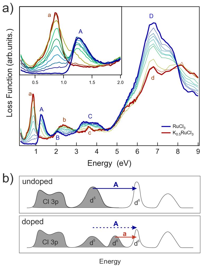
Figure 1a shows the effect of successive K intercalation on the low-energy loss function measured by EELS in transmission, a bulk sensitive probe. The pristine spectrum shows a peak at eV (labeled A), which corresponds to optical gap excitations. The peaks at higher energies (B–D) are due to crystal field and charge-transfer excitations Koitzsch et al. (2016). K intercalation causes drastic changes to the electronic structure, in particular, to the character of the gap. The inset of Fig. 1a expands the low-energy region. We observe that with increasing K content the spectral weight of the original A peak decreases and a new peak at lower energies appears (labeled a). Finally, at saturation, A completely vanishes. In order to find a rationale of these observations we show a schematic picture of the low-energy electronic structure in Fig. 1b. Near the Fermi energy the electronic states are dominated by Ru 4 character. In the undoped case A corresponds to excitations across the Mott gap (). With doping new states are created inside the gap. The fact that A completely vanishes in the experiment could be naturally explained by full electron doping, that is, every Ru3+ (4) ion is reduced to Ru2+ (4) by formation of KRuCl3. Then the occupied states would disappear. However, this stoichiometry is not realized.
Quantitative X-ray photoemission spectroscopy (XPS), core level EELS, and DFT consistently hint at a saturated stoichiometry K0.5RuCl3 (see Supplemental Material). Further, low-energy electron diffraction (LEED) shows a hexagonal pattern as in the pristine sample with modest lattice expansion but without superstructure (see Supplemental Material for details). This information allows us to construct a structural model where the K intercalation takes place between two adjacent Cl layers and occupies interstitial sites (see Fig. 2 and Supplemental Material for details). Most intriguingly, within our DF calculations a ground state with charge order among the two Ru sites, denoted Ru(1) and Ru(2) henceforth, is found. This charge order is not driven by structural symmetry breaking due to the K intercalation. To show this we relaxed the same structure, K0.5RuCl3(a), but with exchanged Ru charge and spin states, i.e., starting from the density and -occupation data of the respective other Ru position (see SFig.1). In this way we obtained a slightly modified geometry, denoted K0.5RuCl3(a’) in STab. I. The electronic state remained essentially unchanged, but with Ru(1) and Ru(2) features exchanged. The total energy of the new solution is only 4 meV per formula unit higher than that of the previous one. This means, the slightly different structural environment of Ru(1) and Ru(2) in K0.5RuCl3(a) allows for charge and spin disproportionation due to the broken structural symmetry; however, it does not determine which of the two Ru sites has a hole in the band and which is non-magnetic. Since the charge order even develops in the case of K0.5RuCl3(b) (see SFig.1), where originally both Ru positions are identical, the order is generic for the considered composition and structural details do not matter.
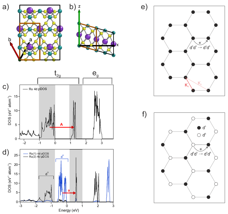
The Ru projected densities of states (DOS) of the isomer from Fig. 2a, b and of prisitine RuCl3 are presented in Fig. 2c, d. The calculations support the schematic picture in Fig. 1b. New states are created inside the Mott gap by potassium doping. The striking difference between RuCl3 and K0.5RuCl3 consists in a splitting of the occupied and of the unoccupied bands in the latter case, caused by the charge order. The empty sub-band of RuCl3 can be identified as driving force of the charge order: Upon doping this band to half-filling, it is split into an occupied, Ru(1) dominated and an unoccupied, Ru(2) dominated part. Further, the observed reduction of the gap size from A = 1.2 eV in RuCl3 to a = 0.8 eV in K0.5RuCl3 can be understood by the splitting of the occupied band into an upper Ru(1) sub-band at the Fermi level and a lower Ru(2) sub-band. The distance of the latter to the first empty, Ru(2) band is fixed by the term.
The described situation is summarized in Fig. 2e, f and explains the complete suppression of A upon half doping. In the undoped case, the gap is defined by the Mott excitation . In the doped case, this transition is blocked, as all sites are surrounded by sites. The gap excitation a is now associated with the charge fluctuation . Features B–D of the undoped material have been assigned recently to interband transitions with strong charge transfer character of C and D Koitzsch et al. (2016) or, alternatively, to multiplets (A–C) and charge transfer excitations (D) Sandilands et al. (2016b). While further work is needed for a unified theoretical description, we note that the initial state concentration still amounts to 50 % under full doping and, thus, should be present in the doped spectrum but likewise reduced in intensity by 50 %. This is indeed observed (see the charge transfer feature d in comparison to D in Fig. 1).
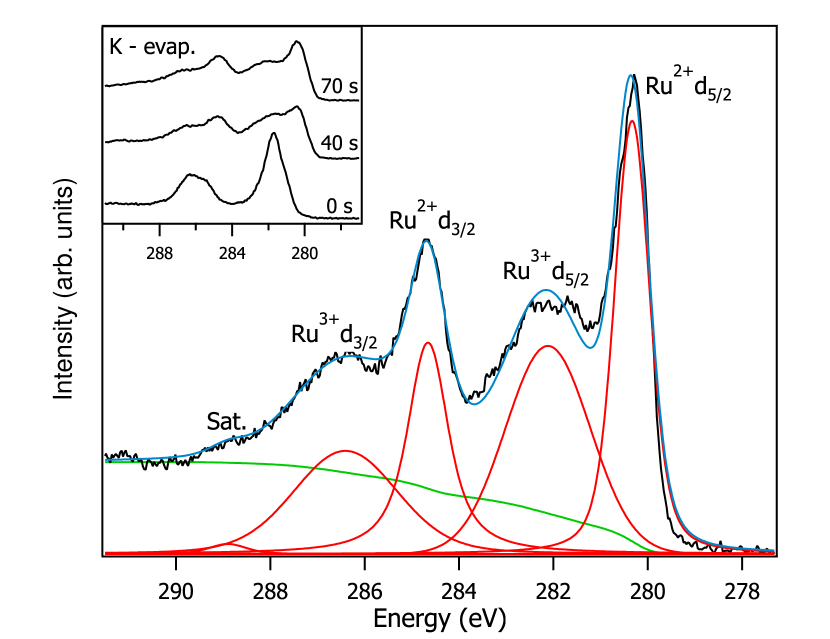
Further quantitative support for the calculated charge-ordered ground state is provided by XPS core level spectra of the Ru 3 states, Fig. 3. The Ru 3 line is spin-orbit split into and components. With doping a narrow lower-energy peak appears for both components. Quantitative analysis of these spectra is complicated and a complete fitting is not possible (see Supplemental Material). However, we modeled the spectral shape of a high resolution scan of the fully doped sample (Fig. 3, main panel) by five Voigt peaks (two spin–orbit components for each of the two Ru sites plus one small charge transfer satellite at the high energy side) and a standard Shirley background. The intensity ratio of the spin–orbit components is fixed to and the Ru2+ : Ru3+ ratio to . Peak positions and total width have been allowed to vary freely. This fit results in reasonable agreement with experiment, which implicitly confirms a K0.5RuCl3 stoichiometry of the fully doped sample and the Ru2+ : Ru3+ ratio. The measured energy separation between Ru2+ and Ru3+ of agrees well with the corresponding DF value of .
Nominally, K doping of -RuCl3 to K0.5RuCl3 reduces Ru(1) from Ru3+ (4d5) to Ru2+ (4d6), while Ru(2) remains in a (4d5) configuration. However, our Mulliken-type analysis tells a different story: the charge difference between Ru(1) and Ru(2) amounts to only 0.15 and the K electron is mainly supplied to the Cl ions being adjacent to the K layer. Almost 50 % of the Ru electrons form a broad hybrid band with Cl states between 5.5 and 2.0 eV. Reversely, the narrow and bands contain an appreachiable weight of Cl between 10 % and 60 %, i.e., they are formed by - molecular states of the appropriate symmetry (see Supplemental Material for details). Thus, a better description of the charge-ordered state would be K1+[RuCl3]0[RuCl3]1-, where [RuCl3]0 has one hole in the -- states and [RuCl3]1- has filled -- states. Note, that each Cl ion is shared among one Ru(1) and one Ru(2); thus, all Cl ions carry almost the same charge. For the sake of brevity, we will further stick to the nominal notation.
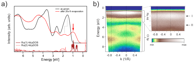
Figure 4 presents the valence band photoemission results. The doped sample shows an additional peak at low energies (red arrow in Fig. 4a). The original Ru3+ related peak is reduced in intensity and shifts somewhat to higher energies. A similar spectral shape was obtained by Rb intercalation, although without any peak shift Zhou et al. (2016). A similar spectral evolution upon alkali metal doping of the Mott-insulator TiOCl has been explained previously by the electrostatic potential of the K+ ions, which effectively localize the doped electron at the closest Ti site Sing et al. (2011). In the present case, at variance, the charge order develops out of a rather homogeneously distributed charge supplied by the K+ ions to the Cl layers.
With increasing evaporation time the principal shape of the low-energy region does not change anymore (see the Supplemental Material). For better comparison of the DF calculations with experiment we have stretched the energy axis by a renormalization factor of 1.25 and shifted the energy by 0.5 eV within the gap. Such factors are often encountered when comparing photoemission data to DF calculations, especially when the screening is bad Koitzsch et al. (2016).
Figure 4b shows the angle dependence of the valence band for the doped sample. The Cl 3 related region shows clear dispersion, similar to the undoped sample Koitzsch et al. (2016). This excludes strong surface deterioration by the intercalation process. The Ru 4 bands, on the other hand, do not show any dispersion, in agreement with previous results for Rb doping Zhou et al. (2016), although a small but finite dispersion is present for the undoped material Koitzsch et al. (2016). Within the charge disproportionation scheme this is readily understood: the Ru3+ are too far apart in the doped sample to maintain a visible dispersion and so are the newly formed Ru2+. The Cl network is homogeneously doped and retains its dispersion.
The charge ordered ground state found in the DF calculations is consistent with EELS, valence band and core level photoemission data. It is depcited in Fig. 2f. Other examples of charge order in half doped Mott insulators are manganites, where the order is of checkerboard type Tokura and Nagaosa (2000); Dagotto et al. (2001). Geometric frustration is present in magnetite (Fe3O4) and causes a complicated charge pattern below the Verwey transition Wright et al. (2002). Transition metal dichalcogenides with similar crystal lattices tend to form charge density waves Rossnagel (2011). The observed real charge difference in charge ordered systems is always much smaller than one due to the large electrostatic energy cost. Here, our DFT calculation yields a charge difference between Ru(1) and Ru(2) of only 0.15 . Nevertheless, the local spin and orbital magnetic moments are distinctly different: , for Ru(1), , for Ru(2). This means, the charge order causes a difference of one order of magnitude between the magnetic moments of the two Ru sites. The charge order develops due to the combination of two facts: (i) the number of electrons which are available for the 4d-3p- states of each -RuCl3 entity is not integer and (ii) a metallic state that would allow to distribute the extra charge equally among both Ru sites is prevented by the very small dispersion of the bands in the vicinity of the Fermi level. Reduction of decreases the gap until it is closed below eV, eV. Concomitantly, the charge order disappears and magnetic moments of comparable size develop at the two Ru positions.
K intercalation offers a possibility to manipulate the charge and spin pattern of the honeycomb lattice in a controlled fashion. This is especially relevant because these patterns appear independently of the potassium lattice. The effect could be useful to study fundamental properties and to create qualitatively new magnetic groundstates. In particular, the zigzag antiferromagnetic order of the parent compound will disappear. The remaining effective triangular lattice for K0.5RuCl3 is geometrically frustrated. Model calculations are needed to elucidate the magnetic groundstate. As the dominating nearest-neighbor Kitaev exchange term vanishes the prominent background seen in INS Banerjee et al. (2016) and Raman Sandilands et al. (2015) should decrease. Also the spin wave spectrum should vanish or, in case an alternative order is established, look different. The suppresion of nearest neighbor Kitaev interactions might allow quantification of higher order terms, which is a prerequisite for a complete theoretical understanding of the QSL in -RuCl3. In practice, the process of intercalation modifies bond lengths and, hence, the exchange parameters, which has to be taken into account for a quantitative description. But even on a qualitative level the originally homogeneous honeycomb lattice of -RuCl3 decomposes into two triangular lattices of and character which may host new magnetic groundstates Yadav et al. (2016); Rousochatzakis et al. (2015). Recently, in a different approach, the Ru honeycomb lattice has been diluted by Ir substitution, which quickly obstructs the antiferromagnetic order Lampen-Kelley et al. (2016).
In summary, we have investigated the electronic structure of potassium intercalated -RuCl3. EELS, PES, and DFT show consistently and independently a stable K0.5RuCl3 stoichiometry in which a charge disproportionation into Ru2+ and Ru3+ takes place. The charge order is accompanied by almost complete quenching of the magnetic moment at every alternate Ru site. This type of combined charge and spin disproportionation on a honeycomb lattice is difficult to achieve otherwise. In principle, double perovskites may have some potential in this direction, but K0.5RuCl3 has the advantage of chemical simplicity. The resulting peculiar state could offer a valuable platform for the investigation of the Kitaev exchange including higher-order interactions and frustrated magnetism in general.
We thank U. Nitzsche for technical assistance and R. Ray and H. Rosner for valuable discussions. This work has been supported by the German Research Foundation (DFG) under SFB 1143.
References
- Balents (2010) L. Balents, Nature 464, 199 (2010).
- Plumb et al. (2014) K. W. Plumb, J. P. Clancy, L. J. Sandilands, V. V. Shankar, Y. F. Hu, K. S. Burch, H.-Y. Kee, and Y.-J. Kim, Phys. Rev. B 90, 041112 (2014).
- Banerjee et al. (2016) A. Banerjee, C. A. Bridges, J.-Q. Yan, A. A. Aczel, L. Li, M. B. Stone, G. E. Granroth, M. D. Lumsden, Y. Yiu, J. Knolle, S. Bhattacharjee, D. L. Kovrizhin, R. Moessner, D. A. Tennant, D. G. Mandrus, and S. E. Nagler, Nat Mater 15, 733 (2016).
- Sandilands et al. (2015) L. J. Sandilands, Y. Tian, K. W. Plumb, Y.-J. Kim, and K. S. Burch, Phys. Rev. Lett. 114, 147201 (2015).
- Banerjee et al. (2017) A. Banerjee, J. Yan, J. Knolle, C. A. Bridges, M. B. Stone, M. D. Lumsden, D. G. Mandrus, D. A. Tennant, R. Moessner, and S. E. Nagler, Science 356, 1055 (2017).
- Majumder et al. (2015) M. Majumder, M. Schmidt, H. Rosner, A. A. Tsirlin, H. Yasuoka, and M. Baenitz, Phys. Rev. B 91, 180401 (2015).
- Sears et al. (2015) J. A. Sears, M. Songvilay, K. W. Plumb, J. P. Clancy, Y. Qiu, Y. Zhao, D. Parshall, and Y.-J. Kim, Phys. Rev. B 91, 144420 (2015).
- Johnson et al. (2015) R. D. Johnson, S. C. Williams, A. A. Haghighirad, J. Singleton, V. Zapf, P. Manuel, I. I. Mazin, Y. Li, H. O. Jeschke, R. Valentí, and R. Coldea, Phys. Rev. B 92, 235119 (2015).
- Cao et al. (2016) H. B. Cao, A. Banerjee, J.-Q. Yan, C. A. Bridges, M. D. Lumsden, D. G. Mandrus, D. A. Tennant, B. C. Chakoumakos, and S. E. Nagler, Phys. Rev. B 93, 134423 (2016).
- Sandilands et al. (2016a) L. J. Sandilands, Y. Tian, A. A. Reijnders, H.-S. Kim, K. W. Plumb, Y.-J. Kim, H.-Y. Kee, and K. S. Burch, Phys. Rev. B 93, 075144 (2016a).
- Koitzsch et al. (2016) A. Koitzsch, C. Habenicht, E. Müller, M. Knupfer, B. Büchner, H. C. Kandpal, J. van den Brink, D. Nowak, A. Isaeva, and T. Doert, Phys. Rev. Lett. 117, 126403 (2016).
- Jackeli and Khaliullin (2009) G. Jackeli and G. Khaliullin, Phys. Rev. Lett. 102, 017205 (2009).
- Kim et al. (2015) H.-S. Kim, V. S. V., A. Catuneanu, and H.-Y. Kee, Phys. Rev. B 91, 241110 (2015).
- Chaloupka and Khaliullin (2016) J. Chaloupka and G. Khaliullin, Phys. Rev. B 94, 064435 (2016).
- Yadav et al. (2016) R. Yadav, N. A. Bogdanov, V. M. Katukuri, S. Nishimoto, J. van den Brink, and L. Hozoi, Scientific Reports 6, 37925 EP (2016).
- Nasu et al. (2016) J. Nasu, J. Knolle, D. L. Kovrizhin, Y. Motome, and R. Moessner, Nat Phys 12, 912 (2016), letter.
- Kitaev (2006) A. Kitaev, Annals of Physics 321, 2 (2006).
- Chaloupka et al. (2010) J. Chaloupka, G. Jackeli, and G. Khaliullin, Phys. Rev. Lett. 105, 027204 (2010).
- Chaloupka et al. (2013) J. Chaloupka, G. Jackeli, and G. Khaliullin, Phys. Rev. Lett. 110, 097204 (2013).
- Kubota et al. (2015) Y. Kubota, H. Tanaka, T. Ono, Y. Narumi, and K. Kindo, Phys. Rev. B 91, 094422 (2015).
- Baek et al. (2017) S.-H. Baek, S.-H. Do, K.-Y. Choi, Y. S. Kwon, A. U. B. Wolter, S. Nishimoto, J. van den Brink, and B. Büchner, Phys. Rev. Lett. 119, 037201 (2017).
- Wolter et al. (2017) A. U. B. Wolter, L. T. Corredor, L. Janssen, K. Nenkov, S. Schönecker, S.-H. Do, K.-Y. Choi, R. Albrecht, J. Hunger, T. Doert, M. Vojta, and B. Büchner, Phys. Rev. B 96, 041405 (2017).
- Hentrich et al. (2017) R. Hentrich, A. U. B. Wolter, X. Zotos, W. Brenig, D. Nowak, A. Isaeva, T. Doert, A. Banerjee, P. Lampen-Kelley, D. G. Mandrus, S. E. Nagler, J. Sears, Y.-J. Kim, B. Büchner, and C. Hess, ArXiv e-prints (2017), arXiv:1703.08623 [cond-mat.str-el] .
- Leahy et al. (2017) I. A. Leahy, C. A. Pocs, P. E. Siegfried, D. Graf, S.-H. Do, K.-Y. Choi, B. Normand, and M. Lee, Phys. Rev. Lett. 118, 187203 (2017).
- Zheng et al. (2017) J. Zheng, K. Ran, T. Li, J. Wang, P. Wang, B. Liu, Z. Liu, B. Normand, J. Wen, and W. Yu, ArXiv e-prints (2017), arXiv:1703.08474 [cond-mat.str-el] .
- Kitaev (2003) A. Kitaev, Annals of Physics 303, 2 (2003).
- Rousochatzakis et al. (2015) I. Rousochatzakis, J. Reuther, R. Thomale, S. Rachel, and N. B. Perkins, Phys. Rev. X 5, 041035 (2015).
- Winter et al. (2016) S. M. Winter, Y. Li, H. O. Jeschke, and R. Valentí, Phys. Rev. B 93, 214431 (2016).
- Sizyuk et al. (2016) Y. Sizyuk, P. Wölfle, and N. B. Perkins, Phys. Rev. B 94, 085109 (2016).
- Zhou et al. (2016) X. Zhou, H. Li, J. A. Waugh, S. Parham, H.-S. Kim, J. A. Sears, A. Gomes, H.-Y. Kee, Y.-J. Kim, and D. S. Dessau, Phys. Rev. B 94, 161106 (2016).
- Koepernik and Eschrig (1999) K. Koepernik and H. Eschrig, Phys. Rev. B 59, 1743 (1999).
- (32) http://www.FPLO.de.
- Sandilands et al. (2016b) L. J. Sandilands, C. H. Sohn, H. J. Park, S. Y. Kim, K. W. Kim, J. A. Sears, Y.-J. Kim, and T. W. Noh, Phys. Rev. B 94, 195156 (2016b).
- Sing et al. (2011) M. Sing, S. Glawion, M. Schlachter, M. R. Scholz, K. Goß, J. Heidler, G. Berner, and R. Claessen, Phys. Rev. Lett. 106, 056403 (2011).
- Tokura and Nagaosa (2000) Y. Tokura and N. Nagaosa, Science 288, 462 (2000).
- Dagotto et al. (2001) E. Dagotto, T. Hotta, and A. Moreo, Physics Reports 344, 1 (2001).
- Wright et al. (2002) J. P. Wright, J. P. Attfield, and P. G. Radaelli, Phys. Rev. B 66, 214422 (2002).
- Rossnagel (2011) K. Rossnagel, Journal of Physics: Condensed Matter 23, 213001 (2011).
- Lampen-Kelley et al. (2016) P. Lampen-Kelley, A. Banerjee, A. A. Aczel, H. B. Cao, J.-Q. Yan, S. E. Nagler, and D. Mandrus, ArXiv e-prints (2016), arXiv:1612.07202 [cond-mat.str-el] .