Halogen Bonding in Nucleic Acid Complexes
Abstract
Halogen bonding (X-bonding) has attracted notable attention among noncovalent interactions. This highly directional attraction between a halogen atom and an electron donor has been exploited in knowledge-based drug design. A great deal of information has been gathered about X-bonds in protein-ligand complexes, as opposed to nucleic acid complexes. Here we provide a thorough analysis of nucleic acid complexes containing either halogenated building blocks or halogenated ligands. We analyzed close contacts between halogens and electron-rich moieties. The phosphate backbone oxygen is clearly the most common halogen acceptor. We identified 21 X-bonds within known structures of nucleic-acid complexes. A vast majority of the X-bonds is formed by halogenated nucleobases, such as bromouridine, and feature excellent geometries. Noncovalent ligands have been found to form only interactions with sub-optimal interaction geometries. Hence, the first X-bonded nucleic-acid binder remains to be discovered.
keywords:
DNA, RNA, ribosome, -hole, sigma-hole, electrostatic potential, Protein Data Bank, drug designIOCB CAS CR] Institute of Organic Chemistry and Biochemistry of the Czech Academy of Sciences, Flemingovo nam. 2, 16610 Prague, Czech Republic \altaffiliationCurrent address: Department of Theoretical and Computational Biophysics, Max Planck Institute for Biophysical Chemistry, Am Fassberg 11, D-37077 Göttingen, Germany \abbreviationsIR,NMR,UV
1 Introduction
Bringing a new drug to the market consumes enormous intellectual and financial resources. It typically takes more than a decade from the discovery of an active compound (lead) to the final approval of a drug, which is based on it. The drug discovery and development declined from the trial-and-error approach when the number of recognized diseases steeply rose. Nowadays, the workflow stems from knowledge gained in a variety of studies believing that this can speed-up the whole drug development.
The number of known drug targets is slightly higher than 300 1. This amount is not extraordinarily high bearing in mind the number of recognized human genes (less than 20,000 2). Among the targets, there are mostly proteins and only a few nucleic acids (NAs). Given that NAs are ubiquitous biopolymers with a myriad of cellular functions though, they represent a clinically prominent class of targets 1.
Many strategies have appeared to optimize the lead compound into a therapeutic substance. The use of halogens is in this sense traditional. Xu et al. estimated that about 25 % of approved drugs are halogenated, and the portion is similar in all stages of drug discovery and development 3. Halogen atoms modulate physicochemical properties of the molecular scaffolds; they affect the polarity and hydro/lipophilicity, which in turn changes membrane and blood brain barrier permeation of the molecule. Also, carbon-halogen covalent bond is difficult to metabolize, so the halogenation prolongs the lifetime of the active compound, but at the same time might increase its liver toxicity 4.
Apart from nonspecific effects, it was about a decade ago recognized that halogens might partake in a structurally specific and directional noncovalent interaction called a halogen bond (X-bond)5. The X-bond is an interaction between a halogen and a Lewis base or an electron-rich moiety. The electron density donors may be represented by electronegative atoms such as oxygen, nitrogen, sulfur, but also by aromatic rings or conjugated -systems.
The X-bond has been found in many protein-ligand complexes including pharmaceutically relevant ones (for review see Refs. 6, 7, and 8). There have been only a few studies on X-bonds, where NAs played a role. The distinguished exceptions are the efforts of Shing Ho and co-workers. They focused on halogen bonding in so-called Holliday junction, a four-stranded (branched) complex of deoxyribonucleic acid (DNA) 9, 10. Using bromouridine as a building block, they directed a DNA into one of the several nearly isoenergetic conformers 11. The DNA model system also demonstrated that among the halogens it is the bromine that has the most favorable entropy/enthalpy compensation. Consequently, bromine was claimed an optimal element for X-bonding in DNA Holliday junctions 12, 13.
To the best of our knowledge, no ligand has been reported so far to form an X-bond in any NA complex. The lack of information on X-bonding in NAs is somewhat surprising because NAs are naturally rich in electronegative atoms which make them (in theory) prospective X-bond acceptors. It seems that there is a missing relation between the worlds of NAs and X-bonds. This work aims at building such a relation. Hence, we continue with the introductions of the two worlds trying to find some overlap between them and highlight the pharmaceutical significance. Later, we analyze known structures of NAs and reveal main features of their complexes. Finally, we discuss all of the few examples of low-molecular compounds whose halogens are involved in interactions with electron donors.
2 Halogen Bonding Features
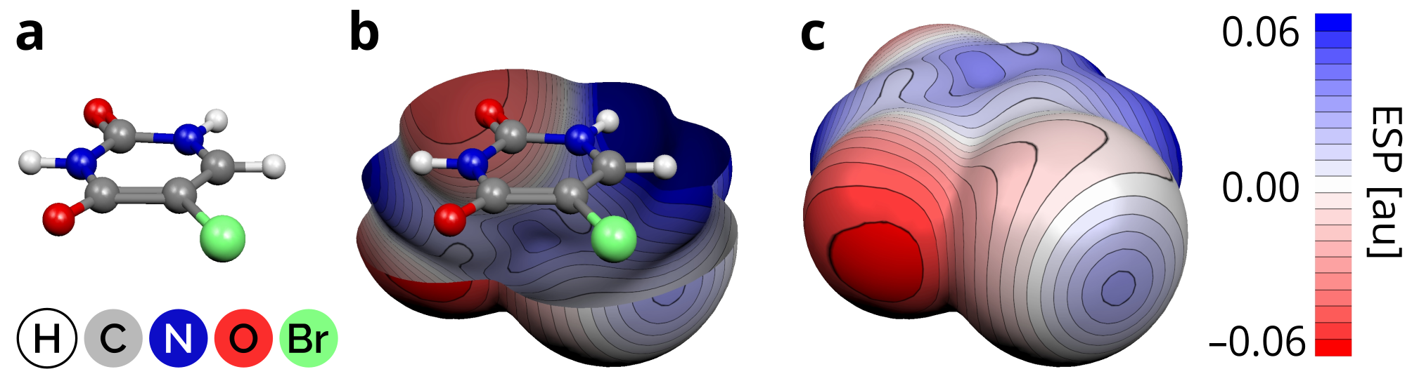
The attraction of halogens with other electronegative atoms was observed as early as the 1950s in the crystallographic studies of Hassel et al. 14 although the synthesis of X-bonded complexes dates back to 19th century 15. The puzzling nature of the attraction between two electron-rich chemical groups was attributed merely to charge transfer effects until Politzer et al. came up with a simple model explaining many of the X-bond features 16. Based on quantum chemical calculations of the molecular electrostatic potentials (MEPs) they proposed that the surface of halogens contains regions of both positive as well as negative electrostatic potential (ESP) 16. The positive region was labeled a sigma-hole (-hole) (Fig. 1), and interestingly enough this label appeared only 15 years after its first evidence 17. The X-bond has been exploited in many areas of chemistry and material science (reviewed e.g. in Refs. 5 and 18) and a great amount of work has been done on the theoretical aspects of X-bonds too 19, 6.
X-bonds are similar in strength to the more common hydrogen bonds (H-bonds). The X-bond stabilization energy typically amounts to 5–25 kJ/mol 20, 21 and it increases with the increasing atomic number of the halogen involved 22. The reason for this is the increasing size and magnitude of the halogen -hole 23. Another factor is the halogen polarizability 24, which also increases with the halogen atomic number. Contrary to traditional view, modern theoretical studies suggest that the role of charge transfer in the X-bond stabilization is modest 25. The strongest X-bonds are found in complexes of iodinated molecules; brominated and chlorinated molecules form weaker X-bonds. Fluorine is the least polarizable and the most electronegative halogen. It possesses positive -holes in rare cases and mostly in inorganic molecules 26, so it is of lower importance for biological applications.
The structural trends of X-bonds in biomolecules have been inferred from several Protein Data Bank (PDB) 27 surveys and theoretical analyses 28, 29, 7, 30. All of the studies focused on protein-ligand X-bonds. The X-bonds are mostly established with the protein backbone. Its carbonyl oxygen is the most frequent electron donor 29. No preference for backbones of -helices, -sheets or loop structures has been found 31. Lu et al. reported that about one-third of protein-ligand X-bonds involve an aromatic ring of the amino acid side chains 29. It was also reported that the X-bonds might not disrupt existing networks of H-bonds 32. Instead, an orthogonal pattern of X- and H-bonds is preferred involving the same electron donor atom 33. Another effect of ligand halogenation is a shortening of proximal H-bonds 34.
3 Nucleic Acids as Drug Targets
The deoxyribonucleic and ribonucleic acids (DNA and RNA) appear in living organisms in several forms. Whereas the DNA adopts only a few conformational classes, the structural diversity of RNA is much broader. DNA mostly occurs in the B-form helical conformation. A key RNA motif is the A-form double helix. However, loop regions, bulges, and other forms of mismatched nucleobases often disrupt the motif. Such a conformational variability is mirrored in the rapidly expanding variety of RNA functions recognized, especially in the last two decades. Apart from the classic roles of ribosomal, transfer and messenger RNAs, RNA was also shown to store genetic information, regulate gene expression, or act as enzyme 35, 36.
Many NA-binders are halogenated. The DNA alkylating agents often contain halogen atoms because halogens facilitate the alkylating reactions on NAs. This way, anticancer cisplatin and its analogues 37, 38 covalently modify DNA. The reaction products block replication or transcription processes. The effect of alkylation is non-specific in terms of DNA sequence and also cell type. The modifications occur in both normal and cancer cells, but the higher proliferation rate of the cancer cells makes these drugs effective.
Alternatively to covalent modifications, various classes of compounds bind to double-stranded DNA (dsDNA) in a noncovalent manner. Examples include the anticancer anthracycline type antibiotics daunomycin and adriamycin, and the polypeptide antibiotic dactynomicin, which works mainly by intercalating into DNA. The intercalation, described as an insertion of a planar molecule between consecutive base pairs, interferes with DNA-processing enzymes 39. Other small molecules bind to dsDNA interacting with base pair functional groups on the floor of the minor or major groove. Much of the research has concentrated on the DNA minor groove recognition leading to improved sequence selectivity of the compounds 40, 41.
Important classes of drugs target ribosomal RNA (rRNA). In fact, most of the RNA-targeting drugs on the market act on ribosomes 42. The ribosome is a biomachine which synthesizes proteins. As such, it represents a critical center of cellular life. The differences between bacterial and eukaryotic ribosomes have allowed developing specific antibacterial agents that are often based on natural products 43, 44, 45. There is a variety of binding locations within the ribosome; the most frequent sites are the peptidyl-transferase center on the 50S subunit and the decoding center on the 30S subunit. Aminoglycosides and tetracyclines are the best-known classes of small molecules whose primary target is the 30S subunit. Oxazolidonones instead exert their antibacterial action by binding to the 23S rRNA present within the 50S subunit. Macrolides and related compounds (lincosamides and streptogramins), as well as chloramphenicol and clindamycin also target the 50S subunit. Several ribosome-binding molecules have been prepared containing a halogen atom stemming mostly from the pioneering case study of chloramphenicol 46.
In the past decade, other non-coding RNAs (ncRNAs) have emerged as prospective drug targets 42, 47. Highly conserved ncRNAs provide new opportunities to expand the repertoire of drug targets to treat infections. Viral regulatory elements located in untranslated regions of mRNA often form folded structures that harbor potential binding sites for small molecules. The absence of homologous host cell RNAs makes them attractive for the development of innovative antiviral compounds.
There are a plethora of ncRNAs under intense pharmaceutical research due to their ability to interact with low-molecular ligands. For instance, human immunodeficiency virus type-1 (HIV-1) Trans-activation response (TAR) RNA plays an essential role in HIV-1 replication through its interaction with the viral trans-activator of transcription. Such interaction might be disrupted by ligands (reviewed in Refs. 48 and 49), where some of them contain halogen atoms 50.
Another example is an internal ribosome entry site (IRES) from Hepatitis C virus (HCV) 51. The IRES RNA contains several independently folding domains that are potential targets for the development of selective viral translation inhibitors. A diverse set of ligands including oligonucleotides, peptides as well as small molecules have been reported to block IRES function by distinct mechanisms 52. Like in the case of other ncRNAs, the drug candidates occasionally contain halogens. Nevertheless, no drug targeting a ncRNA has been approved for the market.
Many lines of evidence are linking mutations and dysregulations of ncRNAs to neurodegenerative disorders 53, 54. The presence of expanded CNG repeats in 5’ and 3’ untranslated regions is related to important diseases such as Myotonic dystrophy type 1, spinocerebellar ataxia type 3, and fragile X-associated tremor-ataxia syndrome. For example, the pathological expansion of CAG repeats (35 consecutive CAG codons) in huntingtin exon 1 encodes a mutant protein whose abnormal function determines Huntington’s disease. Finding compounds able to bind pathogenic CNG repeats with high specificity may be a valuable strategy against these devastating diseases. Their design is still limited by the lack of structural information, although some small molecules have emerged through various strategies 55, 56.
Both DNA and RNA can also adopt a non-canonical higher-order structure called G-quadruplexes (G4s) that are involved in regulating multiple biological pathways such as transcription, replication, translation and telomere structure 57. The building blocks of G4s are guanosine-rich quartets that self-associate into a square-planar platform through a cyclic Hoogsteen H-bonded arrangement. G4s are found in oncogene promoters, in telomeres, as well as in introns of mRNAs. These regions have been recognized as potential targets for anticancer drugs 58. A large number of small molecules are able to bind the quadruplex structures. They are characterized by polycyclic heteroaromatic scaffolds, or by cyclic/acyclic non-fused aromatic rings. Thus far, only a few molecules have been found to selectively bind the telomeric G4s, although their therapeutic potential appears high 59.
4 Nucleic Acids as X-bond Acceptors
In the X-bond, the -hole on a halogen represents a Lewis acid that interacts with a Lewis base. NAs seem to offer an abundance of basic chemical groups. The backbone contains phosphate groups with two oxygens carrying a charge of . Likewise, ribose and deoxyribose contain oxygens with lone electron pairs that could serve as -hole acceptors too. Further, the nucleobases form H-bonds that could be potentially replaced or augmented by X-bonds.
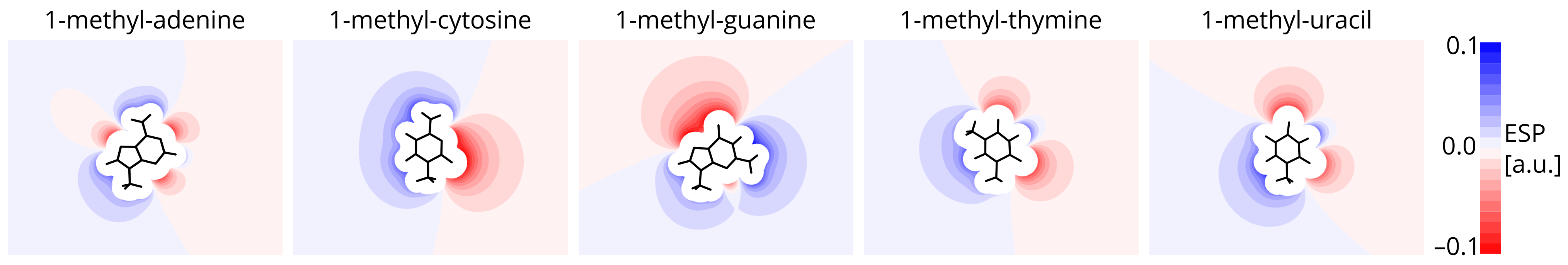
Nucleobases themselves show certain electrostatic diversity, where the negative sites may play a role of halogen acceptors. Fig. 2 depicts MEPs of five most common nucleobases projected onto the plane of their aromatic systems. Cytosine and guanine contain areas of more negative ESP than the other nucleobases. Thymine and uracil resemble each other having two negative sites on the oxygens separated by a small positive site. Perhaps the most heterogeneous ESP is around adenine which exhibits six areas of zero ESP near the molecular surface, as compared to four (G, T, U) and two (C). Apart from this, all of the nucleobases are aromatic, which allows them to act as electron donors via their -electrons above and below their rings (not shown).
In nucleic acids, the situation is more complicated than in nucleobases: the three-dimensional structure of DNA and RNA is electrostatically diverse due to the presence of the phosphate backbone. For instance, the DNA minor groove was shown to be more electronegative than the major groove 60. Thanks to its plasticity, RNA may create folds with even more unusual electrostatic characteristics. Indeed, regions of strong negative electrostatic potentials were exploited in designing efficient TAR binders 61. Electrostatic interaction is also the main driving force of aminoglycoside binding to the ribosome 62. Overall, NAs seem to offer favorable electrostatics to attract positive -holes on halogens. It remains elusive, how is such ability employed in ligand recognition and NA self-assembly.
5 Structural Survey Yields Two Data Sets
To understand interaction preferences of halogens we analyzed known NA structures. We started with a broad set of X-ray structures from the PDB (September 2016) 27. Apart from X-ray structures, the PDB also contains NA structures determined by nuclear magnetic resonance (NMR) techniques. For two reasons, we deliberately omitted those from our analyses. First, we wanted to be consistent with the strategies adopted by previous structural surveys of X-bonds in protein-ligand complexes. Second, there are indications that it may be difficult to assess the quality of the deposited NMR structural ensembles 63, which could complicate the geometric characterization.
Within the X-ray structures, we searched for complexes containing a nucleic acid and a halogen atom (Cl, Br, or I). Fluorine was excluded from the search due to its extremely low ability to form X-bonds in biological systems. The selected structures comprise nucleic acids and their complexes with other nucleic acids, low-molecular ligands, and/or proteins. To get reliable geometric characteristics, only data with the resolution better than 3.0 Å were considered. Note that the previous PDB surveys of X-bonds in protein complexes used the same resolution threshold 28, 30.
From the PDB, we obtained 672 files which were subsequently filtered. We excluded structures containing halogens in the form of ions. Following the recommendation of the International Union of Pure and Applied Chemistry 64, we selected X-bonds as the contacts between halogens and electronegative atoms (N, O, P) shorter than or equal to the sum of van der Waals (vdW) radii 65 (Tab. 1). The interactions involving at least one of the two interacting atoms with the crystal occupancy lower than 0.5 were omitted. Because of the X-bond directionality23, we also required the angle of R–XY to be higher than 120∘. Same or similar geometric criteria were used previously to define biological X-bonds with proteins 28, 29, although the wider angular range was used in other studies as well 7, 30.
| N | O | P | |
|---|---|---|---|
| Cl | 3.30 | 3.27 | 3.55 |
| Br | 3.40 | 3.37 | 3.65 |
| I | 3.53 | 3.50 | 3.78 |
We also searched for the X-bonds that are formed with the aromatic systems of the nucleobases. To this aim, we considered only halogen contacts closer than 5 Å to the aromatic plane that make an angle between the plane normal vector and the X–C bond smaller than 60∘.
In the end, we obtained a set of 21 X-bonds that satisfy the data quality, chemical and geometric criteria. This amount is a rather low. Scholfield et al. reported 760 protein complexes with an X-bond in 2012, which stands for about 1 % of the 80,000 protein structures deposited in the PDB at that time. The 21 X-bonds here represent about 0.2 % of ca 9,000 structures containing NA.
Hence to better capture possible geometric properties of halogen interactions, we collected a more extended set of complexes with a longer interaction distance. An arbitrary threshold of 4 Å was chosen such that it is higher than the X-bond length but still short enough to hint for an attractive interaction. Within the article, the interactions are referred to as linear contacts and they comprehend the X-bonds as well. We found 72 linear contacts. Table 2 summarizes various subsets of the PDB query.
| Cl | Br | I | sum | |
|---|---|---|---|---|
| Files | ||||
| PDB search count | 402 | 204 | 66 | 672 |
| Contains contact(s) | 43 | 162 | 34 | 239 |
| Contains contact(s) with NA building block | 31 | 135 | 22 | 188 |
| Contains linear contacts(s) with NA building block | 22 | 29 | 2 | 53 |
| Contains X-bond(s) with NA building block | 2 | 17 | 2 | 21 |
| Interactions | ||||
| Number of contacts | 611 | 1,319 | 205 | 2,135 |
| Contacts with NA building block | 43 | 315 | 41 | 399 |
| Linear contacts with NA building block | 22 | 48 | 2 | 72 |
| X-bonds with NA building block | 2 | 17 | 2 | 21 |
6 The X-Bonds Favor Nucleo-basePhosphate Pattern
Within the X-bond set, the variety of interacting partners is low; 20 X-bonds involve a halogenated nucleobase, one X-bond is formed by cisplatin chlorine. The set contains 19 X-bonds with a phosphate oxygen as the electron donor; further, there is one X-bonds with an aromatic ring, and one with cytosine oxygen. Overall, only two X-bonds do not concur with the dominant nucleobasephosphate interaction pattern.
The X-bond geometries are close to ideal. The X-bond lengths are shorter than the sum of the vdW radii by about 8 %, which conforms with the contractions reported for the protein X-bonds 7. About 90 % of the X-bonds are straighter than 160∘.
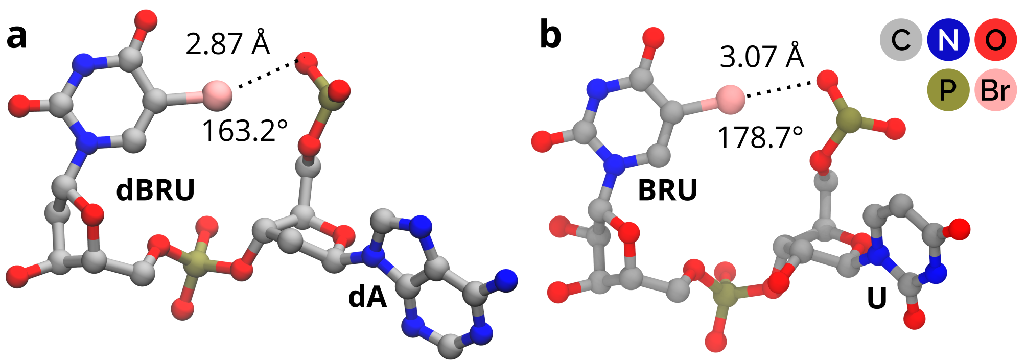
The shortest X-bond in the set belongs to a structure of a Holliday junction; the X-bond is found between a bromodeoxyuridine and a phosphate oxygen of two neighboring residues (Fig. 3a) (PDB: 2org, resolution 2.0 Å) 11. The X-bond was shown to stabilize particular DNA assembly in competition with H-bond. The straightest X-bond also involves a brominated uridine and phosphate oxygen (Fig. 3b) (PDB: 2bu1, resolution 2.2 Å) in a complex of RNA and phage MS2 coat protein 66 and the overall geometry is remarkably similar to the one found in Holliday junction. Whereas the study on Holliday junction fully appreciated the role of X-bonding, in the latter case the specific interaction of the bromine remained unrecognized.
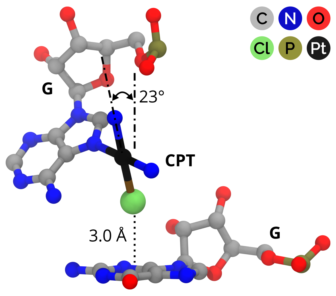
What is important, we have identified no noncovalent ligand involved in X-bonding. The only non-nucleobase residue that participates in an X-bonds is cisplatin covalently bound to an adenine (PDB 5j4c, resolution 2.8 Å67). It is also the only halogen donor that forms an X-bond with a -system, although the interaction with one of the guanine nitrogens would alone classify the interaction as X-bond. The X-bond features superb geometric characteristics (Fig. 4). Unlike proteins, in nucleic acids, the halogen- interaction competes with interactions more often. Especially in dsDNA, it is hardly conceivable that there is a space for a halogen to attack the nucleobases from above or below of their aromatic planes. The situation in RNAs might be more favorable, but the single occurrence of such X-bond is hard to generalize.
7 Interactions Longer Than X-Bonds
Table 3 summarizes counts of various types of interactions, and the statistics of the interaction geometries of the set of 72 halogen linear contacts.
In the NA complexes, the geometric quality of the halogen interactions increases in the order of Cl Br I. The interaction angles are more linear for heavier halogens. Especially the contacts of chlorine are rather bent with the median angle of 141∘. The medians of the interaction lengths (Tab. 3) decrease with the increasing atomic number of the halogen. The surveys on protein-ligand X-bonds 28, 29 revealed the opposite trend, i.e. the increasing length of X-bonds with the increasing atomic number of the halogen involved. In this work (but also in Ref. 28), the statistical sample might be insufficient to provide reliable statistics, which is true especially for the two iodine X-bonds.
| Halogen | Count | Length [Å] (mediqr) | Angle [deg] (mediqr) |
|---|---|---|---|
| chlorine | 22 | 3.490.31 | 1418 |
| bromine | 48 | 3.450.30 | 16522 |
| iodine | 2 | 3.070.06 | 1733 |
| Electron Donor | |||
| N | 21 | 3.470.28 | 14011 |
| non-backbone O | 9 | 3.690.42 | 13525 |
| backbone O | 42 | 3.410.45 | 1668 |
| Halogenated Residue | |||
| Halogenated nucleotide | 51 | 3.430.43 | 16523 |
| Low-molecular ligand | 21 | 3.500.29 | 1407 |
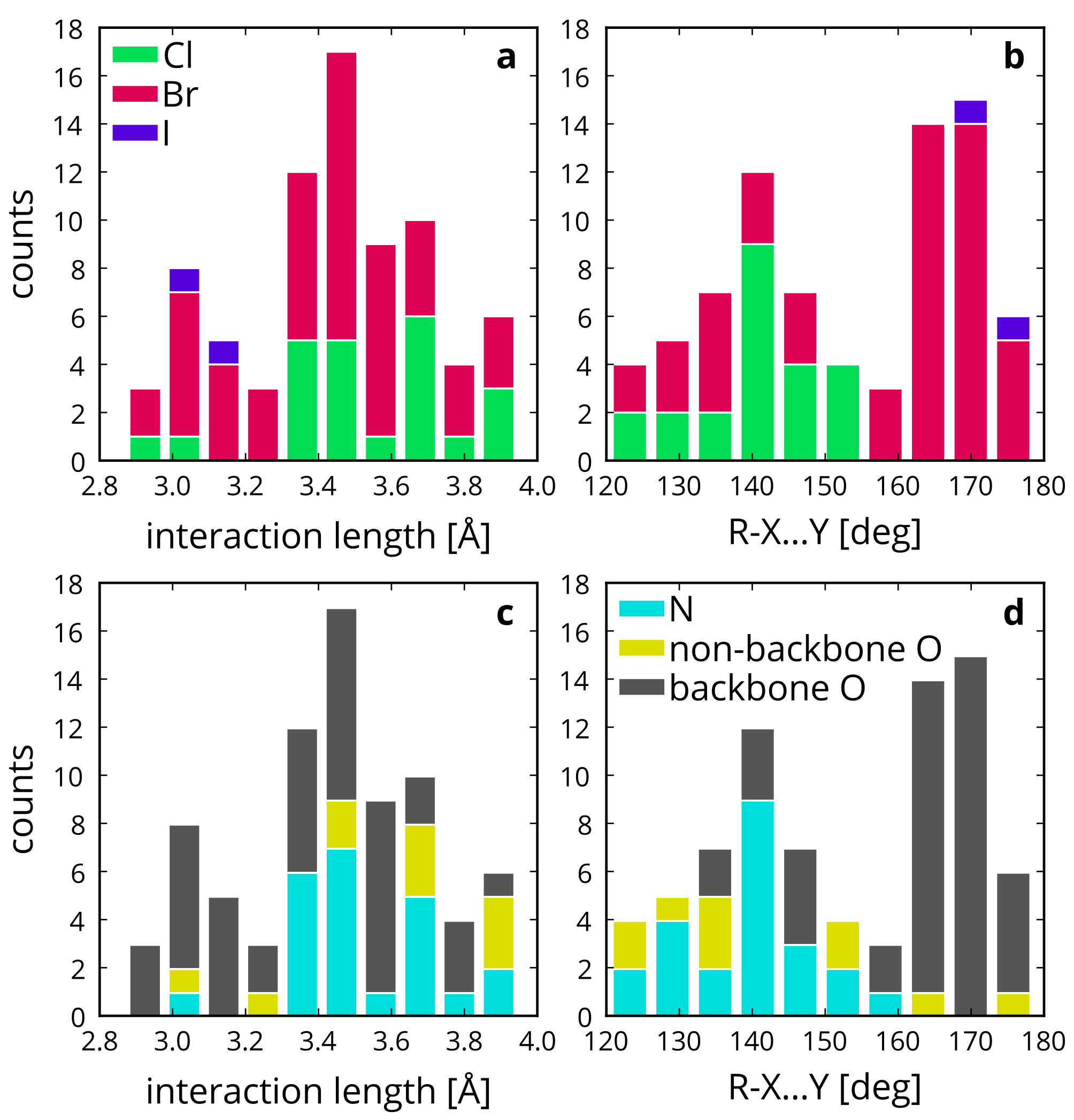
Fig. 5 shows the histograms of the interaction lengths and angles. In the histogram of lengths, the minor peak near 3.0 Å, which is comparable with the sum of the vdW radii, stands for almost ideal X-bond length. There is a major peak near 3.5 Å too. Two peaks also appear in the histogram of interaction angles; around 140∘ and 170∘.
Each of the halogen subsets contributes to the histograms with a different weight. Chlorine- and bromine-containing complexes span the whole range of interaction lengths. The two iodine complexes feature short interactions. The situation with angles is different. Chlorine complexes appear only in the region of lower interaction angles. The highest angle in the chlorine subset is 154∘, so chlorine interactions are likely weak. On the other hand, bromine complexes are scattered across the whole range of angles (Fig. 5b) with a cumulation around 170∘ (Fig. 6). The two iodine X-bonds are very straight (X-bond angle higher than 170∘) suggesting a strong interaction.
We analyzed the electron donors of halogen interactions found in the NA complexes. The backbone oxygen atom is the most common one. We identified 51 linear contacts of halogens with oxygen (71 %), and 21 with nitrogen (29 %). Although we included phosphorus as an electron donor in the search, no linear contact was found. The actual occurrence of the oxygen and nitrogen in NAs is roughly 2:1 (O:N), which likely contributes to the dominance of the linear contacts with oxygen. Most of the nitrogen interactions were found with chlorine, whereas bromine preferentially interacts with oxygen. 82 % of the all interacting oxygens belong to the phosphate backbone. Unlike other oxygens and nitrogens in NAs, the phosphate oxygens carry a negative charge, which explains their higher propensity to halogen -holes.
According to the electron-donating atoms, the geometric quality of the interactions increases in the order of non-backbone oxygen nitrogen backbone oxygen (Tab. 3). The interactions which employ a backbone oxygen are typically shorter and straighter than the others.
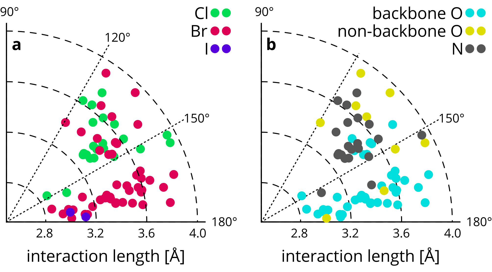
We conclude that different interaction atoms likely occur in different interaction geometries. It is also apparent on a projection of the interaction geometries to the polar coordinates (Fig. 6). We observed no correlation between interaction lengths and the corresponding angles.
8 Halogenated Ligands Show Sub-Optimal Interaction Geometries
We did not find any X-bonded noncovalent ligands in NA complexes. Nevertheless, in the set of linear contacts we have found 15 unique ligands – 14 chlorinated and one brominated. The remaining interactions involve halogenated nucleobases as halogen donors. The halogen interactions of ligands are longer and less linear compared to the interactions of halogenated nucleobases (Tab. 3). The interaction angles deviate notably from linearity, which suggest that such interactions do not play a critical role in the NA-ligand recognition.
From the pharmaceutical point of view, the low-molecular ligands are of higher interest than the halogenated building blocks. Below we discuss all of the instances among the linear-contact data set which involve a low-molecular ligand. Structural formulas of the ligands discussed are shown in Fig. 7.

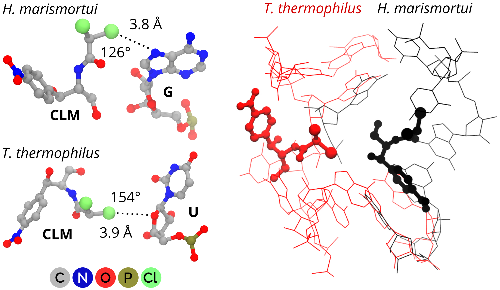
Several halogenated compounds bind to the bacterial ribosome. Chloramphenicol (1) 46 is a classic antibiotic compound that targets the ribosomal A-site crevice in the 50S subunit (PDB 1nji, resolution 3.0 Å68; 4v7w, resolution 3.0 Å69). 1 contains two aliphatic chlorines that are activated by the nearby carbonyl group. Two distinct binding orientations in two different ribosomal system were proposed (T. thermophilus 68 and H. marismortui 69) (Fig. 8). The lengths of both chlorine interactions are beyond the respective sums of the vdW radii (about 3.3 Å, Tab. 1), and quite bent. One of the chlorines approaches either the ribose in-ring oxygen (3.9 Å, 154∘), or a guanine nitrogen (3.8 Å, 126∘), respectively. The geometries suggest that the halogens interactions contribute weakly to the complex stability.
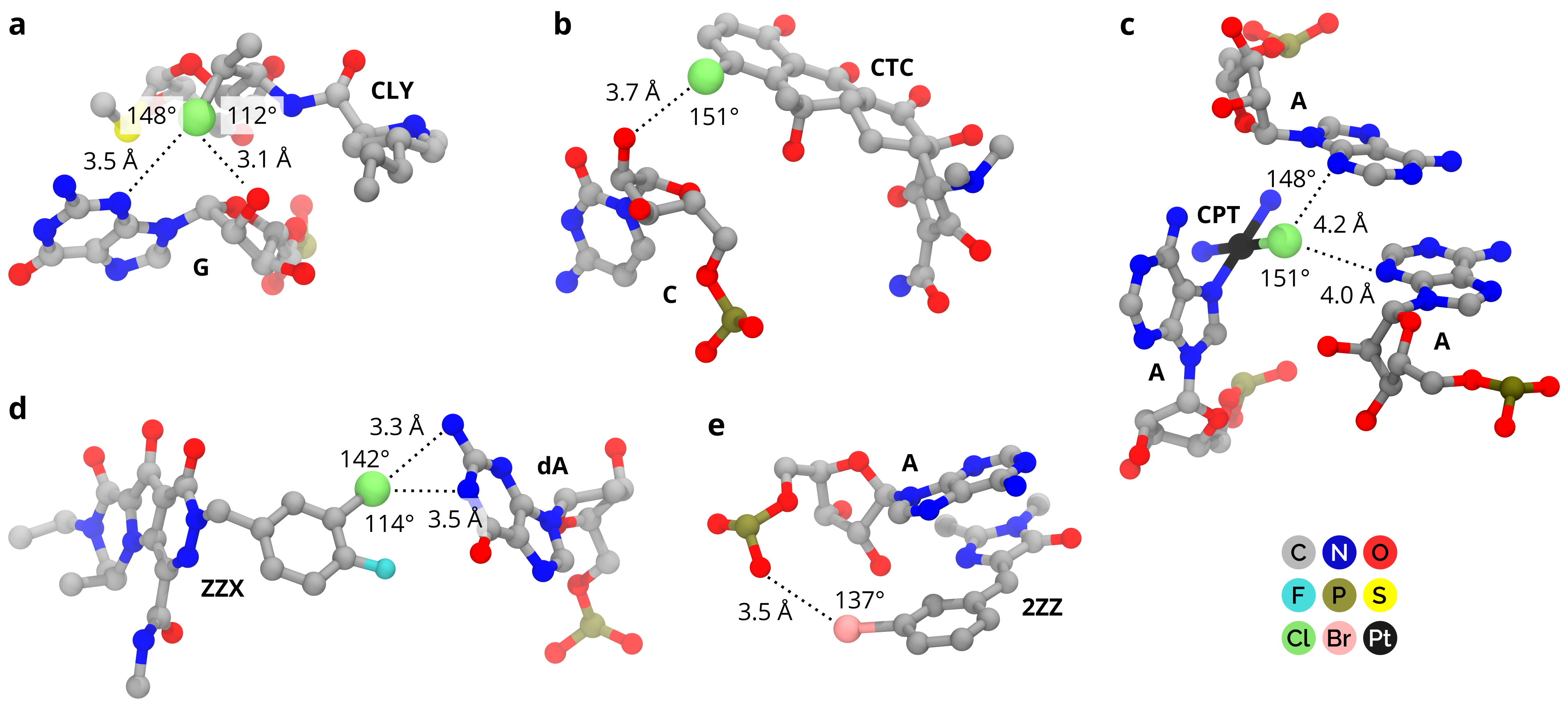
Clindamycin (2) 70 is another halogenated ribosome binder 71 (PDB 1yjn, resolution 3.0 Å) that binds into the 50S subunit (Fig. 9a) and forms linear contacts. 2 contains one chlorine that directs towards the sugar edge of guanosine. There is an interaction with guanine nitrogen (3.5 Å, 148∘). At the same time, there is a shorter but more bent contact with sugar O2’ oxygen (3.1 Å, 112∘).
7-chlorotetracycline (3) 72 belongs to a class of tetracycline antibiotics that bind into the ribosomal 30S subunit. This class acts by preventing correct processing of aminoacyl-tRNA. Although no complex of 3 with the ribosome satisfies the data-set criteria, a complex with an RNA aptamer does. The aptamer was designed to bind 3 with sub-nanomolar affinity (PDB 3egz, resolution 2.2 Å73). It features an interaction between a chlorine and a ribose oxygen of a cytidine (Fig. 9b) (3.7 Å, 151∘). Due to the sub-optimal geometry, the role of halogen interaction in complex stabilization is likely marginal. Moreover, there are many other intermolecular interactions between the 3 and the aptamer, such as a stacking of a planar part of 3 with a guanine, and an oxygen-magnesium coordination (not shown). Those reduce the relative importance of the halogen contact even more.
Recently, crystal structures of T. thermophilus ribosomes with cisplatin 37 (4 in Fig. 9c) were resolved (PDB 5j4b, resolution 2.6 Å; 5j4c, resolution 2.8 Å67). Nine molecules of cisplatin are covalently bound to the ribosome in place of one of the chlorines. Two of the cisplatin residues were identified to interfere with the mRNA tunnel and the GTPase site – the places critical for the ribosome functions 67. One cisplatin out of the nine forms an X-bond with a guanine -system (Fig. 4). Another cisplatin directs with its chlorine in between two stacked adenines with the shortest distance to a nitrogen of 4.0 Å and angle 151∘. Another nitrogen is about 4.2 Å far away and forms the angle of 148∘ (Fig. 9c).
Our data set contains several ligands that bind into the prototype foamy virus (PFV) intasome, i.e. a complex of a viral integrase and DNA. Such a system serves as a model for HIV-1 integrase inhibition. Several inhibitors were developed with a halogenated phenyl ring (5 and 6) that form X-bonds with NAs 74, 75, 76. No difference in the interaction geometries was found between 5 and 6 analogs. 5-analog interactions show the best geometric characteristics among the PFV ligands (but generally still modest); the chlorine on 5 interacts with two adenine nitrogens (Fig. 9d). While the contact length of 3.3 Å is comparable with the sum of vdW radii, the angle of 142∘ is far from the ideal 180∘. The other contact is 3.5 Å-long and even more bent (angle about 114∘). We add that an aromatic fluorine activates the neighboring chlorine by making its -hole more positive 23. The effect of fluorination on the ligand binding affinity is not straightforward, however 77.
We found only one brominated ligand with a contacts shorter than 4 Å. A brominated imidazole analog (7) binds into a G-quadruplex formed within an RNA aptamer called Spinach (PDB 4q9q, resolution 2.45 Å) 78. The Spinach module allows a green-fluorescence-protein-like functionality by selectively activating fluorescence of 7. 7 binds in a planar conformation with a strong stacking interaction with adenine. Besides this, it forms a contact with a phosphate oxygen of 3.5 Å under the angle of 137∘ (Fig. 9e).
9 Summary and Outlook
The role of X-bonding in drug development has been emphasized for a decade. Nonetheless, the knowledge-based design of halogenated drugs that feature an X-bond with their biomolecular targets is still a tedious task. Although there have been published several pioneering studies that involved protein targets 79, 80, 77, the nucleic acids still lack a successful application.
In this work, we focused on X-ray structures of biomolecular complexes containing halogen interactions with nucleic acids. The general ability of nucleic acids to provide electron donating groups to X-bonding has been confirmed here. Using criteria recommended by IUPAC for X-bond definition, we found 21 NA complexes with the X-bond. We also analyzed interactions of the halogen atoms longer than X-bonds.
All but one of the X-bonds involved a halogenated nucleobase (preferably uridine). Halogenated nucleotides are often utilized in X-ray crystallography, which explains their high occurrence in the NA complexes. The incorporation of heavy atoms such as bromine into the structure helps to overcome the phase problem. Moreover, the halogenated nucleotides alter the physico-chemical properties of the NAs thus modulate the crystallization conditions; in the classical theory this is due to their stacking properties 81, but recently X-bonding has also been shown to play a role 11. What is more, halogenated nucleobases may be also used in radiation anti-cancer therapy as radiosensitizers 82. Although, the exact mechanism of the increased radiosensitivity is not known, the changes in the ionization potential brought by the halogens or global structural changes associated with modified nucleobase pairing have been proposed 83, 84.
An important feature of X-bonds in NA complexes is their preference towards backbone oxygens. This is true also for the longer halogen interactions. The phosphate oxygens tend to form straighter and shorter X-bonds than other electron donors, possibly due to their negative charge.
In NAligand noncovalent recognition, a variety of interactions plays a role. H-bonding and London dispersion-driven stacking interactions belong to the classic interactions, complemented by salt bridges, water-network disruptions, or metal-ion interactions. Strictly speaking, no noncovalent ligand has been found to employ X-bonding in the NA binding (according to the IUPAC recommendations). No X-bonded NA ligand found is an intriguing fact, which may be explained in two ways:
i) The X-bonding ligands may be inefficient in NA recognition. If so, the halogenated ligands that bind into NAs bind due to other kinds of interactions that are stronger than X-bonds. Our data support this hypothesis. All of the halogen interactions of ligands found in the survey here show sub-optimal geometries. Given the high X-bond directionality 85, the stabilization energy of a complex reduces notably when deviating from the ideal geometry. Hence, the contribution to the stabilization of the NA complexes is likely not dominant. Of course, there are exceptions where X-bonding is the driving interaction. For instance, it was proven that a single X-bond/H-bond exchange might transform the DNA conformation completely 11.
ii) Medicinal chemists may under-appreciate the role of X-bonding in the drug-design strategies. Our data support also this hypothesis, because we found only a few halogenated ligands: 20 chlorinated, 8 brominated and one iodinated.
The current study on its own is unable to dissect which reason is more likely. The starting set of PDBs was biased towards the lighter halogens, which are, however, less suitable for strong X-bonds. For a better understanding of the role of X-bonding, structural and functional studies are required, especially of the less-frequent brominated and iodinated compounds.
The effects of halogens are not limited to the X-bonding, though. The halogenation affects the global electron distribution and consequently many of the molecular properties. For instance, Fanfrlík et al. demonstrated that solvation/desolvation may compensate for favorable -holelone pair interaction in halogen-to-hydrogen substitution in a protein-ligand complex 86. Also, a PDB survey of two protein families revealed shortening on H-bonds proximal to ligand halogens 34. This corresponds with the notion that halogenation increases the acidity of proximal H-bond donors. Still, these effects in NAs are difficult to inspect with the limited statistical sample of X-bonds here, and of halogenated NA ligands in general.
Nevertheless, the current analyses may help in designing novel X-bonded NA binders. Such binders should contain a bromine or iodine to form a strong X-bond. The ligand binding should be directed to the vicinity of sugar-phosphate backbone, for example to groove or bulge regions. Consequently, such a ligand should likely not contain extensive aromatic systems that are prone to intercalation into canonical helices. Systematic halogenation of known pharmacologically active compounds may be an optimal strategy to identify new agents with enhanced activity, presumably supported by the specific X-bonds.
10 Biographies
Michal H. Kolář received his Ph.D. in 2013 from Charles University in Prague, and from the Institute of Organic Chemistry and Biochemistry, Academy of Sciences of the Czech Republic. With Pavel Hobza he focused on theoretical and computational description of noncovalent interactions. He is a recipient of the Humboldt Research Fellowship for Postdoctoral Researchers. From 2014 he worked with Paolo Carloni in Forschungszentrum Jülich, Germany, on RNA-ligand recognition. In 2016 he moved to Helmut Grubmüller’s group at the Max Planck Institute for Biophysical Chemistry, Göttingen, Germany, pursuing his interest in non-coding RNAs and computer simulations.
Oriana Tabarrini is a Professor of Medicinal Chemistry at the Department of Pharmaceutical Sciences (University of Perugia, Italy). After the degree in Pharmaceutical Chemistry and Technology she has worked for several years with research grants from pharmaceutical industries. In 2002 was promoted to Associate Professor. Her research has mainly aimed at developing small molecules as pharmacological tools and potential chemotherapeutics with particular focus on nucleic acid binders as antivirals and more recently for the treatment of neurodegenerative disorders. She has published over 85 research articles in leading peer-reviewed journals, including some invited reviews and patents. She has also received several project grants and is an ad-hoc reviewer for several top journals.
11 Abbreviations
- BRU
-
5-bromouridine
- CLM
-
chloramphenicol
- CLY
-
clindamycine
- CPT
-
cisplatin
- DNA
-
deoxyribonucleic acid
- dsDNA
-
double-stranded DNA
- G4
-
G-quadruplex
- H-bond
-
hydrogen bond
- HCV
-
Hepatitis C virus
- HIV-1
-
human immunodeficiency virus type 1
- IRES
-
internal ribosome entry site
- NA
-
nucleic acid
- ncRNA
-
non-coding RNA
- PDB
-
Protein Data Bank
- PFV
-
prototype foamy virus
- RNA
-
ribonucleic acid
- TAR
-
trans-activation response RNA element
- X-bond
-
halogen bond
12 Corresponding Author Information
Email: michal@mhko.science
References
- Overington et al. 2006 Overington, J. P.; Al-Lazikani, B.; Hopkins, A. L. How many drug targets are there? Nat. Rev. Drug Discovery 2006, 5, 993–996
- Ezkurdia et al. 2014 Ezkurdia, I.; Juan, D.; Rodriguez, J. M.; Frankish, A.; Diekhans, M.; Harrow, J.; Vazquez, J.; Valencia, A.; Tress, M. L. Multiple evidence strands suggest that there may be as few as 19 000 human protein-coding genes. Human Mol. Genet. 2014, 23, 5866–5878
- Xu et al. 2014 Xu, Z.; Yang, Z.; Liu, Y.; Lu, Y.; Chen, K.; Zhu, W. Halogen bond: its role beyond drug–target binding affinity for drug discovery and development. J. Chem. Inf. Model. 2014, 54, 69–78
- Hernandes et al. 2010 Hernandes, M. Z.; Cavalcanti, S. M. T.; Moreira, D. R. M.; de Azevedo, J.; Filgueira, W.; Leite, A. C. L. Halogen atoms in the modern medicinal chemistry: hints for the drug design. Curr. Drug Targets 2010, 11, 303–314
- Cavallo et al. 2016 Cavallo, G.; Metrangolo, P.; Milani, R.; Pilati, T.; Priimagi, A.; Resnati, G.; Terraneo, G. The halogen bond. Chem. Rev. 2016, 116, 2478–2601
- Ford and Ho 2015 Ford, M. C.; Ho, P. S. Computational tools to model halogen bonds in medicinal chemistry. J. Med. Chem. 2015, 59, 1655–1670
- Scholfield et al. 2013 Scholfield, M. R.; Zanden, C. M. V.; Carter, M.; Ho, P. S. Halogen bonding (X-bonding): a biological perspective. Protein Sci. 2013, 22, 139–152
- Wilcken et al. 2013 Wilcken, R.; Zimmermann, M. O.; Lange, A.; Joerger, A. C.; Boeckler, F. M. Principles and applications of halogen bonding in medicinal chemistry and chemical biology. J. Med. Chem. 2013, 56, 1363–1388
- Duckett et al. 1988 Duckett, D. R.; Murchie, A. I.; Diekmann, S.; von Kitzing, E.; Kemper, B.; Lilley, D. M. The structure of the Holliday junction, and its resolution. Cell 1988, 55, 79–89
- Lilley 2000 Lilley, D. M. Structures of helical junctions in nucleic acids. Q. Rev. Biophys. 2000, 33, 109–159
- Voth et al. 2007 Voth, A. R.; Hays, F. A.; Ho, P. S. Directing macromolecular conformation through halogen bonds. Proc. Natl. Acad. Sci. U. S. A. 2007, 104, 6188–6193
- Carter and Ho 2011 Carter, M.; Ho, P. S. Assaying the energies of biological halogen bonds. Cryst. Growth Des. 2011, 11, 5087–5095
- Carter et al. 2013 Carter, M.; Voth, A. R.; Scholfield, M. R.; Rummel, B.; Sowers, L. C.; Ho, P. S. Enthalpy–entropy compensation in biomolecular halogen bonds measured in DNA junctions. Biochemistry 2013, 52, 4891–4903
- Hassel and Hvoslef 1954 Hassel, O.; Hvoslef, J. The structure of bromine 1,4-dioxanate. Acta Chem. Scand. 1954, 8, 873–873
- Guthrie 1863 Guthrie, F. XXVIII. On the iodide of iodammonium. J. Chem. Soc. 1863, 16, 239–244
- Brinck et al. 1992 Brinck, T.; Murray, J. S.; Politzer, P. Surface electrostatic potentials of halogenated methanes as indicators of directional intermolecular interactions. Int. J. Quantum Chem. 1992, 44, 57–64
- Clark et al. 2007 Clark, T.; Hennemann, M.; Murray, J. S.; Politzer, P. Halogen bonding: the -hole. J. Mol. Model. 2007, 13, 291–296
- Gilday et al. 2015 Gilday, L. C.; Robinson, S. W.; Barendt, T. A.; Langton, M. J.; Mullaney, B. R.; Beer, P. D. Halogen bonding in supramolecular chemistry. Chem. Rev. 2015, 115, 7118–7195
- Kolář and Hobza 2016 Kolář, M. H.; Hobza, P. Computer modeling of halogen bonds and other -hole interactions. Chem. Rev. 2016, 116, 5155–5187
- Řezáč et al. 2012 Řezáč, J.; Riley, K. E.; Hobza, P. Benchmark calculations of noncovalent interactions of halogenated molecules. J. Chem. Theory Comput. 2012, 8, 4285–4292
- Kolář et al. 2014 Kolář, M. H.; Deepa, P.; Ajani, H.; Pecina, A.; Hobza, P. Characteristics of a -hole and the nature of a halogen bond. In Halogen Bonding II; Metrangolo, P., Resnati, G., Eds.; Springer International Publishing: Cham, 2014; pp 1–25
- Politzer et al. 2010 Politzer, P.; Murray, J. S.; Clark, T. Halogen bonding: an electrostatically-driven highly directional noncovalent interaction. Phys. Chem. Chem. Phys. 2010, 12, 7748–7757
- Kolář et al. 2014 Kolář, M.; Hostaš, J.; Hobza, P. The strength and directionality of a halogen bond are co-determined by the magnitude and size of the -hole. Phys. Chem. Chem. Phys. 2014, 16, 9987–9996
- Riley and Hobza 2008 Riley, K. E.; Hobza, P. Investigations into the nature of halogen bonding including symmetry adapted perturbation theory analyses. J. Chem. Theory Comput. 2008, 4, 232–242
- Řezáč and de la Lande 2017 Řezáč, J.; de la Lande, A. On the role of charge transfer in halogen bonding. Phys. Chem. Chem. Phys. 2017, 19, 791–803
- Metrangolo et al. 2011 Metrangolo, P.; Murray, J. S.; Pilati, T.; Politzer, P.; Resnati, G.; Terraneo, G. Fluorine-centered halogen bonding: a factor in recognition phenomena and reactivity. Cryst. Growth Des. 2011, 11, 4238–4246
- Berman et al. 2000 Berman, H. M.; Westbrook, J.; Feng, Z.; Gilliland, G.; Bhat, T. N.; Weissig, H.; Shindyalov, I. N.; Bourne, P. E. The protein data bank. Nucleic Acids Res. 2000, 28, 235–242
- Auffinger et al. 2004 Auffinger, P.; Hays, F. A.; Westhof, E.; Ho, P. S. Halogen bonds in biological molecules. Proc. Natl. Acad. Sci. U. S. A. 2004, 101, 16789–16794
- Lu et al. 2010 Lu, Y.; Wang, Y.; Zhu, W. Nonbonding interactions of organic halogens in biological systems: implications for drug discovery and biomolecular design. Phys. Chem. Chem. Phys. 2010, 12, 4543–4551
- Sirimulla et al. 2013 Sirimulla, S.; Bailey, J. B.; Vegesna, R.; Narayan, M. Halogen interactions in protein–ligand complexes: implications of halogen bonding for rational drug design. J. Chem. Inf. Model. 2013, 53, 2781–2791
- Wilcken et al. 2012 Wilcken, R.; Zimmermann, M. O.; Lange, A.; Zahn, S.; Boeckler, F. M. Using halogen bonds to address the protein backbone: a systematic evaluation. J. Comput.-Aided Mol. Des. 2012, 26, 935–945
- Rowe and Ho 2017 Rowe, R. K.; Ho, P. S. Relationships between hydrogen bonds and halogen bonds in biological systems. Acta Crystallogr., Sect. B: Struct. Sci., Cryst. Eng. Mater. 2017, 73, 255–264
- Voth et al. 2009 Voth, A. R.; Khuu, P.; Oishi, K.; Ho, P. S. Halogen bonds as orthogonal molecular interactions to hydrogen bonds. Nat. Chem. 2009, 1, 74–79
- Poznański et al. 2014 Poznański, J.; Poznańska, A.; Shugar, D. A protein data bank survey reveals shortening of intermolecular hydrogen bonds in ligand-protein complexes when a halogenated ligand is an H-bond donor. PLoS One 2014, 9, e99984
- Kiss 2002 Kiss, T. Small nucleolar RNAs: an abundant group of noncoding RNAs with diverse cellular functions. Cell 2002, 109, 145–148
- Mercer et al. 2009 Mercer, T. R.; Dinger, M. E.; Mattick, J. S. Long non-coding RNAs: insights into functions. Nat. Rev. Genet. 2009, 10, 155–159
- Rosenberg et al. 1965 Rosenberg, B.; Van Camp, L.; Krigas, T. Inhibition of cell division in Escherichia coli by electrolysis products from a platinum electrode. Nature 1965, 205, 698–699
- Weiss and Christian 1993 Weiss, R. B.; Christian, M. C. New cisplatin analogues in development. Drugs 1993, 46, 360–377
- Baguley 1991 Baguley, B. DNA intercalating anti-tumour agents. Anti-Cancer Drug Des. 1991, 6, 1–35
- Baraldi et al. 2004 Baraldi, P. G.; Bovero, A.; Fruttarolo, F.; Preti, D.; Tabrizi, M. A.; Pavani, M. G.; Romagnoli, R. DNA minor groove binders as potential antitumor and antimicrobial agents. Med. Res. Rev. 2004, 24, 475–528
- Barrett et al. 2013 Barrett, M. P.; Gemmell, C. G.; Suckling, C. J. Minor groove binders as anti-infective agents. Pharmacol. Ther. 2013, 139, 12–23
- Thomas and Hergenrother 2008 Thomas, J. R.; Hergenrother, P. J. Targeting RNA with small molecules. Chem. Rev. 2008, 108, 1171–1224
- Hermann 2005 Hermann, T. Drugs targeting the ribosome. Curr. Opin. Struc. Biol. 2005, 15, 355–366
- Poehlsgaard and Douthwaite 2005 Poehlsgaard, J.; Douthwaite, S. The bacterial ribosome as a target for antibiotics. Nat. Rev. Microbiol. 2005, 3, 870–881
- Wilson 2014 Wilson, D. N. Ribosome-targeting antibiotics and mechanisms of bacterial resistance. Nat. Rev. Microbiol. 2014, 12, 35–48
- Ehrlich et al. 1947 Ehrlich, J.; Bartz, Q. R.; Smith, R. M.; Joslyn, D. A. Chloromyeetin, a new antibiotic from a soil Actinomycete. Science (Washington) 1947, 417
- Hermann 2016 Hermann, T. Small molecules targeting viral RNA. WIREs RNA 2016, 7, 726–743
- Mousseau et al. 2015 Mousseau, G.; Mediouni, S.; Valente, S. T. Targeting HIV transcription: the quest for a functional cure. In The Future of HIV-1 Therapeutics; Torbett, B., Goodsell, D. S., Richman, D., Eds.; Springer International Publishing: Cham, 2015; pp 121–145
- Tabarrini et al. 2016 Tabarrini, O.; Desantis, J.; Massari, S. Recent advances in the identification of Tat-mediated transactivation inhibitors: progressing toward a functional cure of HIV. Future Med. Chem. 2016, 8, 421–442
- Mayer and James 2004 Mayer, M.; James, T. L. NMR-based characterization of phenothiazines as a RNA binding scaffold. J. Am. Chem. Soc. 2004, 126, 4453–4460
- Lukavsky 2009 Lukavsky, P. J. Structure and function of HCV IRES domains. Virus Res. 2009, 139, 166–171
- Dibrov et al. 2013 Dibrov, S. M.; Parsons, J.; Carnevali, M.; Zhou, S.; Rynearson, K. D.; Ding, K.; Garcia Sega, E.; Brunn, N. D.; Boerneke, M. A.; Castaldi, M. P.; Hermann, T. Hepatitis C virus translation inhibitors targeting the internal ribosomal entry site: miniperspective. J. Med. Chem. 2013, 57, 1694–1707
- Castel et al. 2010 Castel, A. L.; Cleary, J. D.; Pearson, C. E. Repeat instability as the basis for human diseases and as a potential target for therapy. Nat. Rev. Mol. Cell Biol. 2010, 11, 165–170
- Mirkin 2007 Mirkin, S. M. Expandable DNA repeats and human disease. Nature 2007, 447, 932–940
- Kumar et al. 2012 Kumar, A.; Parkesh, R.; Sznajder, L. J.; Childs-Disney, J. L.; Sobczak, K.; Disney, M. D. Chemical correction of pre-mRNA splicing defects associated with sequestration of muscleblind-like 1 protein by expanded r (CAG)-containing transcripts. ACS Chem. Biol. 2012, 7, 496–505
- Bochicchio et al. 2015 Bochicchio, A.; Rossetti, G.; Tabarrini, O.; Krauss, S.; Carloni, P. Molecular view of ligands specificity for CAG repeats in anti-Huntington therapy. J. Chem. Theory Comput. 2015, 11, 4911–4922
- Lipps and Rhodes 2009 Lipps, H. J.; Rhodes, D. G-quadruplex structures: in vivo evidence and function. Trends Cell Biol. 2009, 19, 414–422
- Han and Hurley 2000 Han, H.; Hurley, L. H. G-quadruplex DNA: a potential target for anti-cancer drug design. Trends Pharmacol. Sci. 2000, 21, 136–142
- Neidle 2009 Neidle, S. The structures of quadruplex nucleic acids and their drug complexes. Curr. Opin. Struct. Biol. 2009, 19, 239–250
- Chin et al. 1999 Chin, K.; Sharp, K. A.; Honig, B.; Pyle, A. M. Calculating the electrostatic properties of RNA provides new insights into molecular interactions and function. Nat. Struct. Mol. Biol. 1999, 6, 1055–1061
- Davis et al. 2004 Davis, B.; Afshar, M.; Varani, G.; Murchie, A. I.; Karn, J.; Lentzen, G.; Drysdale, M.; Bower, J.; Potter, A. J.; Starkey, I. D.; Swardbrick, T.; Aboul-ela, F. Rational design of inhibitors of HIV-1 TAR RNA through the stabilisation of electrostatic hot spots. J. Mol. Biol. 2004, 336, 343–356
- Ma et al. 2002 Ma, C.; Baker, N. A.; Joseph, S.; McCammon, J. A. Binding of aminoglycoside antibiotics to the small ribosomal subunit: a continuum electrostatics investigation. J. Am. Chem. Soc. 2002, 124, 1438–1442
- Nabuurs et al. 2006 Nabuurs, S. B.; Spronk, C. A. M.; Vuister, G. W.; Vriend, G. Traditional biomolecular structure determination by NMR spectroscopy allows for major errors. PLoS Comput. Biol. 2006, 2, e9
- Desiraju et al. 2013 Desiraju, G. R.; Ho, P. S.; Kloo, L.; Legon, A. C.; Marquardt, R.; Metrangolo, P.; Politzer, P.; Resnati, G.; Rissanen, K. Definition of the halogen bond (IUPAC recommendations 2013). Pure Appl. Chem. 2013, 85, 1711–1713
- Bondi 1964 Bondi, A. Van der Waals volumes and radii. J. Phys. Chem. 1964, 68, 441–451
- Grahn et al. 2001 Grahn, E.; Moss, T.; Helgstrand, C.; Fridborg, K.; Sundaram, M.; Tars, K.; Lago, H.; Stonehouse, N. J.; Davis, D. R.; Stockley, P. G.; Liljas, L. Structural basis of pyrimidine specificity in the MS2 RNA hairpin–coat-protein complex. RNA 2001, 7, 1616–1627
- Melnikov et al. 2016 Melnikov, S. V.; Söll, D.; Steitz, T. A.; Polikanov, Y. S. Insights into RNA binding by the anticancer drug cisplatin from the crystal structure of cisplatin-modified ribosome. Nucleic Acids Res. 2016, gkw246
- Hansen et al. 2003 Hansen, J. L.; Moore, P. B.; Steitz, T. A. Structures of five antibiotics bound at the peptidyl transferase center of the large ribosomal subunit. J. Mol. Biol. 2003, 330, 1061–1075
- Bulkley et al. 2010 Bulkley, D.; Innis, C. A.; Blaha, G.; Steitz, T. A. Revisiting the structures of several antibiotics bound to the bacterial ribosome. Proc. Natl. Acad. Sci. U. S. A. 2010, 107, 17158–17163
- Birkenmeyer and Kagan 1970 Birkenmeyer, R. D.; Kagan, F. Lincomycin. XI. Synthesis and structure of clindamycin, a potent antibacterial agent. J. Med. Chem. 1970, 13, 616–619
- Tu et al. 2005 Tu, D.; Blaha, G.; Moore, P. B.; Steitz, T. A. Structures of MLS B K antibiotics bound to mutated large ribosomal subunits provide a structural explanation for resistance. Cell 2005, 121, 257–270
- Duggar 1949 Duggar, B. M. Aureomycin and Preparation of Same. U.S. Patent 2,482,055, 1949
- Xiao et al. 2008 Xiao, H.; Edwards, T. E.; Ferré-D’Amaré, A. R. Structural basis for specific, high-affinity tetracycline binding by an in vitro evolved aptamer and artificial riboswitch. Chem. Biol. 2008, 15, 1125–1137
- Hare et al. 2010 Hare, S.; Vos, A. M.; Clayton, R. F.; Thuring, J. W.; Cummings, M. D.; Cherepanov, P. Molecular mechanisms of retroviral integrase inhibition and the evolution of viral resistance. Proc. Natl. Acad. Sci. U. S. A. 2010, 107, 20057–20062
- Métifiot et al. 2012 Métifiot, M.; Maddali, K.; Johnson, B. C.; Hare, S.; Smith, S. J.; Zhao, X. Z.; Marchand, C.; Burke Jr, T. R.; Hughes, S. H.; Cherepanov, P.; Pommler, Y. Activities, crystal structures, and molecular dynamics of dihydro-1 H-isoindole derivatives, inhibitors of HIV-1 integrase. ACS Chem. Biol. 2012, 8, 209–217
- Raheem et al. 2015 Raheem, I. T. et al. Discovery of 2-pyridinone aminals: A prodrug strategy to advance a second generation of HIV-1 integrase strand transfer inhibitors. J. Med. Chem. 2015, 58, 8154–8165
- Fanfrlík et al. 2013 Fanfrlík, J.; Kolář, M.; Kamlar, M.; Hurný, D.; Ruiz, F. X.; Cousido-Siah, A.; Mitschler, A.; Řezáč, J.; Munusamy, E.; Lepšík, M.; Matějíček, P.; Veselý, J.; Podjarny, A.; Hobza, P. Modulation of aldose reductase inhibition by halogen bond tuning. ACS Chem. Biol. 2013, 8, 2484–2492
- Huang et al. 2014 Huang, H.; Suslov, N. B.; Li, N.-S.; Shelke, S. A.; Evans, M. E.; Koldobskaya, Y.; Rice, P. A.; Piccirilli, J. A. A G-quadruplex–containing RNA activates fluorescence in a GFP-like fluorophore. Nat. Chem. Biol. 2014, 10, 686–691
- Llinas-Brunet et al. 2010 Llinas-Brunet, M. et al. Discovery of a potent and selective noncovalent linear inhibitor of the hepatitis C virus NS3 protease (BI 201335). J. Med. Chem. 2010, 53, 6466–6476
- Hardegger et al. 2011 Hardegger, L. A.; Kuhn, B.; Spinnler, B.; Anselm, L.; Ecabert, R.; Stihle, M.; Gsell, B.; Thoma, R.; Diez, J.; Benz, J.; Plancher, J.-M.; Hartmann, G.; Banner, D. W.; Haap, W.; Diederich, F. Systematic investigation of halogen bonding in protein–ligand interactions. Angew. Chem., Int. Ed. 2011, 50, 314–318
- Bugg and Thewalt 1969 Bugg, C. E.; Thewalt, U. Effects of halogen substituents on base stacking in nucleic acid components: the crystal structure of 8-bromoguanosine. Biochem. Biophys. Res. Com. 1969, 37, 623–629
- Erikson and Szybalski 1963 Erikson, R. L.; Szybalski, W. Molecular radiobiology of human cell lines: V. Comparative radiosensitizing properties of 5-halodeoxycytidines and 5-halodeoxyuridines. Radiat. Res. 1963, 20, 252–262
- Wetmore et al. 2001 Wetmore, S. D.; Boyd, R. J.; Eriksson, L. A. A theoretical study of 5-halouracils: electron affinities, ionization potentials and dissociation of the related anions. Chem. Phys. Lett. 2001, 343, 151–158
- Heshmati et al. 2009 Heshmati, E.; Abdolmaleki, P.; Mozdarani, H.; Sarvestani, A. S. Effects of halogen substitution on Watson–Crick base pairing: A possible mechanism for radiosensitivity. Bioorg. Med. Chem. Lett. 2009, 19, 5256–5260
- Kolář et al. 2014 Kolář, M. H.; Carloni, P.; Hobza, P. Statistical analysis of -holes: a novel complementary view on halogen bonding. Phys. Chem. Chem. Phys. 2014, 16, 19111–19114
- Fanfrlík et al. 2015 Fanfrlík, J.; Ruiz, F. X.; Kadlčíková, A.; Řezáč, J.; Cousido-Siah, A.; Mitschler, A.; Haldar, S.; Lepšík, M.; Kolář, M. H.; Majer, P.; Podjarny, A. D.; Hobza, P. The effect of halogen-to-hydrogen bond substitution on human aldose reductase inhibition. ACS Chem. Biol. 2015, 10, 1637–1642
- Van Der Walt et al. 2011 Van Der Walt, S.; Colbert, S. C.; Varoquaux, G. The NumPy array: a structure for efficient numerical computation. Comput. Sci. Eng. 2011, 13, 22–30
- Hunter 2007 Hunter, J. D. Matplotlib: A 2D graphics environment. Comput. Sci. Eng. 2007, 9, 90–95
13 Graphical TOC Entry
![[Uncaptioned image]](/html/1709.02231/assets/fig-toc.png)